Why is angiography performed?
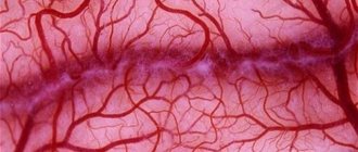
This study allows you to identify the exact location of narrowings and blockages of blood vessels, determine the location of their pathological expansions (aneurysms), the presence of blood clots, atherosclerotic plaques and their prevalence.
Angiography is available to all areas of our body (brain, neck, arms and legs, heart, chest, abdominal cavity), as well as vessels of any size (from the largest - the aorta, to the smallest vessels - capillaries). Unlike duplex scanning, the results of which are largely determined by the quality of ultrasound equipment and the qualifications of the researcher, angiography allows you to obtain the most objective information about the prevalence and severity of vascular disease.
For what conditions is angiography used?
A doctor may recommend angiography as a diagnostic procedure for the following diseases and conditions:
- Atherosclerosis of the branches of the aortic arch (narrowing and blockage of the carotid, vertebral and subclavian arteries, brachiocephalic trunk).
- Aneurysms are pathological dilations of blood vessels, for example, the abdominal aorta.
- Arterial thrombosis and embolism is an acute violation of the patency of the arteries, as a result of their sudden blockage by blood clots.
- Diseases affecting the arteries of internal organs, including the kidneys and intestines.
- Coronary heart disease is a group of diseases that develop due to narrowing or blockage of the arteries of the heart.
- Control of the performed surgical intervention.
- Obliterating atherosclerosis and endarteritis of the vessels of the lower extremities (narrowing and blockage of the arteries of the legs).
- Extravasal compression syndrome is compression of blood vessels from the outside.
- Vascular injury and its consequences.
- Deep vein thrombosis and thrombophlebitis of the superficial veins of the arms and legs.
- PE (pulmonary embolism) is the formation of blood clots in the veins with their subsequent migration with the bloodstream into the arteries of the lungs.
The main diagnostic purpose of the study is to assess the possibility of surgical treatment of the disease, as well as to determine the scope of the planned operation. In some cases, during the diagnostic procedure itself, for example, when angiography of the vessels of the lower extremities is performed, the doctor may also perform therapeutic interventions (see endovascular interventions).
Cost of vascular examination
The price of venography varies depending on the type and method of examination, the contrast agent used, the region and other factors. On average across the country, venography costs 10-15 thousand rubles. Thus, you can undergo an examination of the veins of the lower extremities at a price of 6,000 rubles, CT and MRI - from 5,000 rubles, check the veins of the pelvis from 15,000 rubles.
There are cheaper examination methods, for example, Doppler sonography or duplex scanning of veins. However, they do not provide such accurate data, and if the result is questionable, the doctor may prescribe venography.
Phlebography has a high diagnostic value in identifying many serious pathologies. With the development of medical technologies, images are becoming more accurate and of high quality - this makes it possible to make an accurate diagnosis and determine the appropriate treatment method for the disease, even in the early stages.
How to prepare for this procedure
X-ray contrast angiography is an invasive procedure and involves puncture of one of the large vessels in the leg or arm. After an angiographic examination, medical supervision of the puncture site of the vessel and observation of the patient’s general condition are required. Due to these features, this type of study requires hospitalization of the patient in a hospital (usually no more than 2 days), as well as the implementation of a diagnostic minimum, including the following tests and studies:
- blood group (ABO) and Rh factor,
- study of biochemical blood parameters,
- clinical blood test, general urine test,
- coagulogram,
- blood group and Rh factor,
- examination for viral hepatitis, blood for RW, Form-50,
- FGDS,
- ECG (if indicated by Echocardiography).
Before angiography is performed, the patient should be examined by a physician. The patient may be advised to stop taking certain blood thinning medications a few days before the test (aspirin, Plavix, Chimes, Warfarin). It is recommended to take the last meal no later than 8 hours before the procedure.
The area of the intended puncture of the vessel (groin or axillary area) must be shaved the night before or in the morning on the day of the study, after which it is advisable to take a hygienic shower.
Types of phlebography
Depending on which device is used to study blood vessels, there are several types of venography.
X-ray contrast (classic)
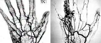
The most commonly used technique. It is carried out using conventional x-rays. The patient is placed on the table, a radiopaque contrast agent is injected intravenously, and a series of photographs are taken (as the contrast spreads through the veins).
During venography of the lower extremities, a tourniquet may be applied to the examined leg to better distribute the contrast.
X-ray contrast venography (venography) - what is it and how does it work? Watch the video:
Ultrasonic
This type of research is used on an equal basis with the previous one due to the speed and safety of its implementation, as well as the equipment of medical institutions. This technique allows you to identify disorders in the early stages and conduct a general assessment of the condition of the veins.
Doppler ultrasound is one of the safest and most effective methods for studying blood vessels and is prescribed to identify blood flow pathologies. We invite you to read articles about ultrasound diagnostics of brachiocephalic arteries and vessels, head and neck, as well as lower extremities.
MRI venography
It is used mainly to capture the veins of the brain. This type of venography is performed without the introduction of radiocontrast, since the tomograph makes it possible to examine the veins of the brain in detail without the use of additional drugs.
MR venography allows you to obtain high-quality, high-precision layer-by-layer images, in which an experienced specialist can notice even a slight deviation from the norm.
CT phlebography
This type of examination uses x-rays, but the information is processed by a computer. After processing, a layer-by-layer image of the area under study is created. Since the image is obtained using X-rays, during CT venography the patient is also injected with contrast . At the end of the examination, the computer produces a three-dimensional image of the vessels, which makes it possible to accurately determine the localization of pathological processes.
If there are contraindications for CT scanning using a contrast agent, the procedure can be performed without contrast. But such images will be less informative and may not provide enough diagnostic information.
How is angiography performed?
The study is performed using a special fluoroscopic machine called an angiograph.
Stage 1
After the patient is delivered to the angiography room, the doctor treats the site of the intended puncture of the vessel with an antiseptic solution based on iodine or ethyl alcohol. After local anesthesia, a percutaneous puncture of the vessel is performed. The patient should notify the doctor in advance if he has allergic reactions to iodine, chlorhexidine, ethyl alcohol, local anesthetics (novocaine, lidocaine) or other medications. Based on the purposes of the study, as well as the prevalence of the disease, the femoral vessels at the level of the inguinal fold or the vessels of the hand in the wrist, in the antecubital fossa or on the shoulder are punctured.
If contrast is injected into the arteries during the study (for example, when angiography of the arteries of the lower extremities is performed), then the procedure is called arteriography, but if the veins are examined - venography.
Stage 2
After installing the introducer (a device that provides constant vascular access during the entire procedure), a conductor (a thin elastic metal probe) is inserted into the vessel through it and advanced directly to the area being examined. A catheter consisting of a plastic tube with an internal lumen is inserted through the guidewire. After the researcher is convinced of the exact location of the catheter, a contrast agent is injected through it, “staining” the vessel.
All manipulations with conductors and catheters are painless and are carried out under X-ray control. During the administration of contrast, the doctor may ask the patient to hold their breath for a few seconds. During vascular contrast, the patient experiences a sensation of warmth or heat in the corresponding part of the body.
Stage 3
After successfully performed angiography, the catheter is removed along with the introducer, and a pressure bandage is applied to the puncture area. The contrast agent introduced into the vessel is excreted from the body through the kidneys within several hours (sometimes up to several days). Angiography lasts on average from 15 to 40 minutes. If therapeutic endovascular interventions are performed, the procedure can last up to 1 hour or more.
Possible complications after angiography
In the vast majority of patients, angiographic examination does not pose any serious danger. The total risk of angiography complications does not exceed 5% and includes the following conditions:
- allergic reactions to antiseptics, local anesthetics or contrast agents used during angiography,
- hematomas in the area of vessel puncture,
- bleeding from the area of vessel puncture,
- contrast-induced nephropathy.
In rare cases, complications such as acute renal failure, myocardial infarction and stroke are possible. Serious complications, as a rule, develop in patients with severe pathology of the relevant organs (kidneys, heart, etc.).
Contraindications
There are not many contraindications to this examination method. Absolute contraindications to x-ray venography are:
- The patient is allergic to iodine - it is included in almost all radiocontrast drugs.
- Acute liver and kidney diseases or exacerbation of chronic ones - this can aggravate the condition of the diseased organ and worsen the general condition of the patient.
- Inflammatory diseases of the veins.
- Thyroid diseases.
Relative contraindications include pregnancy, atherosclerosis of the leg vessels and the patient’s advanced age.
For examination using an MRI machine, the above factors are not contraindications, but in this case the procedure cannot be performed if:
- The patient has metal implants in his body.
- The patient has claustrophobia.
- A patient with a high degree of obesity (weighing over 120 kg).
- The patient is pregnant.
There are almost no restrictions for ultrasound diagnostics , because this method is non-invasive and does not require any preparation from the patient.
A general contraindication for any type of phlebography is the presence of mental disorders in the patient, as there is a risk of inappropriate behavior of the patient.
If the patient has edema, the study is possible, but if the edema is severe, the results may be unreliable.
How to behave after an angiographic examination
After a successful angiography, the patient should spend 6 to 24 hours in a lying position (if puncture of the vessels on the thigh was performed). During this time, he is under observation in the intensive care ward or in the general surgery department under the supervision of on-duty medical personnel. During the specified time, the patient must avoid movements in the hip (or shoulder joint, if access was made through the brachial or axillary artery) on the side of the puncture.
The pressure bandage is removed by a doctor, usually the morning of the next day after the procedure. After removing the bandage, the patient can return to his usual mode of activity, provided he feels well, has normal hemodynamic parameters (pulse, blood pressure) and there are no signs of hematoma in the puncture area. The patient can be discharged from the hospital on the same day.