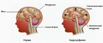
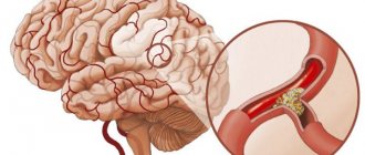
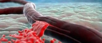
A hematoma is an accumulation of coagulated and liquid blood in a confined space. A hematoma in the head after a blow is formed due to the rupture of a vessel, from which blood escaped into the thickness of the tissue. Depending on its size, a hematoma can compress surrounding tissues, which leads to disruption of its functions.
A hematoma in the head is dangerous because it compresses parts of the brain. This may lead to the death of the victim. Blood accumulation in the head is often treated with surgery rather than conservative treatment.
What it looks like and where it can be located
A child's hematoma usually does not look like a bruise. The color of the skin does not change. Only upon closer examination can small hemorrhages be noticed. In appearance, the hematoma looks like a small ball. This happens due to the accumulation of fluids in the soft tissue layer. When pressed, the ball may roll. Such a hematoma does not form immediately on the child’s head. Liquid accumulates gradually due to poor blood clotting in a newborn baby.
The bruise increases in size within 2-3 days. Its decrease begins only on the 7-10th day. The hematoma completely resolves by the 8th week of the baby’s life.
As for the location, the bruise forms on the cranial bone. It can be located on the side or front of the head, on the back of the head or on the top of the head. The last two options for hematoma localization are the most common.
What is a hematoma and its classification
To understand the classification, symptoms and principles of treatment, it is necessary to understand what a cerebral hematoma is. It is the liquid part of the blood and the clots formed from it, which put pressure on the brain structures (trunk, cerebral hemispheres, cerebellum) and gray matter, causing necrosis and disruption of the central nervous system.
Classification of hemorrhages in relation to the medulla:
- intracranial hematomas (ICH);
- intracerebral hematomas.
By speed of attack:
- acute – has a sudden onset;
- chronic – lasts longer than a week.
By localization:
- frontal lobe;
- temporal area;
- trunk;
- cerebellum.
Types of brain hematomas in relation to the dura mater:
- epidural;
- subdural;
- subarachnoid;
- intracerebral;
- intraventricular.
This classification is of greatest importance for specialists, as it helps to identify symptoms and pathological changes in the body, prescribe the correct treatment and carry out the necessary rehabilitation.
Subdural
This type of hematoma in the head forms in the space between the dura mater and the arachnoid (or arachnoid) membrane. The volume of accumulated blood usually reaches 250-300 ml, significantly increasing the pressure inside the skull. The result of its appearance will be severe disruptions to life, leading to the death of a person in 60% of cases.
A subdural hematoma occurs after a rupture of the vessels supplying the dura mater. The cause of it in 70% is a traumatic brain injury resulting from a traffic accident, a fall from a height, or a blow from a hard object. It has 2 forms, differing in terms of formation:
- Acute – appears in the first 2 days after injury;
- Subacute – symptoms develop after 4 days;
- Chronic – accumulates in the head for a long time (more than 7-10 days).
Acute forms in the absence of adequate treatment (surgery) pose a serious danger to the life of the victim. Chronic hematomas of small sizes are much easier and often go unnoticed due to the lack of visible symptoms. With subdural hemorrhage, a gradual increase in symptoms is characteristic - headache, nausea, vomiting, loss of consciousness. Later, signs of focal damage to the brain substance appear - disruption of the limbs, numbness of a body area, muscle paralysis.
Subarachnoid
Subarachnoid intracerebral hematomas accumulate in the slit-like cavity between the arachnoid membrane and the pia mater of the brain. A distinctive feature of this form is the strong pressure on the nervous tissue. Subarachnoid hemorrhages in 50-70% of cases (according to different authors) lead to permanent disability of a person, in 35-40% - to death, despite adequate treatment.
The cause of this pathology is most often the rupture of a very thinned section of a vessel (aneurysm) or a stroke, as a complication of hypertension. Less commonly, such hemorrhage occurs as a result of injury (most often a fall or blow).
Types of hematoma
There are several types of hematoma on a child’s head. Currently there are:
- Cephalohematoma. Bruising forms between the periosteum and the skull. Such a hematoma occurs infrequently: in approximately 12% of all cases.
- Epidural. A bruise forms between the dura mater and the skull bone. This is a very dangerous and difficult species.
- Intracerebral. With this tissue damage, blood leaks into the brain. In a child, an internal hematoma of the head can cause a serious condition.
- Subdural. With this type of damage, blood gets trapped under the dura mater of the brain.
Causes of intracranial hematomas in newborns
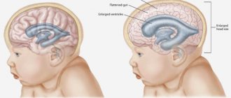
Numerous studies in the field of child neurology and pediatrics have shown that the main cause of the development of intracerebral hematomas in newborns is intrauterine hypoxia and asphyxia during childbirth. It has been proven that numerous hemorrhages in the brain can occur in the absence of any mechanical damage to the head; the presence of hypoxia is sufficient for extensive damage to the fetal vasculature. When there is a lack of oxygen in the baby’s body, serious disturbances in metabolic processes occur, leading to swelling of the brain, changes in the level of intracranial pressure and increased permeability of the vascular wall, which leads to the occurrence of multiple small hemorrhages. When larger vessels are damaged, an intracerebral hematoma is formed.
Additional causes of intracranial hemorrhage are mechanical effects on the fetal head during birth. This phenomenon often occurs in the following cases:
- Discrepancy between the size of the baby's head and the mother's pelvis.
- Pulling the fetus from the birth canal, using traumatic auxiliary devices for this.
- Incorrect insertion of the head into the pelvis.
- Prolonged labor.
- Rapid labor, strong labor activity.
One of the causes of intracerebral hemorrhage in a newborn is rapid labor
The likelihood of a cerebral hematoma in a newborn increases if the child has anatomical and physiological features predisposing to this - thin cranial bones, weak vascular walls, widened sutures between the cranial bones, impaired regulation of vascular tone, an immature blood coagulation system (vitamin K deficiency, hypoprothrombinemia) .
Various intrauterine infections affecting the liver, vascular system and brain of the fetus have a significant impact. Mycoplasma infections are especially dangerous in this regard. An iatrogenic factor (arising as a result of medical intervention) cannot be excluded - improper care of the baby and irrational drug therapy can contribute to the development of a cerebral hematoma.
Symptoms
The presence of a hematoma on a child’s head can be determined by pronounced symptoms, namely:
- Swelling in the area of damaged tissue. When you press it, you can feel the liquid.
- The baby's head becomes asymmetrical. In this case, the place where the hematoma is located has a clear shape due to swelling.
- On examination, minor hemorrhages are noticeable. Outwardly, they resemble small red dots on the damaged area.
- The skin becomes bluish in color. In this case, the child may look unhealthy.
- When pressing on the swollen area, the child experiences pain and begins to cry.
- The baby is lethargic, drowsy, weak, sleeps a lot.
Symptoms
The manifestation of symptoms and first signs of cerebral hematoma will depend on the type of this disease.
The first signs of cephalohematoma appear on the 3rd day after the birth tumor subsides. This is due to the fact that blood slowly accumulates under the periosteum.
Externally, this type of hematoma resembles a lump and is localized in the parietal region. At the same time, the skin remains unchanged. On palpation, the hematoma is elastic, soft, and has a dense cushion at the base.
The child has head asymmetry due to a bump.
On day 20, the skin over the site of the cephalohematoma acquires a yellow tint.
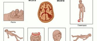
Intracranial hematoma can be identified by the following signs and symptoms:
- drowsiness;
- vomiting or frequent regurgitation;
- there is a noticeable difference in the pathological enlargement of the pupils;
- weakness.
If the amount of blood shed is quite large, then the characteristic symptoms will be convulsions, coma, and lethargic sleep.
Epidural hematomas are very rare in newborns and manifest themselves:
- convulsions;
- mydriasis;
- congestion in the fundus.
Subdural hematomas are characterized by severe headaches, vomiting, bradycardia, rapid breathing, and anisocoria.
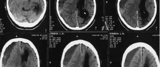
The main methods for studying brain hematomas include:
- General examination of the baby;
- Magnetic resonance imaging;
- Ultrasound diagnostics;
- A blood test is taken for general examinations, as well as to determine the number of platelets.
- EchoEg.
Diagnostics
A hematoma on a child’s head after childbirth is diagnosed by a pediatrician. Several methods are used for this:
- Thorough examination of the newborn baby. The pediatrician can visually inspect the damaged area.
- Blood is drawn for analysis. This allows you to identify the blood clotting factor and establish a hematoma.
- An ultrasound examination is performed. The method allows you to determine the depth and size of the hematoma.
- X-ray. This study makes it possible to identify violations of bone integrity and other damage.
Observation and treatment of birth hematoma on the head of a newborn baby
The bruising subsides gradually on its own. Most often it disappears completely after ten days. It is important to consider the size of the hematoma. Large formations take longer to resolve.
If the child does not show concern and the tumor gradually resolves on its own, then treatment is not applied. In order to exclude the possibility of complications, the child undergoes an ultrasound and sometimes an MRI of the brain.
If the formation does not disperse on its own for a long time, it must be pumped out. If a hematoma is left unattended, it can lead to suppuration and later to ossification. This will lead to deformation of the child's head.
Blood is removed from the hematoma using two needles. One is used to pump out blood, the second to control pressure.
The procedure is simple and does not pose a danger to the child. The effect occurs instantly. After the procedure, the child begins to behave more active and lively.
After removal of the contents of the formation, the child should remain under the supervision of neurologists. If the baby has poor blood clotting, he is given calcium gluconate and vitamin K.
It is important to breastfeed your baby throughout the recovery period. This will help you recover faster.
Birth tumor and hematoma: what is the difference
Often a hematoma on a child’s head is confused with a birth tumor. These are completely different phenomena that have similar characteristics. However, there are obvious differences, including:
- The tumor can be localized on several cranial bones at once, and the hematoma can be localized exclusively on one.
- The tumor neoplasm has a denser structure. During palpation, the liquid inside cannot be felt. This is characteristic of a hematoma.
- Hemorrhage is not typical for tumor formations. Therefore, they dissolve on their own much faster. The hematoma lasts much longer.
Hematoma due to a fall
A hematoma often forms on a child’s head after a fall. In such situations, do not forget that any injury is fraught with serious complications. The bone structures of babies are fragile, and the sutures that connect the bones of the skull are far from perfect. Any injury can lead not only to the development of a hematoma, but also to a fracture, concussion, and also to meningocele. After a child falls, you should immediately see a doctor. In this case, the condition of the baby is of particular importance. It is recommended to carefully examine the bruised area. Warning signs include:
- formation of swelling, edema;
- the skin becomes warmer;
- the child loses consciousness;
- the rate of motor reaction is impaired, which is manifested by restless or inhibited behavior;
- drowsiness and weakness in the baby;
- The newborn constantly spits up milk.
Any injury requires a thorough examination by a doctor and further therapy.
Main reasons
A hematoma on the head of a newborn is a relatively rare occurrence. It occurs in 1-2% of children.
Most often, pathology occurs for two reasons. Firstly, compression of the head occurs during the process of delivery. The baby’s skull bones are quite flexible; they are divided between two fontanelles. This allows the head to be somewhat deformed to fit through the narrow birth canal. The child literally pushes his way through. Therefore, the head is compressed. In most cases, no serious consequences are observed, but sometimes a hematoma forms.
The second reason is the difference in pressure in the womb and in the outside world. The process of birth is a stressful situation for the child’s body. If pressure drops are too sudden or high, the vessels cannot always withstand. This situation is possible with caesarean section and vacuum extraction. In the first case, the child finds himself in the external environment very quickly, and in the second, pressure changes affect him.
A number of factors also contribute to the appearance of hematomas on the head of a newborn:
- large size fruit;
- a narrow pelvis in a woman in labor;
- oligohydramnios;
- weak labor activity;
- short umbilical cord.
The condition of the expectant mother’s body plays an important role in this matter. The likelihood of hematoma formations increases if a woman abused alcohol during pregnancy and did not give up bad habits.
What happens to a hematoma
Once the bruise has formed and enlarged, its growth stops. A hematoma can cause discomfort in a child. Such injuries are often accompanied by pain and can lead to nausea and vomiting.
The bruise may remain unchanged for two weeks. After the specified time, the hematoma begins to decrease in size. At the same time, the child’s well-being begins to improve. The baby is less capricious, eats well and sleeps normally. The hematoma can completely resolve within five days.
What to do
Pediatricians say that a hematoma on the head of a newborn during childbirth is not so rare, and generally does not pose a danger to the child’s health. If the cephalohematoma is small in size, it will resolve without treatment, you just need to ensure maximum rest for the newborn.
Important information: How to quickly get rid of (remove) or cure a bruise on the leg
To clarify its size and exclude other pathologies (bone defects, intracerebral hematomas), an ultrasound, x-ray of the skull bones, and a blood test are prescribed.
The bruise that appears initially grows, then its increase stops. After 1-2 weeks, the tumor begins to decrease in size, the child’s condition returns to normal, excessive drowsiness disappears, and appetite increases. After another week, the hematoma should disappear.
If the bruise is more than 8 cm, then a puncture (puncture) is prescribed with a special needle, through which the liquid is pumped out of the cavity. Then a tight bandage is applied. Children tolerate this procedure well and are quickly transferred to a ward, where they remain under medical supervision for some time. Additional drug treatment may be prescribed to strengthen the capillary walls and improve blood clotting.
If the skin over the cephalohematoma has been damaged, it can become infected and inflamed, and the skin will become red and hot to the touch in the area of the bruise. In this case, a minor surgical operation is indicated as therapy - incision and removal of the hematoma, the use of antibiotics and dressings with disinfectant medicinal solutions.
A large cephalohematoma can be filled with calcium salts, i.e., ossify, subsequently leading to deformation of the skull. An ossified hematoma is treated surgically by excising its calcified fragments and suturing the edges of the wound. The operated child will be under the supervision of a pediatric surgeon and neurologist for up to 1 year.
Care and observation
Within a few weeks, the hematoma should resolve on its own. However, during this period, the baby’s parents must adhere to the following rules:
- You cannot do anything without consulting with specialists. This can only harm the child.
- The condition of the newborn child must be constantly monitored.
- The baby should be examined regularly by a doctor.
- Caring for the child must be carried out with extreme caution. It is not recommended to touch the painful area frequently.
- The baby should eat like a healthy child. You should not allow yourself to overeat.
Treatment
If you discover a cerebral hematoma in an infant, do not panic. It is necessary to find out what type of this disease was detected in the baby. Then follow all the doctor's instructions. And the best thing parents can do is to create conditions for comfort and peace for the baby, even in a hospital room.
After the doctor examines the newborn baby and detects the presence of a hematoma, the baby is sent for additional studies to make an accurate diagnosis and carry out a differential diagnosis. Only after this is treatment prescribed.
Treatment can be surgical (removal of accumulated blood) or medication (in cases of minor blood accumulation). The doctor prescribes vitamins, drugs that improve blood clotting, and antispasmodics. A rehabilitation period is also assigned.
What is the forecast
Doctors do not consider a hematoma on a child’s head that occurs during childbirth to be a very dangerous disease. However, constant monitoring of the baby is required. The swelling should go away on its own. If an operation was performed, then the child’s recovery takes no more than three weeks.
In general, the prognosis is positive. After all, the child’s condition quickly improves, and he becomes completely healthy. There are no problems with this. The main thing is to follow all the recommendations and try not to touch the painful area.
Prevention
All preventive measures can only be taken by the expectant mother.
- Before giving birth, you should take care of a good team of obstetricians who will help the baby be born without any problems. And if complications arise during childbirth, they will provide timely and competent assistance to both the mother and the newborn baby;
- A pregnant woman should try to lead a healthy lifestyle;
- Do not bother yourself with heavy physical activity;
- Use medications with caution.