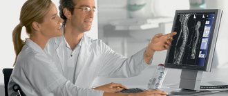
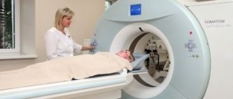
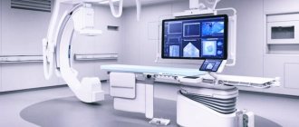
CT angiography of various vessels in St. Petersburg is becoming an increasingly popular appointment for neurologists, vascular surgeons, cardiologists and doctors of other specialties. Often, in order to examine the vessels, ultrasound or X-ray methods are used before CT angiography, but it is the angiography study that is the most informative and indicative, allowing one to accurately identify the cause of vascular disease and begin treatment as soon as possible.
Angiography
|
What is it prescribed for?
Angiography may be prescribed in the following cases:
- Difficulty breathing, shortness of breath and periodic pain in the sternum.
- Chest injuries or previous surgeries in this area. Vascular studies before surgery, if necessary.
- The presence of congenital pathologies, as well as assistance in their diagnosis if they are suspected.
- Prevention and monitoring of patients undergoing drug treatment for diseases of the heart, arteries or veins, if the desired results were not achieved and the symptoms remained or worsened.
Angiography is a fairly broad concept that includes several types:
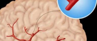
- Cerebral , where the vessels of the brain are examined.
- Phlebography , where the veins of the extremities are examined to study the nature of the blood flow of the veins.
- Fluorescein angiography, where the vessels of the eyeball and adjacent areas are examined.
- Angiopulmonography , where the vascular network of both lungs is examined.
- Thoracic aortography, where the aorta and its branches (heart vessels) are examined.
- Renal arteriography, where the kidneys are examined for the presence of injuries, hematomas and tumors.
Angiography, if carried out in a timely manner, helps prevent a number of diseases, and sometimes detect the presence of blood clots for their timely elimination. Indeed, in such cases, time often passes in a matter of hours.
INDICATIONS FOR PURPOSE OF THE STUDY AND TYPES OF DISEASES THAT CAN BE DIAGNOSED
Catheter angiography is used to study blood vessels in key areas of the body, including a number of organs.
- Brain.
- Neck.
- Heart.
- Breast.
- Belly, for example, kidneys and liver.
- Pelvic organs.
- Limbs.
ANGIOGRAPHY IS USED UNDER A NUMBER OF CONDITIONS AND SUSPECTS, WHEN IT IS NECESSARY TO CARRY OUT CERTAIN ACTIONS.
- Let's say we need to identify anomalies, such as an aneurysm in the aorta , both in the chest and abdomen, or similar pathologies in other large arteries.
- Find atherosclerotic plaques in the carotid artery in the neck, which can restrict blood flow to the brain and cause a stroke.
- Identify small aneurysms or arteriovenous malformations —abnormal connections between blood vessels inside the brain or other parts of the body.
- Detect atherosclerosis , which has narrowed the arteries in the legs, and help prepare for endovascular intervention or surgery.
- Detect disease in the kidney arteries or visualize blood flow to help prepare for a kidney transplant.
- Direct interventional radiologists and surgeons to perform repairs in the affected blood vessels, such as implantation of stents or post-implantation evaluation of the site.
- Detection of damage to one or more arteries in the neck, chest, abdomen, pelvis or extremities in patients after trauma.
- Assess the arteries supplying the tumor before surgery or other procedures such as chemoembolization or selective internal radiation therapy.
- Identify damage or separation in the aorta in the chest or abdominal area - its main branches.
- Show the extent and severity of the consequences of coronary artery disease and organize a plan of action for surgery such as coronary artery bypass grafting and stenting.
- Examine the pulmonary arteries in the lungs to look for pulmonary emboli—blood clots, such as those that travel from leg veins, or pulmonary arteriovenous malformations.
- Conduct investigations for congenital abnormalities in the blood vessels , especially arteries in children, such as malformations in the heart or other vessels due to congenital heart disease.
- Assess other obstructions to blood flow in the vessels.
Types, MR and CT
An invasive method of using angiography consists of introducing contrast (very often iodine is used in its role) and using X-rays the necessary areas of arteries and veins are studied. This angiography is the most accurate and informative.
Although X-ray contrast angiography is considered a somewhat outdated method, it is
actively used in medicine and so far none of the other alternative methods can provide such accurate information as invasive.
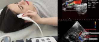
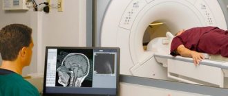
Non-invasive angiography uses ultrasound, computed tomography (CT) and magnetic resonance imaging (MRI) . Of course, such methods of obtaining information also have their advantages.
They help to consider the problem comprehensively, because often vascular diseases are not isolated from other deviations from the norm and pathologies. And sometimes they are signs of more serious diseases. Therefore, non-invasive angiography is a whole complex of studies that makes it possible to determine the influence of many factors on the areas under study.
Indications for angiography and purposes of the study
The main purpose of angiography is to identify anatomical and structural defects of blood vessels. Due to the closed structure of the circulatory system, the contrast agent is distributed throughout the body, which makes it possible to examine every area.
Indications for angiography include disorders of various body functions that may be caused by impaired blood supply. One of the most striking examples is determining the causes of angina, which occurs due to persistent stenosis of the coronary vessels. By obtaining an image “throughout”, it is possible to study the changed area and plan a treatment regimen. Similar goals are pursued when studying the blood supply to the brain. It is especially important to find defects at the site of vessel branching, since this is where pathological changes most often occur. Indirect signs of such disorders are:
- headache;
- disorders of consciousness;
- worsening sleep;
- transient ischemic attacks;
- thrombosis of veins and other vessels.
To conduct the study, a preliminary consultation with the attending physician is necessary, who will carefully analyze the clinical picture in order to exclude unjustified prescription of angiography. Today, the price of such a scan is still high, so doing it if the symptoms are controversial is unwise.
Very often, angiography is prescribed for tertiary syphilis. This is due to the fact that the pathogen has affinity for the vascular wall of the aorta. Over time, it becomes thinner and bulges, which causes an aortic aneurysm. Aortography is a subtype of angiography that allows you to quickly obtain reliable results about the condition of the artery.
The main indications for the study include:
- vascular aneurysms;
- changes in the lumen of arteries or veins;
- vascular malformation;
- violation of vascular patency.
If the goal of the study has not been achieved, then magnetic resonance imaging is prescribed, which has higher accuracy and level of visualization. Thanks to MRI, it is possible to make a reliable diagnosis, but it has a significant drawback - it is extremely expensive compared to angiography.
Classic angiography
Classic angiography is performed in surgical practice when it is necessary to perform operations on blood vessels or internal organs. It is extremely important to timely assess their patency, since otherwise the risk of intra- and postoperative complications increases significantly.
Classic angiography is a simple procedure that can be performed in almost every medical institution, since its implementation does not impose serious requirements on the quality of the equipment used.
Cerebral angiography
The purpose of cerebral angiography is to visualize the vessels supplying the brain and spinal cord. Its difficulty lies in the need to fine-tune the X-ray power. The problem is that bone tissue is capable of absorbing ionizing radiation, so the vessels are not visible on the images.
To obtain accurate results, the use of modern X-ray equipment, high-quality pharmacological X-ray contrast agents, as well as the skill of a radiologist is required. If at least one of the listed factors is not met, then the risk of obtaining biased results increases significantly.
Vertebrology in back treatment
Due to the sedentary lifestyle and weakness of the body of the average modern person, vertebrology is becoming an important subspecies of traumatology. X-rays of the vessels that supply blood to the bone structure of the vertebrae can detect any changes in them.
If narrowing of arteries or veins or a violation of their patency is detected, then during a high-precision operation this defect will be eliminated. This type of study must meet the same requirements as cerebral angiography, since the vessels lie close to bone structures.
What parts of the body are examined?
Most often, angiography is prescribed to examine the following parts of the body:
- Brain . After contrast is administered, X-rays of the head are taken in different projections. The substance is administered twice for a more accurate diagnosis.
- Coronary vessels of the heart . A contrast agent is injected into the femoral or inguinal vein using a catheter. The catheter is advanced to the aorta. After this, contrast is supplied to the left and right coronary arteries alternately.
- Vessels of the extremities . When studying the upper extremities, the substance is injected into the pleural arteries of the left and right arms. To determine the condition of the veins of the lower extremities, contrast is administered either as in the previous case - through the femoral artery, or through the abdominal aorta. X-ray shooting is carried out from several angles and positions.
- Internal organs . A contrast agent is injected into the aorta or into large veins communicating with the organ being examined. Angiography of internal organs is indicated in cases where it is not possible to determine the nature of the disease or there are doubts about the correct location of the vessels.
All studies using radiology and the introduction of a contrast agent are performed under local anesthesia and cause virtually no discomfort to the patient.
Types of angiography
Research activities are divided into several types.
Depending on the area being studied
There are the following types:
- cerebral (brain vessels);
- angiopulmonography (pulmonary vessels);
- portography (portal vein);
- phlebography (renal vein and its trunks);
- coronary angiography (arteries of the heart);
- vertebral (vertebral arteries);
- angiocardiography (heart chambers and main blood vessels);
- carotid (carotid arteries);
- cagraphia (vena cava);
- celiacography (cranial trunk);
- mesentericography (mesenteric artery).
By survey scale
The following varieties are distinguished:
- general (all vessels are examined);
- selective (a catheter injects a contrast agent into specific vessels of a certain organ; the carotid and vertebrobasilar areas of the brain are selectively studied);
- total selective (aimed at examining the capillaries of the pool).
By type of procedure
The following diagnostics exist:
- venography (assesses the functioning of the veins);
- aortography (examines the aorta of organs);
- arteriography (checks the condition of the arteries);
- lymphadenography (assesses the functioning of the lymph nodes).
Indications and contraindications
Angiography is indicated in the following cases:
- Thromboembolism.
- Atherosclerosis.
- Suspected development of cysts or tumors.
- Diseases of internal organs.
- Determination of the presence of diseases of the heart and its blood vessels.
- Diagnosis of retinal pathologies.
- Prevention of complications in the postoperative period.
Contraindications to this research technique:
- The patient is in serious condition.
- The course of any disease in an acute form (acute cardiac, renal, liver failure, etc.).
- Venereal diseases.
- General weakness of blood vessels and their tendency to frequent ruptures and bleeding.
- Tuberculosis.
- Severe mental illness and inability to control the patient.
- Presence of oncological tumors.
- Pregnancy.
Angiography is recommended for those patients who have some difficulties making a diagnosis, but their general condition does not cause serious concern.
Contraindications
There are a number of conditions for which diagnostics are not recommended:
- progressive angina;
- cachexia (depletion of the body);
- tuberculosis;
- third degree hypertension;
- post-infarction state;
- chronic liver and kidney diseases;
- venereal diseases;
- mental disorders;
- pathological anomalies of the cardiovascular system;
- patient's age (too young or mature);
- pregnancy;
- the presence of cancer.
Vascular angiography should not be performed if the patient is inoperable or cannot tolerate the contrast agent.
Preparation rules
Before ordering X-ray studies using contrast, you must:
- Take a general and biochemical blood test to determine the nature of its coagulation.
- If possible, avoid eating food several hours before the procedure (exceptions include diabetics and people with kidney disease).
- Increase the amount of fluid consumed.
- If there is a risk of allergic reactions, antihistamines are used.
- Stopping medications that affect blood clotting.
When performing angiography in children, should be paid to their history of chronic diseases and allergies.
Preliminary preparation
Angiography requires special preparation:
- 2 weeks before the procedure you should not drink alcoholic beverages.
- A week before the procedure, you need to exclude all blood thinners (for example, aspirin) from your medications (if any).
- Five days before the procedure, the patient must undergo additional examinations: fluorography, ultrasound of the heart
, ECG, coagulogram (blood test for clotting), blood biochemistry, blood test for Rh factor, test for HIV, hepatitis, syphilis. - 2 days before angiography, a test is performed to determine the patient’s tolerance to the components of the contrast agent (in particular iodine). If the test shows the presence of an allergic reaction, this is a contraindication and the procedure must be cancelled.
- 1 day before the examination, in the evening, you need to remove hair from the area where the puncture is expected to be made.
- At night (immediately before the procedure) you need to take a sedative. The doctor will recommend which one.
- On the morning of the procedure, the patient is given a cleansing enema for the intestines.
- On the day of the procedure, you should refrain from eating and drinking to avoid nausea and vomiting after contrast is injected into your body.
- Before starting the procedure, you must empty your bladder.
Carrying out the procedure
The angiography algorithm is as follows:
- Introduction of antiallergic drugs.
- Treating the area of the body where the contrast agent will be injected with an antiseptic.
- Administration of a local anesthetic (most often lidocaine is used).
- The skin is incised to provide access to an artery or vein.
- A tube is installed - an introducer.
- A drug is administered that prevents vasospasm (Novocaine is used if there is no allergy to it).
- A catheter is inserted into the hollow tube and moved to the beginning of the vessel being studied (the process is controlled using x-rays).
- A contrast agent is introduced and an image is taken (to obtain more accurate information, the process can be repeated several times).
- Removal of the catheter and introducer.
- Stop bleeding, if any.
- Applying a tight bandage.
To reduce the risk of blood clots and blockages, you must remain in bed for some time after the procedure.
Find out in more detail what it is and what results vascular angiography can achieve in diagnosing diseases from this video:
Research stages
Angiography lasts 30–120 minutes and is performed only in specialized diagnostic centers equipped with modern equipment. During the procedure, a recording is made onto a CD or DVD.
Stages of diagnostics:
- The patient takes a horizontal position on a special angiograph table.
- The point where the puncture will be made is treated with an antiseptic.
- A special probe (2 mm introducer) is installed into the lumen of the artery, which serves as an adapter for other surgical instruments.
- A syringe filled with a contrast agent is attached to the catheter.
- During the administration of the drug, X-ray radiation penetrates into the lumen of the artery through the area being examined. Using rays, a specialist can see the catheter insertion point and the vessels being examined on medical equipment.
- The catheter is brought to the desired vessel and it is filled with a substance.
- The specialist takes pictures and after that all devices are removed from the body.
- The puncture point is sealed with a sterile napkin and pressed closely to the skin to prevent bleeding.
When performing a total selective angiogram (if there is a suspicion of cerebral hemorrhage), targeted diagnostics are performed.
Possible complications after execution
The consequences of angiography include:
- Allergy . Most often occurs due to contrast or anti-clotting drugs.
- Swelling and hematomas . Occur in places of microoperative intervention.
- Bleeding . Since blood thinning substances are introduced into the body, clotting may be low for some time after the procedure.
- Vascular injuries.
- Heart failure . This may occur if the technique of the procedure was violated.
Most complications can be avoided with a detailed examination of the patient's medical history, as well as by following the correct technique. Complications caused by disturbances in the functioning of internal organs must be treated immediately , inpatiently, if symptoms occur in the first hours after angiography.
Rehabilitation treatment and recommendations for patients
The speed of recovery after angiographic studies depends on how extensive they were . General recommendations include:
- Maintaining bed rest and diet.
- No stress or shock.
- Elimination of physical activity during the recovery period, and especially on the limbs, if they were the ones subjected to research.
- Taking antihistamines for preventive purposes.
- See a doctor if you experience discomfort at the catheter insertion site or if your condition suddenly worsens.
Recovery after angiography, if it was performed competently and professionally, lasts several days and, as a rule, does not cause any particular difficulties for patients.
PREPARATION FOR ANGIOGRAPHY AND FEELINGS DURING THE PROCEDURE
Before angiography, the patient needs to remember some conditional points.
You should tell your doctor what medications you are taking and if you have any allergies, especially to iodine, which is part of contrast agents. Also, the specialist must be aware of recent illnesses or other medical conditions that could affect the angiographic examination.
The patient may be asked to remove all or some of their clothes and wear special light clothing. This individual approach is especially often used in private medical clinics. In addition, jewelry, dentures, glasses, and any metal objects will either need to be left at home or removed immediately before the procedure.
Women should always tell their doctor and x-ray technologist if they are pregnant. Many x-ray-based imaging studies are not performed during pregnancy to avoid exposing the fetus to radiation. If x-rays are necessary, precautions will be taken to minimize radiation exposure to the child.
When breastfeeding, during the examination, you need to express milk in advance for subsequent feeding, which will take place after the angiography procedure. This is done for the safety of the child - after using contrast, iodine will be excreted not only in the urine, but also partially penetrate into the milk. Such concentrations may be toxic to the infant.
In some cases, you may be asked not to eat or drink anything for four to eight hours before the procedure. You need to be sure that there are clear instructions from the medical institution in this regard.
If general pain medications were actively used during the study, the patient should not drive for 24 hours after angiography . Therefore, you need to make sure that there is a person present who can arrange delivery to your home. If this is not possible, the patient may be offered a night in hospital.
The research methodology is not particularly difficult for an experienced specialist who has a certain amount of experience.
You will be asked to empty your bladder before starting the procedure. The angiography procedure is usually performed on an outpatient basis. Specialists will need intravenous access.
A small amount of blood must be obtained before the procedure begins to ensure that the kidneys are working and the blood will clot normally. To reduce anxiety and pain during the procedure , a small dose of sedatives may be administered through an IV.
The area of the groin or arm where the catheter will be inserted is shaved, washed, and numbed with local anesthesia. The radiologist will make a small incision (usually a few millimeters) in the skin within the numb area where the catheter will be inserted into the artery. The catheter tube is then passed through the artery into the area to be examined. After which contrast is injected through the same catheter, and several sets of x-rays are taken. The catheter is removed and the incision is closed using gentle pressure for 10-20 minutes. In some cases, a special closing device may be used.
When the examination is completed, you may be asked to wait until the specialists are satisfied that the procedure was normal, then the IV will be turned off. An angiogram can be performed in less than an hour. However, the process may take several hours. When a contrast agent is injected into the vessel, during the procedure it clearly defines the boundaries of the bloodstream, making them bright and white in the pictures.
Patient's feelings
- Under general sedation and local anesthesia, a slight burning sensation will occur at the site where the catheter will be inserted.
- The patient will not feel the catheter moving into the artery. But when the contrast agent is injected, he may feel warmth or a slight burning sensation in the injection area. The most difficult part of the procedure is completed, and the patient can now lie down for several hours. During this time, you should observe and notify the nurse if there is any bleeding, swelling or pain in the area where the catheter entered the skin.
- You can resume a normal diet immediately after the angiographic examination. Active household or professional activities are allowed no earlier than one day after completion of the procedure.
Principles for interpreting results
The principles for interpreting the results are presented in the table below.
| What is observed | What does this indicate? |
| Vessel kennels have intermittent outlines | Thrombosis and atherosclerosis |
| Protrusion of the walls of blood vessels | Congenital changes, aneurysms, thrombosis |
| The contrast does not enter the capillaries, but directly into the vein | Congenital pathologies |
| The contrast agent passes very slowly through the narrowing of blood vessels | Ischemia, inflammation, atherosclerosis |
| Excessive tortuosity of venous arteries and vessels | Heart defect, aneurysm |
| At the beginning of the vessel there is a sharp narrowing | Arteritis, thrombosis |
| Contrast does not apply | Thrombophlebitis, thrombosis, thromboembolism |
| Contrast penetrates into surrounding tissue | Hematoma, traumatic brain injury, aneurysm |
| Narrowing of the lumen between vessels | Arteritis, myocarditis, congenital anomalies |
As can be seen from the evaluation table, often several noticeable deviations from the norm at once indicate the presence of the same disease. To establish a more accurate diagnosis, non-invasive angiography is sometimes used.
Vascular angiography: what it is and how it is done, indications, consequences
You can quite simply describe what angiography means - it is obtaining images of blood vessels. This diagnostic procedure allows you to conduct a full examination of the vessels, determine where they are blocked, identify areas of blood clots, places of narrowing and thinning of the vascular walls. For angiography
a special contrast agent is introduced, which is highlighted during scanning and helps to identify potential or existing pathologies.
Average prices in Russia and abroad
The average cost in Russia for such a procedure ranges from 2,500 to 10,000 rubles . The pricing policy for such studies varies greatly, because the doctor may prescribe any additional studies. Below are the average prices for angiography of various parts of the body.
| Field of study | Price, rub |
| Brain | 2400-8500 |
| Thoracic, abdominal cavity | 2800-3200 |
| Small pelvis | 3100-3600 |
| Soft fabrics | 3200-3600 |
| Lower limbs | 2800-6000 |
| Upper limbs | 2300-5500 |
Prices for angiography abroad largely depend on the equipment and drugs used - sometimes they do not differ at all from Russian ones.
Angiography can be used not only as an independent set of studies, but also as an important part of making an accurate diagnosis . Very often, only with the help of such a procedure can one exclude or identify diseases that could not be identified by other means.
Cost of procedures
Table "Prices for angiographic studies."
| Name | Price, rub |
| MSCT angiography of the heart (CT coronary angiography) without the cost of contrast agent | 12650 |
| MRI of the cervical spine and neck arteries | 6400 |
| MRI of cerebral arteries and veins | 6400 |
| Prices are relevant for three regions: Moscow, Chelyabinsk, Krasnodar. | |
Photo gallery
These images provide angiographic studies.
Renal artery aneurysm
Angiography of cardiac vessels
CT angiography of the lower extremities
MR angiography of the cervical spine
MR angiography of the brain
Angiography of cerebral vessels
