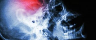
MRI of the head is an informative and modern diagnosis without harm to the body. If you suffer from severe headaches that do not go away over a long period, then medical attention is required. There is no need to delay treatment and endure pain; it is better to do a full MRI of the head or parts of the head as soon as possible. This will help identify the disease and begin effective treatment as soon as possible.
Many are afraid to do an MRI and in vain, because it is safe and absolutely painless .
In what cases is MRI of the brain performed? Types of research.
Typically, patients are referred for brain MRI by neurologists and neurosurgeons. You can, of course, sign up for the study yourself, but if you are bothered by certain symptoms, it is better to first visit a doctor, he will prescribe the necessary types of diagnostics.
Magnetic resonance imaging helps diagnose many diseases of the central nervous system:
- hydrocephalus (“dropsy of the brain”);
- various developmental anomalies;
- infectious processes;
- causes of seizures in patients with epilepsy;
- tumors of the brain and spinal cord;
- intracranial hemorrhages;
- stroke;
- causes of visual and hearing impairment;
- multiple sclerosis, Alzheimer's disease, Parkinson's disease;
- arterial aneurysms, arterial occlusions (blockages), venous thrombosis.
Get a consultation with a doctor
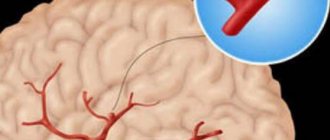
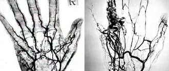
MRI of the cerebral arteries is performed with contrast. A special drug is injected intravenously, which will “paint” the blood vessels and allow them to be clearly outlined in the photographs.
Using functional magnetic resonance imaging, you can evaluate the activity of different areas of the brain. This helps to obtain valuable information in a number of pathologies, such as neurodegenerative diseases.
Result of MRI
Basically, the result of an MRI either confirms or refutes a certain diagnosis. Therefore, it is recommended to be prepared in advance for any diagnostic outcome. In any case, only knowing the name of the disease can you fight it. Therefore, even if the result seems negative to you, do not be upset or fall into despair. A correct diagnosis is half the way to recovery.
The MRI result is usually interpreted by the attending physician. But if you want to hear several points of view (this is true for complex or rare diseases), nothing prevents you from consulting and providing diagnostic results to several specialists.
CT or MRI?
Both studies work approximately the same way and create highly informative images with layer-by-layer sections of a certain area of the body, three-dimensional images. But during a CT scan, X-rays are used, and during an MRI, a strong magnetic field is used.
Computed tomography better visualizes the bones of the skull and vertebrae, and intracranial hemorrhages. In addition, it is cheaper and simpler than MRI, so it is more preferable for quickly examining people who have suffered a traumatic brain injury or a stroke.
MRI images clearly show ligaments and tendons, nerve fibers, and tissue of the brain and spinal cord. This study is better suited than CT for diagnosing benign and malignant neoplasms of the nervous system.
Come to an appointment with specialist doctors at the Medicine 24/7 clinic, they will assess your situation and tell you whether you need to do an MRI of the head and cerebral vessels, or undergo other diagnostic procedures.
We will call you back
Leave your phone number
Contraindications
You cannot do an MRI of the head if you have the following contraindications:
- the presence of electronic devices and metal objects in the patient’s body (pacemaker, hearing aid, etc.);
- first trimester of pregnancy;
- mental illnesses that may interfere with the study.
A contrast study should not be done in the following cases:
- chronic renal failure;
- pregnancy;
- individual intolerance to contrast media.
How to prepare for the procedure?
Magnetic resonance imaging does not require special preparation. Some clinics give patients advice on what to eat the night before and on the day of the test. If your doctor does not advise you to do so, you can eat and take your medications as usual.
Before the procedure at the Medicine 24/7 clinic, you will be consulted by a doctor. If you suffer from any diseases, allergies, or have recently undergone surgery, you must inform your doctor about this. If a patient has kidney problems, blood tests may be needed to evaluate kidney function. In some cases, radiography may be required to check whether there are metal implants or foreign bodies in the patient's body.
When going for the procedure, you do not need to wear any metal jewelry - they will still have to be removed before the study, because a strong magnetic field is used during MRI. Do not wear clothes with metal buttons or fasteners.
What does it show?
The performance of the brain greatly affects the entire body. In the presence of any disease in the brain, the functioning of internal organs and the body as a whole is disrupted. The procedure diagnoses the following pathologies:
- Inflammatory processes;
- Infections;
- Various head injuries;
- Concussion;
- Cyst in the head;
- Tumor (malignant and benign);
- Thrombosis;
- Dizziness of unknown nature;
- Stroke;
- Anomaly in the development of structures and the vascular system.
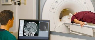
How is an MRI of the brain performed?
If an MRI of the brain is being performed without contrast, you will immediately be asked to go into the office and lie down inside the machine. If contrast is required, you will first be given gadolinium intravenously.
You must lie still during the examination to ensure clear images. The diagnostic table moves, while the device takes a series of images. The doctor is in the next room and communicates with the patient through a microphone.
How long does a brain MRI take? On average, the study lasts 45 minutes, the total time depends on the need to use contrast and perform a functional MRI.
How they do it
The patient enters the treatment room in loose clothing, as they should not cause discomfort to the person being examined. In some diagnostic centers it is provided individually to everyone.
The subject lies down on a movable table, where the head is secured with special straps to prevent the patient from moving, as this is prohibited in order to obtain the most accurate information. The table slides inside the ring, which begins to rotate. Every 1-2 millimeters (depending on the device), the tomograph makes sections.
During diagnosis, the patient may experience side effects: nausea, dizziness and an unpleasant taste in the mouth. The examination lasts from 10 to 40 minutes.
How safe is a head MRI? Are there any contraindications?
Magnetic resonance imaging does not use X-rays, so it is safer than x-rays and computed tomography. However, there are some contraindications.
First of all, this is the presence of any metal objects in the patient’s body, such as pacemakers, defibrillators-cardioverters, metal clips on blood vessels, embolic coils, some types of cochlear implants, and foreign bodies.
There have been many studies on the safety of MRI during pregnancy. No risks were found, however, in pregnant women this study is carried out with caution and only in cases where it cannot be avoided. Magnetic resonance imaging is especially not recommended in the first trimester, when the organs of the unborn child are forming.
During an MRI of the brain, only the head is inside the machine, however, difficulties may arise for people suffering from claustrophobia.
The procedure is problematic if the patient suffers from a mental disorder and is in a state of agitation and cannot lie still. Most devices are not designed for people weighing more than 150 kg.
The main contraindications for MRI with contrast are the patient's allergy to the drug and severe renal impairment.
Take care of yourself, book an MRI of your brain now
Contraindications for use
There are relative and absolute contraindications. In the first case, conducting an examination is undesirable, but its help is resorted to in case of vital necessity. If the patient has absolute contraindications, then the use of diagnostics is strictly prohibited. Absolute contraindications include cases when the patient has:
- neuro- or pacemaker;
- electronic or ferromagnetic implant in the middle ear;
- a prosthesis or cochlear implant in the inner ear;
- metal implants, ferromagnetic fragments;
- insulin pump;
- heart valve prosthesis;
- Ilizarov apparatus.
Relative contraindications include the following:
- a clip that was installed due to removal of the gallbladder;
- tremors and conditions when a person is unable to hold his breath for a long time;
- pregnancy period;
- fixed braces and dentures, as well as stents and vena cava filters;
- heart failure;
- claustrophobia;
- coronary artery bypass grafting;
- pain that does not allow a person to remain motionless for a long time.
How much does a brain MRI cost?
Prices for MRI of cerebral vessels in Moscow vary, depending on the pricing policy of a particular clinic, the model of the device (more modern and high-precision ones are usually more expensive), and related medical services (consultation with a doctor, blood tests, radiography). In our Medicine 24/7 clinic, you can conduct a routine examination and MRI of the brain with contrast in Moscow using one of the latest generations of equipment and get a consultation from an experienced doctor cheaper than in many other medical centers.
For more information, to schedule an initial consultation and to undergo a head MRI at our clinic, please contact us by phone
Preparation
Preparing for a CT scan of the brain does not require much effort: it is enough not to drink or eat for 2-4 hours before the procedure, since it is performed on an empty stomach.
Immediately before the examination, it is important to remove all metal objects and jewelry from the patient, as they may interfere with the diagnostic process.
If the study is carried out with contrast, it is important to warn the doctor about any allergic reactions, especially to iodine. A contrast agent is injected intravenously for clearer visualization. If you want to clarify the condition of the blood vessels, you can undergo a CT scan of the brain vessels separately.
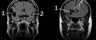
Decoding the received data
Decoding of the results is carried out immediately after the study and takes about half an hour. To accurately determine where to get an MRI of the brain, you should check the availability of licenses and other permits of the selected diagnostic center. After the examination, the decrypted results are given to the patient or transferred to his attending physician.
The transcript of the image contains information about:
- the amount of fluid in the spinal cord canal;
- blood flow speed;
- activity of the cerebral cortex when exposed to various stimuli;
- level of tissue diffusion.
MRI of the brain is a painless and harmless procedure that cannot cause headaches or other ailments in patients. However, with the help of this type of diagnosis, the doctor can get a clear clinical picture, determine the cause of pain and establish the correct diagnosis.