Indications for examination
Among the indications:
- stenosis (narrowing);
- occlusion (blockage);
- atherosclerosis;
- cardiovascular pathologies;
- aneurysms;
- thrombosis of blood vessels in the legs;
- retinal diseases;
- study of liver function;
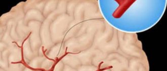
- identification of tumors, blood clots, fusions of arteries and veins in the brain;
- surgery on the heart or brain is planned;
- determination of the extent of vascular damage during wounds and traumas.
Traumatic brain injury
If there is a risk of vascular damage due to traumatic brain injury, MR angiography is prescribed. A modern research method makes it possible to diagnose false aneurysms and disorders in patients with gunshot wounds in the first few days after resuscitation. The examination is carried out to exclude dissection and damage to the carotid and vertebral arteries.
Atherosclerosis, inflammation of the walls, pathological vasodilatation
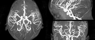
Using this method, you can determine the slightest disturbances in the structure of the walls of blood vessels. A visualization of the arterial circle of the brain is created, obtained by exposing the examination area to radio waves in an external magnetic field. Angiograms clearly show the posterior, anterior, middle and carotid cerebral arteries, as well as the basilar artery. MR angiography of intracranial arteries allows you to quickly see disturbances in vascular architecture, as well as problems with blood flow caused by atherosclerosis, tumors, and aneurysms. At the same time, an examination of the arteries of the neck can be carried out. The procedure is aimed at identifying pathology in the structure of the vascular walls. MR cerebral angiography of cerebral vessels provides a complete picture of blood flow functions.
Aortic dissection
The technique makes it possible to determine the exact location of the dissection and where the blood flow is changed. A contraindication to diagnosis is the presence of a metal valve in the patient.
External vessel compression syndrome
Magnetic resonance, or MRI, angiography is prescribed for external vessel compression syndrome. In a diagnostic center, a contrast agent is injected into the patient's artery and three-dimensional images of the vessels that have undergone deformation are taken.
Congenital heart defect
Angiography is the main method for studying patients with congenital heart defects. During the procedure, the pressure gradient and oxygen saturation of the blood are measured. As a result, flows, resistances and shunts within the heart are calculated, and the function and anatomy of this organ is accurately assessed.
Principle of CT angiography of the brachiocephalic arteries
A computer multislice tomograph consists of a conveyor table, a working unit (gantry) and equipment that allows you to process and visualize the examination results.
The gantry contains a beam tube that emits a fan-shaped stream of RG rays and a block of capture sensors. Radiation passing through the human body is partially absorbed by tissues. At the same time, different organs weaken the intensity of the photon flux to varying degrees. Sensors record changes and transmit information to the computer processor. Vascular examination is carried out with mandatory X-ray contrast enhancement. An iodine-containing drug is administered intravenously, which makes it possible to separate the arteries in the images from the surrounding tissues and study the level of blood flow in them. Contrast is administered directly during the procedure. Its volume is calculated in accordance with the patient’s body weight.
Multislice tomography allows you to create a three-dimensional picture of the vasculature based on several thousand two-dimensional sections, the thickness of which is 0.5-0.7 mm. This is enough to detect small pathological foci, which is often found in pathologies of the cardiovascular system. Another significant advantage is visualization of not only the lumen, but also the vessel wall. The effectiveness of MSCT significantly exceeds that of X-ray contrast angiography and, especially, Dopplerography. Only MRI of the vascular apparatus can be considered a similar method in terms of effectiveness.
Methodology and preliminary preparation
The technique is based on the influence of a magnetic field and electromagnetic waves. As a result of the study, multidimensional images of the vessels of a certain area are obtained. Particularly valuable is the diagnosis of blood vessels in the neck and brain. The duration of MR angiography is 30-40 minutes.
The procedure can be performed with or without the administration of a contrast agent. Before angiography, an allergy test for a contrast agent is performed. It is not recommended to eat food 8 hours before the procedure. The doctor must be warned in advance about taking medications. Before the procedure, you must remove all metal-containing products.
Among the preparatory activities:
- eliminate alcohol 15 days before the procedure;
- the woman needs to make sure that she is not pregnant;
- take blood clotting tests;
- do an ultrasound of the heart;
- do an enema.
The study is carried out with an empty bladder.
What do the pictures show?
The described modern study of cerebral vessels can determine a huge number of different pathologies in their functioning and structure. Based on the results of the diagnostics, the following health problems can be detected:
- Dangerous pre-stroke condition of a person, its course and possible consequences.
- Atherosclerotic plaques can also be examined with an MRI device. If the entire circulatory system was subject to various pathological changes, if its size is very small.
- Thrombosis of the arteries of the head and neck.
- Complications that occur after a bruise, concussion or after a traumatic open craniocerebral injury.
- Hematomas in the brain, reflecting in 3D.
- Brain contusion.
- Various tumors of a malignant or benign nature.
The images show vascular thrombosis and vasculitis.
Magnetic resonance scanning is available even when diagnosing the most minor malforation. It is also possible to recognize the development of such problems and aneurysms without any problems. This is the main advantageous feature of modern resonant contrast.
To work with tumors, to exclude or, conversely, confirm them, contrast is used. This is very convenient and effective if the formation is hidden behind the arteries. At the same time, contrast makes it possible to determine the presence of metastases. A specialist can determine their location and size as accurately as possible. In some cases, it is possible to most accurately suggest the histological origin of the pathology.
The patient's condition after the procedure and possible complications
Bed rest is recommended during the day. The attending physician monitors the patient’s general condition (examination of the intervention area, temperature measurement). If the person’s condition is satisfactory, the bandage is removed on the second day and sent home. The risk of complications is minimal. For most people, the study is not dangerous.
Possible problems:
- allergy to contrast agent, anesthesia;
- in the presence of chronic diseases there is a risk of acute renal failure and myocardial infarction;
- bleeding from the place where the puncture was made.
What will MR angiography show?
MRI of arteries, sinuses and veins reflects a wide range of vascular abnormalities. Tomography in angio mode is capable of well visualizing:
- arteriovenous malformations;
- stenosis of the vascular bed
- thrombosis;
- vascular occlusion;
- bifurcations and kinks of blood vessels;
- vascular parietal tumors;
- acute circulatory disorder.
MR angiography allows you to find the cause of swelling of the legs and distinguish varicose ulcers from thrombotic ulcers. A non-invasive method for diagnosing blood vessels in medical centers in St. Petersburg is used quite often for planning surgical interventions.
Features of angiography of various organs
The method allows you to study vessels of any size (from the smallest capillaries to the aorta), organs and systems of the body. The procedure is carried out before surgery.
Angiography of cerebral vessels
This is the most common way to diagnose disorders in the blood supply to the brain. Visualization helps to identify pathological processes, cysts, tumors, and micro-stroke.
Cerebral MR angiography of blood vessels is prescribed for:
- dizziness and nausea for unknown reasons;
- prolonged headache that cannot be relieved with medication;
- preparation for neurosurgery on the brain.
Angiography of the heart
Indications for the procedure are:
- angina pectoris in a progressive stage;
- myocardial infarction;
- heart rhythm disturbances.
Angiography of the lower extremities
Diseases of the veins and arteries of the lower extremities are widespread. MR angiography is performed if the following diseases are present or suspected:
- thrombophlebitis;
- thrombosis;
- endarteritis;
- atherosclerosis.
Angiography of the neck
To assess the condition of the vascular stack, localize disorders and clarify the diagnosis, neck angiography is prescribed. The procedure is indicated for bruises and injuries, headaches, dizziness, disturbances of consciousness and sleep. With its help, Takayasu's disease, stenosis and developmental anomalies, vascular neoplasms, compression of blood vessels by scars and tumors are determined.
3. How is the procedure of MR angiography of the cerebral arteries performed?
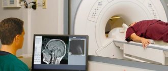
When going for diagnostics using an MRI scanner, you need to expect that the procedure will take a significant amount of time - from 30 minutes to 1.5 hours.
Special preparation for the study is not required, however, the doctor must warn the patient about the need to remove all metal objects, including dentures, and also asks to inform about the presence of implants in the body, their location, volume, and the material from which they are made.
The patient is placed horizontally on the surface of the tomograph, after which the head is fixed with a special helmet. For the accuracy of the diagnosis, any head movements must be excluded.
If the patient is prone to claustrophobia, he will be asked to take sedatives.
The operation of the tomograph is accompanied by rather sharp sounds, so special protective headphones are put on the ears of the person being examined. The scanning unit makes rotational movements around the patient's head and transmits a three-dimensional image to the monitor. The doctor deciphers the data received and draws conclusions about the state of the patient’s vascular system of the brain.
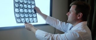
The results of MRI diagnostics are given to the patient in the form of a report, which may contain a printed scan of the image or a disk with files.
About our clinic Chistye Prudy metro station Medintercom page!
Contraindications for the procedure
- Renal, liver and heart failure during the period of decompensation.
- Poor blood clotting.
- Pregnancy and lactation period.
- A number of mental illnesses.
Important!
MRI technology does not allow simultaneous visualization of arteries and veins. Therefore, conventionally called MR angiography consists of two different methods:
- MR venography;
- MR arteriography.
MR venography
– is a diagnosis of sinuses and intracranial veins. No contrast is applied. Can be performed independently or in combination with MRI of the brain. MR venography allows you to identify aneurysms, vascular malformations, thrombosis, various developmental and localization anomalies, tumor formations growing into blood vessels, etc.
MR arteriography
is a visualization of the arteries, with which you can identify blockages, their location, and the degree of threat to health and life. Allows you to identify hemorrhage, thrombosis, vascular spasms, etc. This technique is especially important because it allows you to control the treatment of those patients who are at very high risk.
What is magnetic resonance angiography
MR angiography of blood vessels is a technology that involves scanning the circulatory network of the brain with the rays of a magnetic resonance imaging scanner, with the introduction of a contrast agent into the blood. It is carried out on tomographs with a magnetic field whose intensity is higher than 0.5 Tesla (Tesla is a unit of measurement of magnetic induction).
MRI of the brain with angiographic mode allows you to visualize a detailed three-dimensional image of the circulatory system of the brain, as well as the projection of each individual vessel, which allows you to identify even the smallest anomalies. Based on the examination results, the patient is given an accurate diagnosis and appropriate treatment is prescribed.
At the end of the procedure, the patient is given images, a decrypted specialist’s report, and, if necessary, it is possible to write the information to disk.
Important! Using the properties of a magnetic field instead of X-rays avoids negative effects on the health of the subject, and makes angiography a safe alternative to radiography.
Preparation for MRI of cerebral vessels
The study does not require special preparation. You should notify your doctor in advance about the presence of:
- claustrophobia;
- allergies to drugs;
- chronic pathologies;
- metal implants in the body.
Before entering the diagnostic room, you must remove all jewelry and metal accessories and leave your mobile phone outside the door.
You can download the document for review: MRI studies (general preparation rules)