Quite often, with ophthalmological diseases, patients are faced with the appearance of a veil in the eyes and blurred images when focusing vision. This phenomenon interferes with the usual rhythm of life. When a veil appears in the eyes, not only the clarity, but also the clarity of the image is disrupted. Sometimes the veil in the eyes resembles a translucent film, which greatly interferes with looking at objects. With some pathologies, a person may experience a so-called red veil before the eyes, which is accompanied by a distortion of color rendition. However, any manifestation of blurriness indicates the onset of the development of a serious ophthalmological disease.
Causes
Fog in the eyes can appear for many reasons. This can be either an ophthalmological disease or a consequence of systemic internal disorders or simple fatigue. Next, we will look at the key factors influencing the appearance of this symptom. Why double vision is described here.
Physiological
Blurred vision has two main causes. It can be:
- a sharp change in blood pressure. In the case when blood pressure changes sharply, the clarity of the image may blur until complete temporary darkening in the eyes. At first glance, this can happen if you make sudden movements, for example, suddenly sit down or, on the contrary, stand up. Also, a person’s blood pressure depends on weather conditions and a sharp change in the clarity of vision can be explained by a sudden change in weather;
- Low serum glucose levels. Low blood glucose levels are primarily a negative result from excessive physical exertion or poor working conditions (professional activity). In addition, professional athletes often have a blurry look. In such a situation, the patient is recommended to take a short break from work, if possible, and stop overloading the body for a while. It is worth noting that none of these reasons threaten a person’s life.
The most important thing is to seek help from an ophthalmologist if odd vision occurs.
Pathologically
There are quite a few reasons for the occurrence of pathological blurred vision. Eye diseases that cause the appearance of veils and blurriness are:
- Destruction of the vitreous body is a pathological condition that develops as a result of structural disorders of the vitreous body, thereby provoking compaction and its destruction. The vitreous body is one of the main components of the dioptric system of the visual organs. It is this system in the structure of the eyes that provides a continuous flow of light rays to the retina. You can learn about the causes of retinal detachment in this material.
The prognosis for this disease is favorable, but in advanced cases partial loss of vision is possible.
- Refractive errors of the eyes. This pathological condition includes progressive myopia and farsightedness in children and adults. The sudden appearance of a veil in such situations is primarily associated with a violation of the focus of the visual system. But when wearing contact lenses or glasses, the veil disappears. Find out about false myopia by following the link.
- Presbyopia. This disease can be attributed to age-related changes. In people over 40 years of age, vision deteriorates when viewing images closely. The pathology develops as a result of a violation of the optical system of the eye lens. The first signs of vision loss pathology are observed when reading books or newspapers. Treatment consists of spectacle therapy. Preventive measures for farsightedness are collected here.
- Dry eye syndrome. Dry eyes are often the cause of white puffiness before the eyes. The cornea of the eye, deprived of moisture, begins to dry out, resulting in fogging of the endothelium of the cornea - this leads to blurred vision. Regular use of moisturizing eye medications will solve this problem.
- Optic neuritis. In other words, this is an inflammatory process of the main nerve of the eyes, which leads to a sharp decrease in visual acuity and the appearance of blurred vision. This pathology is temporary and with effective therapy the veil goes away.
Causes of double vision
Temporary episodes of double vision can occur for a variety of reasons, including drinking too much alcohol, being overly tired, or chronic lack of sleep. These causes of short-term diplopia are usually not a cause for concern.
If symptoms persist for a long time or continue to return, causes may include:
- stroke;
- head injury;
- corneal unevenness;
- cataract;
- eye surgery;
- dry eyes;
- damage to the cranial nerves;
- brain tumor or cerebral aneurysm.
A head injury, tumor, stroke or related condition can cause sudden onset of double vision. After reviewing the process, the optometrist refers the patient to a specialist, such as a neurologist or neurosurgeon, for further testing and treatment.
Corneal irregularities
Double vision is caused by factors such as keratoconus (cone-shaped cornea) and corneal dystrophy (deterioration of structure, thinning). Twins are very difficult to manage. Corneal irregularities can be corrected by wearing special contact lenses. Some people with this condition may require surgery - corneal transplantation or implants.
Dry eyes
This is the next cause of double vision. Tired, dry eyes, such as those with Sjögren's syndrome, can cause double vision due to insufficient tears. Many people suffering from this pathology of dry eyes can benefit from moisturizing eye drops and eye vitamins.
Refractive surgery
Any manipulation intended to improve vision may lead to symptoms due to changes in the curvature of the cornea. This causes the light rays to scatter instead of focusing correctly. The problem usually resolves within a few weeks or months as the eye recovers from surgery. In this case, you may need to use eye drops for some time. In some cases, a second laser vision correction procedure is recommended.
Cataract
Cataracts can cause double vision, but this is usually observed in only one eye. This is because clouding of the lens, which sits behind the pupil, can cause light rays to scatter in different directions, creating multiple incomplete images, especially when a person looks into the light. Cataract surgery usually corrects this problem.
Cranial nerve damage
Double vision may be caused by paralysis or partial dysfunction of one or more of the muscles that control the position and interaction of the eyes. The disease is caused by:
- diabetes;
- head injury;
- tumor;
- multiple sclerosis;
- meningitis;
- high blood pressure;
- blockage of an artery or aneurysm.
The condition requires seeing a doctor and treatment. In addition to conservative therapy, surgical methods may be used and special glasses may be prescribed.
Fluoroquinolones
In addition to the above reasons, fluoroquinolones are not the last factor affecting the eyes. They can cause double vision. The researchers analyzed 171 cases involving systemic fluoroquinolones (antibiotics used to treat bronchitis, pneumonia, sinusitis and other illnesses caused by infections occurring between 1986 and 2009. In 53 patients, the antibiotic regimen was stopped, and that's when the dichotomy went away. One of the side effects The effects of fluoroquinolones is tendon dysfunction, which, when occurring in the muscles around the eyes, can cause this problem.
Instead of seeing a double image clearly, a person may only notice a partial or ghostly, blurry outline around or to the side of what they are looking at (especially if it is a light source). Double vision with cataracts is usually seen in one eye. Silhouettes around objects may occur after corneal transplant surgery. Some of these problems disappear when the problem is resolved, such as after cataract surgery.
Strabismus
When two eyes move and focus correctly and accurately at the same time, we see only one image. If the two eyes focus differently, double vision may occur. Some people are born with eyes that don't work together, a condition called strabismus. Organs can face inward or face outward. One eye is even able to rise and the other to fall.
If a person has strabismus, he sees two objects instead of one because each eye projects different images at the same time. The brain usually adapts by closing or ignoring information from one eye. This is called suppression. Surgery or vision therapy helps many people with strabismus overcome double vision.
Loss of vision during pregnancy
A decrease in the percentage of vision during pregnancy is observed in 40% of women. Deterioration of vision is characterized by sudden physiological and hormonal disruptions in the body of the expectant mother. As a result, the elasticity of all organs, including the eyes, decreases.
Why does vision deteriorate during pregnancy?
- The fetus consumes all the beneficial vitamins and nutrients that a woman consumes.
- Structural changes in the cornea of the eyes, due to severe swelling and fluid retention in the body.
- Increased intraocular pressure (only during contractions).
- Chronic eye diseases, which are accompanied by impaired visual acuity of a woman.
If there is a sharp drop in vision during pregnancy, you should immediately consult an ophthalmologist.
Impaired removal of excess fluid from the body is a primary problem, which causes visual impairment. Since the cornea of the eyes is oversaturated with moisture, its shape thereby changes.
Neurological diseases
The mechanism of vision is carried out with the help of certain nerve fibers included in the autonomic nervous system, and is controlled and regulated by neurons of the brain centers belonging to the central nervous system. Disturbances in the functioning of the central nervous system and peripheral system can affect visual acuity and the ability to see in general. Disruption of cerebral blood supply due to ischemia, stroke, aneurysm, atherosclerosis leads to clouding of the eyes.
Thus, blurred vision may be a symptom of:
- poisoning;
- traumatic brain injury;
- vegetative-vascular dystonia;
- cerebral atherosclerosis;
- aneurysms;
- hypertensive crisis;
- diabetes mellitus
Also, the reason that the image in the eyes has become cloudy and blurry can be a deficiency of vitamins, especially vitamin A (necessary for nourishing the tissues of the eye) and group B (necessary for the normal functioning and good conductivity of nerve fibers, including the eye), lack of nutrients substances.
Blurred vision sometimes indicates the onset of a dangerous disease such as multiple sclerosis. This symptom appears earlier than others and indicates damage to the nerve fibers of the eye. The patient experiences changes in visual acuity throughout the day and limited visual field.
Blurred vision and loss of consciousness can be caused by prolonged fasting or an overly strict diet. In this state, the body experiences a severe deficiency of nutrients, glucose and vitamins. This leads to the fact that the energy resource of the central nervous system is greatly limited, and vision decreases. The longer this condition lasts, the more dangerous the processes occurring in the body that lead to visual impairment.
Long-term starvation, vitamin deficiency and nutritional deficiency lead to the fact that degenerative processes become irreversible.
Diagnostics
Only an ophthalmologist can diagnose the exact cause of the appearance of a veil before the eyes. The main methods for diagnosing blurred vision are:
- visual inspection of the affected area using a slit lamp;
- tonometry – determination of intraocular pressure indicators;
- fundus examination;
- Ultrasound examination of the eyeballs.
In the absence of pathological signs in the visual organs, the ophthalmologist refers the patient to a neurologist to determine diseases of the nervous system.
Treatment
Spectacle therapy
When choosing a method of vision correction - glasses or contact lenses, most people choose the most accessible and simple method - glasses.
But in fact, spectacle therapy is used very often, since it is an effective and affordable method that has practically no contraindications. Techniques for producing eyeglass lenses are being modernized, and the way they are selected is being improved and detailed. Benefits of eyeglass therapy:
- convenient and practical method of use that does not require any skills;
- allowed to use with painted eyelashes;
- lack of full contact with the surface of the eyeball;
- glasses can be used for a long time without regular changes;
- You can choose the color of the frame and lenses to your liking.
But there are also disadvantages of eyeglass therapy:
- glasses may not suit every person, as a result of which the appearance may not be satisfactory;
- It is impossible to lead an active lifestyle with glasses;
- some restrictions on the use of sunglasses;
- You should always carry glasses with you;
- if chosen incorrectly, vision problems can only get worse;
- appearance of image distortions when removing glasses;
- choosing glasses is quite difficult when vision is more than 2 diopters;
- Stylish and modern glasses are not affordable for everyone.
Astigmatism
Astigmatism is a refractive error that causes distorted images to be projected on the retina, affecting near and distance vision.
Astigmatism can occur alone or in combination with myopia or hypermetropia and is usually stable throughout life.
Main symptoms:
- Getting distorted images (most common symptom)
- Problems with changing distance/near vision.
- Difficulty viewing small details, both up close and at a distance.
- Headaches, eye pain, or dizziness as a result of muscular effort as the eye tries to compensate for the defect by accommodating the lens (the eye's natural lens, whose elasticity allows focusing). This especially happens in cases of a combination of astigmatism and hypermetropia.
For more information about this pathology, click on this link.
How to proceed?
- Astigmatism can be corrected with glasses or contact lenses.
- If the patient wants to do without optical correction, there are surgical treatment methods.
- Refractive surgery includes various methods, depending on the specifics of each diagnosis: Excimer laser: used to target the middle layers of corneal tissue.
- Incisional technique (arcuate keratotomy): consists of making incisions on the surface of the cornea and is used for high-degree astigmatism.
- Toric intraocular lenses: phakic (implanted between the cornea and the lens) and pseudophakic (replace the lens). Widely used to correct high degree astigmatism.
Contact lenses
Eye lenses are a modern, painless vision correction. Therefore, young people most often choose contact lenses. The lenses are completely invisible to the eyes, there is a choice.
Benefits of contact lenses:
- the quality of vision in lenses does not change during sudden temperature changes and precipitation;
- there is an opportunity to engage in active sports;
- excellent vision correction for anisometropia even more than 2 diopters;
- you can wear sunglasses;
- the use of disposable lenses will not cause even the slightest harm to the organ of vision and will not provoke the development of inflammatory processes.
Disadvantages of contact lenses:
- When performing water procedures, lenses must be removed to prevent the development of infection;
- If the rules of hygiene and use of optics are not followed, complications may develop, including complete loss of vision.
Laser vision correction
Today there are many ways to correct vision using laser. These primarily include:
- Photorefractive keratectomy is the most effective and oldest method of laser vision correction, which is used in the first stages of eye diseases. During the operation, the top layer of the cornea is removed with further smoothing of structural changes. The recovery course after surgery lasts from 4 to 5 days. Is laser correction of farsightedness effective in the article?
- Subepithelial keratomileusis - this technique is used for a thinned cornea of the eye. The operation involves the formation of a valve from Bowman's membrane, artificial corneal epithelium and stroma. The valve is fixed and secured using a temporary lens. The rehabilitation course is only a couple of days.
- Laser keratomileusis is the safest and most modern method of eye correction. The entire operation is divided into two stages. The first involves cutting the upper layer of the cornea. This makes it possible to eliminate serious pathological changes in the organs of vision. The second stage involves removing structural changes in the deep layers of the cut area. The entire process in most cases goes without complications, recovery after surgery lasts 2-3 days.
Stages of laser vision correction.
Gymnastics for the eyes
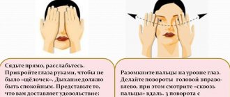
Vision exercises are a good way to relieve stress and fatigue. Let's consider the main ones:
- take a comfortable position on the sofa, without any distractions. Close your eyes and relax. Then begin performing circular eye movements for 5 minutes. First in one direction, then in the other;
- sit up straight, close your eyes tightly for 5-10 seconds, then rarely open your eyes. Repeat this exercise 10 times. Such actions help strengthen the muscles of the eyelids, improve blood circulation;
- For this exercise you need to make a small dot from plasticine with glass. Then fix your attention on some object behind the glass, look at the given image for several seconds, then sharply shift your gaze to a point on the glass. In the future, it may be possible to complicate the load - focus on four objects at different distances. Most of the exercises are included in gymnastics for both farsightedness and myopia.
Gymnastics is not a panacea and does not save you from the need for conservative or surgical treatment.