Leber's amaurosis is a hereditary disease that damages the nerve endings in the retina and is accompanied by systemic disorders (brain, urinary system). The condition has specific symptoms, so the doctor can suggest a diagnosis immediately after examining the child.
Such children need to constantly consult with a geneticist or pediatrician. The sooner the disease is detected, the sooner the doctor will begin symptomatic therapy.
Causes
The main cause of the disease is the inheritance of a pathological gene from parents to child. During embryogenesis, cones and rods (nerve endings) located on the retina of the eyes are formed incorrectly.
About 90 mutations are known that lead to the disease. As a result, the following violations occur:
- changes in the synthesis of pigments in cones and rods;
- decreased absorption of vitamins involved in the metabolism of the visual organs;
- disturbance of embryogenesis, as a result of which the number of photoreceptors and pigments on the retina decreases.
At the moment, scientists can isolate a pathological gene that is located in the genome of the mother or father of the child. Therefore, if you have an eye disease, it is recommended to consult a geneticist before planning a pregnancy.
Geneticists have discovered the gene responsible for Leber's neuropathy
Specialists of the Medical Genetic Research Center named after. acad. N.P. Bochkova (MGNC) together with colleagues from the Research Institute of Eye Diseases and the Helmholtz Center (Germany) discovered the DNAJC30 gene, mutations of which lead to Leber's hereditary optic neuropathy (LEONL). An article dedicated to the new DNAJC30 gene was published in The Journal of Clinical Investigations, the press service of the Moscow State Research Center reported.
NOPD is the most common mitochondrial disease in adults. It is classified as orphan (one case per 30 thousand people), affecting men 8 times more often than women. The average age of onset of the disease is 28 years, and it manifests itself as loss of central vision in both eyes.
According to ophthalmologists, about 20% of patients with the clinical picture of NOPD were previously left without molecular confirmation of the diagnosis; a gene mutation could not be detected in them. This was the reason for the start of an international study.
The study, which started in 2021 in the laboratory of mitochondrial genomics at the Institute of Neurogenomics of the Helmholtz Center in Munich under the leadership of Dr. Holger Prokisch , involved an international group of biochemists, geneticists, and ophthalmologists from 34 institutes. All ophthalmological studies were carried out by specialists from the Russian Research Institute of Eye Diseases, and genotyping of patients, checking the inheritance of the mutation, and studying the frequency of its occurrence in the population were carried out in the laboratory of hereditary metabolic diseases of the Moscow State Research Center.
Of the 33 patients participating in the study, 16 were identified in Russia, the rest in Romania, Germany, Italy and Ukraine. Patients with an unconfirmed diagnosis underwent exome sequencing (all genes that encode proteins) and identified changes in a new gene, the function of which had not previously been studied.
The product of this gene, the DNAJC30 protein, is a chaperone (proteins that help form the correct conformation of other proteins) of the mitochondrial respiratory chain. Its absence due to a gene mutation leads to the accumulation of non-functional proteins in the mitochondria, which disrupts the respiratory chain of cell mitochondria and leads to the development of the clinical picture of the disease.
“Imagine a shoe with laces - this is exactly what one of the main protein complexes in mitochondria looks like. Laces always wear out faster and need to be changed on time. Polina Tsygankova, a senior researcher at the Laboratory of Hereditary Metabolic Diseases of the Moscow State Research Center .
Patients with a mutation in the DNAJC30 gene are more likely to experience the phenomenon of vision restoration - 80% of them can regain it. Moreover, such a mutation is typical for Eastern European populations (up to 0.45% of the population can be its carriers) and is inherited in an autosomal recessive manner - the parents of the patients are healthy carriers of the mutation, and the child who received one damaged copy of the gene from each of them , gets sick.
Leber's classification of amaurosis
There are 16 types of the disease, which are the most common for this diagnosis. They are divided according to the chromosome on which the damage occurs and according to the type of inheritance:
- autosomal recessive, which manifests itself only if the mutant gene was inherited from both parents;
- autosomal dominant, always producing clinical symptoms when it enters the child’s genetic material.
When making a diagnosis, the doctor indicates not only the name of the defective gene, but also the type of mutation that appeared in the child. This is due to the fact that the disease is multifaceted and depends on the location of damage to the organ of vision. Often mutations on the same gene lead to different clinical symptoms.
Genetics: how, to whom and when the disease is transmitted
Mitochondrial inheritance pattern of Leber syndrome
Leber's hereditary optic atrophy is mediated by a DNA mutation in the mitochondria, which a person (mostly male) always receives from the mother, since only the egg cell transfers its mitochondria to the developing embryo (mitochondria from the father's sperm are not transferred).
Although the vast majority of patients with Leber disease have homoplasmic mutations, 10-15% of mutations are heteroplasmic. Tissue-specific segregation may be responsible for differences in interindividual phenotypes. Some studies suggest that the risk to patients is minimal if heteroplasm is less than 60%. Sons of mothers who have heteroplasma levels ≤80% are less likely to suffer from the disease.
The issue under discussion is the appearance of Leber syndrome in female carriers of mutations, which, depending on the genetic background, have significantly lower penetrance than males. Some studies suggest that the cause of differential penetrance is a modifying X-linked gene, leading to the manifestation of the disease in women only in the homozygous state. The second putative factor is X-inactivation of the “wild-type” X chromosome.
Symptoms of Leber's amaurosis
Clinical symptoms depend on the gene on which the mutation occurred and the degree of damage to the visual organs. During an external examination of the patient, violations are most often not detected. Parents may become suspicious of them over time, as the child grows up. When he learns to talk, he may independently complain about the lack of color perception or distortion of the surrounding reality.
Sometimes the following signs of illness occur immediately after birth:
- trembling of the eyes that occurs 3 months after birth;
- reduced pupil reaction to bright light or its complete absence;
- congenital blindness;
- rubbing the eyes with fingers;
- strabismus;
- farsightedness;
- a sharp decrease in visual acuity.
When the child reaches the age of ten, the condition worsens sharply. Most patients completely lose vision function. A violation of the internal structure of the eyes develops:
- increased intraocular pressure, which leads to glaucoma;
- clouding of the lens, forming cataracts;
- central nervous system disorder, mental retardation;
- inflammatory condition of the kidneys;
- hearing loss up to complete deafness.
Theodor Carl Gustaf von Leber is a German ophthalmologist, whose name became part of the names of individual nosological forms - Leber hereditary optic neuropathy (LHON, Leber hereditary optic neuropathy) and Leber optic amaurosis (LCA, Leber congenital amaurosis). These diseases have different pathogenesis and clinical manifestations, so they must be clearly distinguished. This review will focus only on Leber hereditary optic neuropathy (LEONL).
At the age of 31, Dr. Leber published a paper in which he described 15 cases from 4 families with hereditary optic atrophy (the main clinical symptom of OPA) [1]. For many years after this, scientists and doctors tried to figure out the nature of inheritance of this disease, until the American biochemist Douglas Wallace discovered a mutation in mitochondrial DNA (mtDNA) associated with NOPD in 1988 [2]. To date, 18 pathogenic mtDNA mutations associated with NOPD have been described, and the same number of mutations, the pathogenicity of which has yet to be proven or disproved. In addition, the molecular pathogenesis of the disease is becoming increasingly clear, which in turn contributes to the development of therapy for NOPD. The cause of NOPD is associated with mutations in mtDNA only. In addition, this disease is the most common mitochondrial pathology. The frequency of NONL is approximately 1:50,000, but carriers of mutations are approximately 5 times more common - 1:10,000 [3].
Clinical picture of the disease
The manifestation of the disease is usually observed in adolescence and the first period of adulthood. It has been noted that in men, mutations manifest themselves clinically on average 5 times more often. As a rule, the disease does not affect the eyes simultaneously, but at intervals of several weeks or months. The onset of the disease is characterized by a sharp painless decrease in central vision, and absolute centrocecal scotomas appear. The development of Leber's disease is characterized by two stages - acute and atrophic [4]. In the acute phase, a triad of signs is distinguished: 1) hyperemia and blurring of the boundaries of the visual disc; 2) peripapillary microangiopathy; 3) absence of fluorescein transudation around the optic nerve head (OND) during fluorescein angiography of the retina. The atrophic stage begins with blanching of the entire optic disc or its temporal half within a few weeks or months. Families are characterized by wide variations in clinical manifestations and number of cases. Atypical cases of the clinical course of the disease include an increase in visual acuity, a decrease in the size of the central scotomas and the transition of a scotoma from absolute to relative while maintaining the pattern of partial atrophy of the optic nerves in the fundus [5].
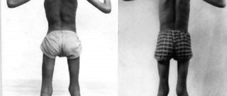
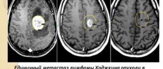
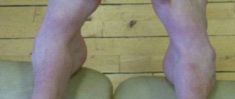
Sometimes a decrease in visual acuity in NOPD occurs together with other neurological symptoms [6]. These may be tremor, ataxia and spastic paresis. Polyneuropathy is manifested by disturbances in tactile and vibration sensitivity in the distal parts of the lower extremities. Achilles reflexes may be reduced or completely absent. Since tremor is also detected in 20% of cases with NOPD, a genetic relationship can be assumed. Cases of NOPD in combination with spastic paraparesis, ataxia, and sensitivity disorders of the polyneuric type have been described. Computed tomography scans of the patients revealed diffuse damage to the white matter of the brain, which indirectly indicated a possible relationship with multiple sclerosis. In addition to neurological disorders, patients with NOPD may experience some osteoarticular changes. In isolated cases, cardiac disorders are observed, manifested on the ECG by prolongation of the Q
-
T
, deep
Q
and high
R
. Sensorineural hearing loss is rare.
Molecular genetics of the disease
Since NOPD is associated with mutations in mtDNA, this determines the nature of inheritance of the disease: the mutation is inherited by all children of the sick woman (mitochondrial inheritance). It is worth noting that mitochondrial inheritance, to a first approximation, can be confused with X-linked recessive inheritance - in both cases, the children of the sick woman are affected. The difference lies in the sex ratio of the affected offspring: with mitochondrial inheritance, all children have an equal chance of getting the disease, while with X-linked recessive inheritance, the mutation is clinically manifested only in males. It is worth noting, however, the incomplete penetrance of mutations associated with NOPD—the mutation carrier will not always have certain clinical manifestations of the disease. There are also sporadic cases - a mutation that has arisen in mtDNA de novo,
can also confuse the understanding of the nature of inheritance. Thus, pedigree analysis is an integral part of making a diagnosis.
Molecular genetic analysis can detect pathogenic mutations in mtDNA only in half of the cases clinically diagnosed as NONLD. But when a mutation is detected, in 95% of cases it is one of the three so-called primary mtDNA mutations (m.3460G>A, m.11778G>A and m.14484T>C). The remaining percentage consists of “secondary” mutations, the number of which is growing every year [7]. Cases have been described when in one family it is possible to detect the presence of two pathogenic mutations at once - m.11778G>A and m.3460G>A [8]. In addition, “primary” NOHL mutations can be detected together with the m.1555A>G mutation associated with sensorineural hearing loss (SNHL, sensorineural hearing loss) [9], deletions associated with progressive external ophthalmoplegia (PEO, progressive external ophthalmoplegia) [10 ], as well as with “secondary mutations” that aggravate the clinical course of the disease [11]. Mutations such as m.3376A>G and m.3697G>A can lead to the simultaneous manifestation of symptoms of NOPD and MELAS in a patient [12].
NOPD is one of the few mitochondrial diseases for which a correlation has been established between the expression of a pathogenic mutation and membership in a specific phyletic lineage (mtDNA haplogroup). Thus, the m.11778G>A and m.14484T>C mutations are often associated with haplogroup J, while the m.3460G>A mutation is often associated with haplogroup K. It has been shown that the m.3460G>A mutation, found in Siberia, associated with haplogroups - derivatives of macrohaplogroup M, which is most often represented among the indigenous inhabitants of Siberia (Altaians, Tuvans, Buryats). At the same time, the m.11778G>A mutation, according to published data, is expressed against the background of haplogroups of the TJ cluster [13].
Since several mtDNA are present in a cell at the same time, mutating independently, a situation called heteroplasmy arises (a condition in which a cell has different variants of mtDNA), a state in which many pathogenic mtDNA mutations occur. Moreover, the level of heteroplasmy, as a rule, determines the phenotypic expression of the mutation. However, the majority of NONL mutations (approximately 85%) are characterized by a state of homoplasmy, i.e., when all mtDNA variants are identical. In the work of N.V. Volodko et al. (2006), in particular, a large family was characterized, including 5 generations, where it was possible to identify the m.3460G>A mutation, which arose de novo
in the 3rd generation. When assessing the level of heteroplasmy, it was shown that this mutation was 27% when it appeared and after a generation, reaching a level of more than 80%, it manifested itself clinically [13]. Since knowledge of the heteroplasmy level of a mutation is necessary to predict the onset of its clinical manifestation, the development of accurate methods for assessing heteroplasmy is becoming an increasingly urgent task. We specifically use a droplet digital polymerase chain reaction approach to determine the absolute copy numbers of mutant and wild-type mtDNA [14].
Such an uneven distribution of patients with NOPD by gender made us think about the influence of the X chromosome on the phenotypic manifestation of mtDNA mutations. In addition, it is possible that the incomplete penetrance of this disease is also due to the modifying effect of some nuclear genes. Thus, linkage analysis between gene loci (linkage analysis) revealed two loci - Xp21-Xq21 and Xq25-27.2, presumably influencing the manifestation of the disease. Experiments on cytoplasmic hybrids containing mutations m.3460G>A, m.11778G>A and m.14484T>C showed that 17β-estradiol activates mitochondrial biogenesis. Since estrogen receptors are localized in the mitochondria of human retinal ganglion cells, studies in cybrids partly explain the observed sex distribution of patients with NONLD [15].
From genotype to phenotype
Changes in the mtDNA sequence (mutations of structural genes) lead to changes in the polypeptide sequence of proteins synthesized from these mtDNA in mitochondria. Such proteins, which are part of oxidative phosphorylation complexes, disrupt the functioning of these complexes, which ultimately affects the energy production of the mitochondria. Most mutations in NONL affect ND -
genes, i.e. genes whose products are involved in the assembly of complex I (NADH-ubiquinone reductase). In this regard, the main biochemical defect observed in NOPD is a decrease in the activity of complex I. Mutations in the genes encoding subunits of complexes III, IV, and V likely act by the same mechanism. Mutations in tRNA genes, theoretically, disrupt the translation process of all mtDNA genes, thus leading to a decrease in the activity of all oxidative phosphorylation complexes. As a result, any mtDNA mutations should reduce ATP synthesis in mitochondria.
In 1995, J. Rizzo and in more detail P. Riordan-Eva and co-authors proposed the so-called “axoplasmic stasis” hypothesis, according to which cells of the retinal ganglion layer play a central role in the development of the disease [16, 17]. As a result of a decrease in the amount of ATP, the energy-dependent transport of mitochondria to the axon periphery deteriorates. Since mitochondria are formed only in the soma of the neuron, their lifespan is from 1 to 2 weeks, some of them do not have time to reach the distal parts of the axon, which increases the lack of ATP at its periphery. This is especially true for unmyelinated, long, narrow fibers with a high ratio of cell membrane area to cell volume and a high level of activity, such as the cells of the retinal ganglion layer. Thus, a vicious circle arises in which mtDNA mutations lead to a decrease in ATP production, which in turn negatively affects the axonal transport of mitochondria to the distal parts of the axon, and this leads to an even greater peripheral decrease in ATP production. Eventually, along with increased production of reactive oxygen species (ROS), this can trigger apoptosis. This can explain the “all or nothing” phenomenon observed with NONL - a clinically healthy person turns into a practically blind person within a few days. However, one of the mysterious issues of NONL is the nature of intercellular interactions, which allow a large number of cells to be involved in the process of death at the same time. How can we explain how, after two to three decades of normal function, most of the approximately 1.2 million optic nerve axons die simultaneously? This requires the presence of a common reservoir of certain factors or the ability to quickly exchange apoptotic signals. One of these types of cellular junctions that provide rapid propagation of the wave of apoptosis may be gap junctions of axonal extensions of cells of the retinal ganglion layer. Indeed, research suggests that gap junctions, by allowing cells to exchange ROS and calcium ions, are responsible for the centripetal spread of neuronal death, and that chemical agents that close gap junctions can limit the propagation of the apoptotic wave. Another mechanism for the rapid propagation of a signal to trigger apoptosis could be a decrease in the amount of ATP, which plays the role of a neurotransmitter in the interaction of cells of the nervous system [18].
Within the framework of this hypothesis, the incomplete penetrance of NONL can be explained by individual anatomical features. For example, the inherently increased number of ganglion cell axons in the optic disc and peripapillary region initially impedes the transport of mitochondria to axon terminals. In addition, the structure of the lamina cribosa may also cause differences in penetrance, since the point at which it crosses the optic nerve is the narrowest point of the latter.
Prospects for treatment
Despite increasing understanding of the nature of the molecular pathogenesis of NOPD, therapy remains inadequate. The main approaches to the treatment of this disease can be divided into drug treatment and experimental methods of gene therapy. This review will not discuss drug therapy, since there is no evidence of real benefit to patients after using certain drugs [19]. The exception appears to be ibedenone, a coenzyme Q10 analogue that is undergoing clinical trials [20, 21].
Experimental methods are aimed at etiological therapy and, although in most cases they are at the stage of testing in cell lines or animal models, significant progress is being seen. Using an allotropic expression strategy, a vector system was created that included the ND4
, not containing m.11778G>A.
Before transfection, all mtDNA in cytoplasmic hybrids was represented by mutant variants, as a result of which the ATP level was reduced by 60%. After transfection, it was possible to achieve a 3-fold increase in the level of ATP production [22]. A similar strategy was used by other researchers who achieved the expression of the ND4
connected to a part of the
COX10
encoding the signal sequence of the corresponding protein.
As a result, it was possible to achieve almost complete restoration of ATP levels [23]. Using NADH oxide reductase (NDI1) from Saccharomyces cerevisiae as the electron donor of the respiratory chain, the growth of cells with m.11778G>A on a nutrient medium with galactose was restored, although the ATP level could not be restored completely [24]. A vector system was constructed that combines the ND1
and part of the
COX10
, which is necessary for delivering the former to mitochondria. In a cell line containing the m.3460G>A mutation, a significant decrease in ATP synthesis was observed in a state of homoplasmy, but after the introduction of the vector, it was possible to achieve almost complete restoration of the initial ATP level [25]. To suppress the m.14459G>A mutation, a modified version of the TALEN genome editing system was used. Based on this approach, the so-called mitoTALENs were constructed - proteins that can be imported into mitochondria and recognize mtDNA. mtDNA with m.14459G>A was subjected to targeted elimination, which resulted not only in a decrease in the amount of mutant mtDNA, but also in an increase in the activity of complex I oxidative phosphorylation [26]. Next, mitoTALEN was applied to mouse oocytes carrying the m.14459G>A mutation. It was found that the copy number of mutant mtDNA decreased 24 h after mitoTALEN transfection [27]. It is worth noting that the m.14459G>A mutation was in the cell line in a state of heteroplasmy—the simultaneous presence of both mutant and wild-type mtDNA.
In another original study, a recombinant AAV2 viral vector was constructed that specifically enters mitochondria by delivering a nested genetic construct. Transfection of this construct into a cell culture containing mtDNA with m.11778G>A (MT-ND4) resulted in normalization of ATP levels [28]. "Reverse system" containing a mutant variant of the ND4
(m.11778G>A), was delivered into the vitreous cavity of mice. As a result, degeneration of the retinal ganglion layer and optic nerve atrophy were found in mice, which characterizes NOPD in humans. Experiments on animal models have also shown that DNA delivered into mitochondria by recombinant AAV2 viruses is stably maintained over a number of generations, is stably expressed, and is not integrated into mtDNA [29]. After a series of final tests, the design was used for the first phase of clinical trials (NCT02161380) in patients with NOPD [30–32]. The study authors concluded that there were no serious safety concerns with the approach in the first 5 clinical trial participants. Follow-up studies of these and additional participants planned over the next 4 years are needed to confirm the preliminary findings. Although the development of a gene therapy drug is still a long way off, the data obtained from John Guy's laboratory are impressive and encouraging.
The work was carried out within the framework of the federal target program “Research and development in priority areas of development of the scientific and technological complex of Russia for 2014–2020” with financial support from the state represented by the Ministry of Education and Science of Russia, agreement No. 14.575.21.0108 of November 27, 2014. Unique identifier RFMEFI57514X0108.
The authors declare no conflict of interest.
Information about authors
Mazunin Ilya Olegovich
- Ph.D. biol. sciences, head lab. molecular genetic technologies of the Institute of Living Systems of the IKBFU. I. Kant e-mail
Diagnosis of Leber amaurosis
Diagnosis of the patient’s condition is carried out in several stages:
- Anamnesis collection. This is data obtained from the words of the child’s parents. Most often, the disease is hereditary, so the doctor may suspect the diagnosis if the father or mother has it.
- General examination of the patient. Immediately after birth, clinical symptoms are rarely detected. Amaurosis develops gradually. When the child begins to walk and talk, symptoms become most obvious.
- Fundus examination. The doctor instills a solution into the child’s eyes, which disrupts the accommodation process. At first, retinal disorder is not observed. Over time, the doctor may see a disruption in the functionality of the retina and clouding of the lens.
- Measuring intraocular pressure. It increases upon reaching 7-10 years. Therefore, this technique is carried out only at this age.
Treatment of Leber's amaurosis
At the moment there is no treatment that would completely eliminate the disease. Tests are being carried out in the field of genetic engineering to introduce a specific gene into the retina of the eyes. Some categories of patients showed improvement in their condition.
Symptomatic treatment is carried out:
- vitamin therapy;
- remedies for cataracts;
- drops to reduce intraocular pressure;
- injections into the eye area that have vasodilating properties to treat farsightedness.