The brain is the main organ of the central nervous system of the human body. It consists of a colossal number of neurons, between which synaptic connections are established. It is the interaction of neurons that forms complex electrical impulses that allow us to process information, feel emotions and simply live, since the brain is also responsible for the functionality of organs.
The organ is divided into various sections, but the system functions as a single whole. Its slightest failure can cause pathologies of both the physiological and emotional spectrum. MRI is used to monitor pathological changes in the brain. What do you need to know about the procedure, how exactly is it performed, and what possible risks should the patient be aware of?
Cerebrovascular diseases
Vascular pathologies of the brain are the cause of 15% of deaths in economically developed countries. Such diseases occupy leading positions in the structure of population mortality.
Doctors pay great attention to the diagnosis of cerebral vascular diseases that can lead to acute disorders of cerebral circulation. These include:
- vascular tumors (angiomas);
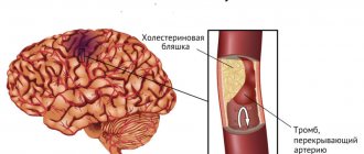
- atherosclerosis;
- narrowing of the lumen (stenosis) of blood vessels;
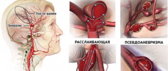
- aneurysms (pathological dilatation of arteries);
- vasculitis (inflammation of the walls of blood vessels);
- arteriovenous malformations;
- dural-venous fistulas;
- arterio-sinus anastomosis;
- thrombosis and embolism;
- vascular development abnormalities;
- vasospasm.
Information about the characteristics of the blood supply is necessary for drawing up prognoses and a treatment plan after a stroke, traumatic brain injury, tumor diseases, pathologies of the organs of vision and hearing, epilepsy, dementia, etc.
What does an MRI show?
- Thrombophlebitis;
- cysts;
- the presence of sclerotic plaques in the vascular lumens;
- vascular aneurysm;
- liquor imbalance;
- tumors (including vascular);
- abnormal structure;
- compression of capillaries and veins by scars;
- hydrocephalus;
- stratification or protrusion of blood vessels;
- epilepsy;
- ischemia, hemorrhagic injuries;
- areas damaged by multiple sclerosis;
- multiformation of blood vessels due to abnormal structure;
- brain and cranial injuries, their degree;
- lesions after strokes and heart attacks;
- inflammatory or infectious lesions (for example, encephalitis, meningitis).
And also, any changes in the brain and vascular system, their functionality is assessed. At the same time, the dynamics of deviations is traced.
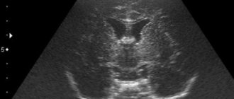
MRI of the brain and blood vessels helps identify tumor neoplasia. Damage is determined by darkening and gaps. Cerebral fluids are grayish streams, black cavities are intracerebral sinuses. The location of lesions after a stroke allows one to diagnose the form of the disease and prescribe an effective treatment regimen.
Types of MRI of the brain with blood vessels
Schematic representation of a cerebral aneurysm
For MR angiography (this type of magnetic resonance imaging is called MRA) without contrast, special modes are used that help clearly visualize the structure of cerebral vessels, identify the slightest changes in their anatomy and topography, determine the lumen and presence/absence stenosis, distinguish between fast and slow blood flow. If necessary, the study can be carried out together with contrasting for an even more detailed study of the structures.
There are three types of MR angiography depending on the pulse sequences:
1. Time-of-flight MRA is used to study the arteries of the brain. Specialists obtain sections directed perpendicular to the blood flow.
2. Phase contrast angiography is used to study the arteries of the neck and cerebral veins. Using the method, you can estimate the speed of blood movement.
3. 4D angiography is the fastest of all MRAs. Helps separate venous and arterial blood flow. Used to study malformations and fistulas.
The types are often combined and complemented by MRI of the arteries of the neck. The optimal type of MRI of the brain with blood vessels is chosen by the attending physician. A radiology doctor may recommend using contrast if regular images are not informative enough.
Technique
Before performing an MRI of the brain, all jewelry, metal rivets, and dentures are removed. If you intend to use a contrast agent during the procedure, you must have your blood tested in advance for allergies.
After preparation, the patient is asked to lie on the retractable part of the tomograph. The medical staff secures it with bolsters and belts. Equipment is attached to the patient's head that will receive and send back radio waves. If contrast is used, the agent is administered intravenously before the start of the study. If desired, the patient can wear sound-proofing earplugs.
After turning on the equipment, a picture of the brain, its parts and structures is displayed on the monitor screen. Diagnostics allows us to detect even small neoplasms, existing disease processes and their rudiments.
How is MRI of cerebral vessels done?
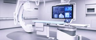
Modern tomographs make it possible to obtain clear images of cerebral vessels.
The procedure for conducting MRA is as follows:
- The subject lies down on the tomograph platform. To ensure complete immobility, the laboratory assistant fixes his body with belts and the position of his head with bolsters. The conveyor moves until the brain area is in the center of the tomograph frame. The device makes noise during operation, so the laboratory assistant suggests that the person use earplugs or headphones to listen to music.
- The x-ray technician goes into another room, checks that the speakerphone is working properly, and reminds the patient to lie still while the scan is taking place. MRI with contrast differs in that native images are taken first, and then the drug is administered and the study is resumed. Conventional angiography takes about 15 minutes, contrast prolongs the procedure to 30-35 minutes.
- Upon completion of the scan, the patient is returned his personal belongings and asked to wait for the results.
The radiologist's report is issued in printed form, and the images are recorded on a data medium. The preparation time for protocols ranges from 15 to 60 minutes. The doctor interprets the images, but does not have the right to make a diagnosis, prescribe therapy or make prognoses. This is done by the attending physician. If necessary, the radiologist gives explanations to the patient about the images or recommends which specialist to contact.
The results may be sent by email if you indicated the address in the documents before taking the study. The original report can be collected on any other day, but in this case the patient will not be able to receive an explanation from the radiologist.
Diagnostic clinic "Magnit" provides. At the request of the patient, another doctor can analyze the results obtained, make his own conclusion or supplement the existing one. Those who have been diagnosed in other medical institutions can also get a “Second Opinion”. The service is appropriate when the patient does not trust him, in controversial clinical cases, in severe pathologies with a long history, as well as when assessing dynamic changes. A “second opinion” helps reduce the risk of errors when making a diagnosis.
Dystrophic changes in the brain on MRI
Cerebral angiography
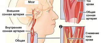
When the blood supply is disrupted, oxygen and trophic starvation of cells (ischemia) develops. This leads to degenerative processes and is accompanied by dysfunction. The severity of the latter is variable, depending on whether blood is completely blocked or a partial influx remains. Dystrophic changes can be local or diffuse. Total brain damage is recorded in meningitis, encephalitis, focal changes are typical for cysts, small ischemic processes, and the formation of post-traumatic scars.
Clinical manifestations may include:
- headache;
- high blood pressure;
- the appearance of paresthesia (a feeling of numbness or tingling in the limbs), loss of sensitivity;
- deterioration of vision (up to blindness, which indicates damage to the optic nerve), memory, decreased intellectual abilities;
- insomnia;
- hyperkinesis (uncontrolled muscle contractions) and convulsions.
As the pathology progresses, paresis and paralysis are expected, so it is important to do an MRI of the brain vessels at the first symptoms of trouble. Single lesions can be detected in young men and women and do not always indicate pathology. The doctor’s tactics are dynamic observation and repeated magnetic scanning after 3-6 months, which will allow not to miss the development of any serious disease, for example, multiple sclerosis. Over the age of 60-65 years, lesions are found in almost all patients, which is explained by natural aging. These changes are irreversible, but the progression of the process can be slowed down with adequate treatment.
Provoking factors include:
- chronic alcohol and nicotine intoxication;
- stressful situations;
- irrational work and rest regime;
- obesity;
- low physical activity;
- persistent increase in blood pressure;
- diabetes;
- hypercholesterolemia.
The type of dystrophic disorders in the gray and white matter of the brain on MRI will depend on the nature of the pathological process.
MRI of cerebral vessels - indications and contraindications
MRI of head vessels, 3D model
Scanning is carried out on the recommendation of a doctor. The results of the study may be required to draw up a complete clinical picture of an already diagnosed disease, study the consequences of an injury, identify the causes of chronic poor health, and confirm the doctor’s suspicions.
MRI of cerebral vessels should be done if a person has complaints about:
- repeated fainting;
- progressive deterioration of vision or hearing;
- constant dizziness;
- headaches of unknown origin;
- noise, especially pulsating, in the ears or head (ringing, whistling, squeaking);
- convulsions;
- disturbances of sensitivity and movement in the limbs, trunk, face;
- sleep disorders (insomnia, problems falling asleep, waking up at night);
- frequent nosebleeds;
- memory impairment;
- loss of balance, lack of coordination of movements;
- acute exophthalmos (protrusion of the eyeball);
- signs of intracranial hypertension.
An examination may be required at the stage of planning an operation, after surgery, to monitor the disease over time (tumors, atherosclerosis, thrombosis, etc.), as well as to monitor the effectiveness of prescribed therapy. Usually the procedure is carried out as planned.
Contraindications for MRI of cerebral vessels is the presence of metal implants in the patient (pacemaker, hemostatic clips, prostheses, etc.). If there are foreign bodies made of titanium (plate, dentures) in the area being examined, it is necessary to provide a document that confirms the material. The corresponding statement can be obtained from the clinic where the operation was performed.
Scanning results can be negatively affected by braces and hearing correction devices. These must be notified to the X-ray technician prior to the procedure.
The following conditions are considered relative contraindications:
- first trimester of pregnancy;
- weight more than 120 kg;
- fear of confined spaces;
- neurological disorders accompanied by uncontrolled body movements;
- severe pain syndrome.
If you are claustrophobic or overweight (if the patient's body weight exceeds the carrying capacity of the tomograph conveyor), you can perform the study on an open-type device, however, such devices have less accuracy (compared to contour ones).
In case of acute pain syndrome, hyperkinesis (when a person cannot control movements), it is suggested to undergo diagnostics in a hospital setting under sedation.
It is necessary to take into account how MRI of cerebral vessels is performed: with contrast, the number of contraindications does not increase, with the exception of women during pregnancy. The latter category of patients cannot use even safe drugs based on gadolinium chelates. The indicators are usually well tolerated, but to reduce autonomic reactions the patient needs a light meal 45 minutes before the scan.
Prescriptions and contraindications
An MRI of the brain is done if there are internal injuries or external injuries to the skull (fractures, internal deformities). Vessels must be examined in case of high intracranial pressure, severe vegetative vascular dystonia. An MRI technique is required after a mini-stroke, to determine the presence of blood clots, or with constant causeless pain in the head. Tomography is indicated for:
- suspected neoplasms in the tissues of the brain or the organ itself;
- examination of nerve tissue;
- to assess the extent of brain disease after heart attacks and strokes;
- examination for the likelihood of various forms of vascular anomalies;
- suspected multiple sclerosis or cerebral hemorrhage;
- monitoring the patient after surgery to identify complications or abnormalities.
People with congenital heart defects and vasculitis must be examined. MRI of the brain is done for athletes and people with bad habits, as they increase the risk of diseases of the vascular system.
On a note!
The research method is used to monitor the course of pathologies and determine the effectiveness of treatment.
For children, MRI examinations are performed, in addition to the main indications, for fainting, muscle cramps, partial loss of hearing or vision. If children are lagging behind in development, there is a sharp change in mood. The MRI technique is indicated for epilepsy.
Contraindications
Contraindications for the study include insulin pumps, all implants and stimulators. MRI is not recommended for patients with heart failure, claustrophobia, or an existing allergy to the contrast agent. It is not advisable to perform diagnostics on people who have tattoos with metal particles on their bodies. The MRI technique is not used when hemostatic clips are in the brain. Relative research prohibitions include pregnancy.
Preparation for MRI of cerebral vessels
Visualization of individual cerebral vessels with MR venography
The examination does not require special actions from the patient the day before. There is no need to stop medications, follow a diet or a special daily routine.
When examining blood vessels with contrast, it is worth having a snack three quarters of an hour before the scan. Nursing mothers need to stock up on breast milk for 2 feedings, which they will have to skip after the procedure.
You should come to the clinic 10-15 minutes before the appointed time to fill out documents and prepare for the scan. You need to have a passport, a referral from a doctor, reports and photographs from previous similar studies, if any, with you. A person must warn the x-ray technician about:
- all your illnesses;
- drug intolerance;
- presence of claustrophobia;
- the presence of metal and electronic devices in organs and tissues (pacemakers, prostheses, etc.);
- tattoos.
Before scanning, the patient removes all jewelry and clothing with metal elements, including glasses, piercings, dentures, and hair clips. Electronic devices (phone, watch, hearing aid) will also have to be left outside the MRI room.
Before the scan begins, the laboratory assistant will give the patient a button for emergency communication. It must be pressed in case of any discomfort (dizziness, nausea, impaired consciousness, panic, etc.). As soon as the person gives a signal, the study will be paused and the radiologist will come to the rescue. The button looks like a rubber bulb and calls a laboratory assistant in a matter of seconds.
What brain pathologies does MRI show?
The distribution of white and gray matter and pathological processes within the brain are illustrated in detail by MRI. Magnetic scanning is one of the high-precision non-invasive methods of instrumental diagnostics for neurosurgeons, neurologists, and, less often, psychiatrists. Analysis of changes in the two main components of the brain - gray and white matter - is important for clinical diagnosis and therapy of a huge number of diseases: epilepsy and episyndrome, stroke, Alzheimer's disease, malignant and benign neoplasms, multiple sclerosis, infectious and inflammatory processes, post-traumatic injuries, etc.
Distribution of gray and white matter in the brain
Gray matter
The gray matter of the brain is responsible for most of the functions of higher nervous activity and is represented by neuron bodies, glial cells, a cluster of dendrites, thin tiny blood vessels - capillaries - and unmyelinated axons. The main histological structures are centers, each of which controls some action: the act of urination, defecation, heartbeat, etc. Neurosurgeons consider the gray color to be conventional; rather, this substance has an earthy tint. The composition of the main structures of the brain is clearly distinguishable, mainly due to differences in water and protein content. This allows one zone to be differentiated from another on tomograms. Pathological processes localized in the gray matter lead to disturbances in perception, speech, emotions, memory, sensory sensitivity, will, muscle movements, etc.
White matter
The white color is caused by bundles of nerve fibers covered with a myelin sheath. The main purpose of this brain structure is the transmission of impulses from the main centers to the periphery (lower links of the nervous system).
MRI results of cerebral vessels
Such scanning of arteries and veins provides information in the form of images in three planes, and using 3D modeling, three-dimensional images can be obtained. By assessing the contours on the slides, the radiologist will notice any deviations from the norm and describe them in the report. During image analysis, the following can be detected:
- neoplasms (indirectly) and features of their blood supply;
- damage (ruptures) of blood vessels;
- areas of pathological narrowing of the lumen and tortuosity of the course of arteries or veins;
- localization and size of blood clots, emboli;
- signs of atherosclerosis;
- thinning and deformation of arterial walls (aneurysm);
- acute ischemia;
- consequences of stroke;
- inflammatory changes in the walls of blood vessels;
- arteriovenous malformations;
- cavernous angiomas;
- vasospasm, etc.
Visualization of blood vessels provides a deeper understanding of the processes occurring in brain cells. The procedure allows you to determine the true cause of migraines, dizziness, memory problems, and reveal the pathogenesis of serious brain diseases. The information obtained during MRA is extremely important for selecting treatment tactics and monitoring its effectiveness.
Advantages of magnetic resonance imaging over other methods
An MRI examination gives results much more accurately than x-rays, echoencephalography (EchoEG), ultrasound and other diagnostic options. It allows you to obtain maximum data about existing tumors, diseases, post-traumatic and post-stroke changes. Unlike CT and X-rays, in this case the body is not irradiated.
Only soft tissues are visualized in the finished images. The bones of the skull are not visible, so they do not interfere with analysis and decoding.
The contrast agent used in MRI diagnostics is much less likely to cause allergic reactions compared to X-ray contrast agents used for radiography.
Contraindications
- Installed pacemakers and other electronic devices that are disrupted by the surrounding electromagnetic field.
- Fixed dentures with metal elements present in the mouth, crowns containing metal, braces and other orthodontic structures. The metal they contain is heated by a magnet and deteriorates, damaging surrounding tissue.
- Tattoos applied to the skin, made with metal-containing ink. Due to the heat caused by the electromagnets, burns may occur in these areas. In the absence of information about the pigment. used when applying a tattoo, it is better not to risk it and do a CT scan, ultrasound or x-ray. Examination is also prohibited for any metal piercing that cannot be removed.
- MRI examinations with contrast are not performed during pregnancy or if contrast agents are intolerant. Such an examination is not prescribed for severe kidney pathologies that make it difficult to eliminate gadolinium.
Magnetic resonance imaging is a safe and highly informative procedure that detects pathologies in the early stages. Therefore, in case of migraine-like phenomena, coordination problems, a sharp decrease in hearing and vision, fainting, or progressive memory deterioration, you must definitely go to the clinic and be examined. The price of MRI diagnostics is low and quite affordable for Muscovites and residents of Moscow Region.
Get an MRI of the brain >>>