Diagnostics
All patients with suspected diffuse changes in the pituitary gland are examined according to a single plan, which includes sequential clinical, laboratory and instrumental methods.
Clinical diagnosis
First, the specialist collects complaints. Then it checks visual acuity, changes in its fields, the condition of the fundus and the presence of oculomotor disorders. Then a neurological examination, which consists of an assessment of reflexes and speech contact.
Laboratory methods
General blood and urine tests are prescribed, as well as a biochemical analysis of blood and the functioning of the coagulation system. After this, a radioimmune study of hormones in the blood.
Evaluate the content:
- prolactin;
- somatotropic hormone (STH);
- adrenocorticotropic (ACTH);
- thyroid stimulating (TSH);
- somatomedin;
- triiodothyronine (T3);
- thyroxine (T4);
- cortisol;
- sex hormones.
Instrumental methods
When examining the pituitary gland, MRI and ultrasound (ultrasound) are mainly used. However, MRI remains more informative. His work is based on reading the tissue reflection of magnetic waves that he himself creates. He takes layer-by-layer photographs, allowing you to see the sections.
Ultrasound is based on the ability of ultrasound beams to penetrate tissue. This method is safer in terms of radiation exposure. But with such a study, all rays from all organ tissues that cover the object under study are superimposed.
Undoubtedly, there are ways to examine only one organ, for example, the special position of the patient during the procedure. But ultrasound is used as an additional diagnostic method.
On this topic
- central nervous system
What are the dangers of a porencephalic cyst of the brain?
- Olga Vladimirovna Khazova
- May 27, 2021
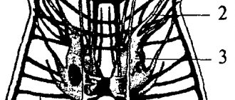
X-ray examination is also used as an additional method. Lateral craniography is performed and the condition of the sella turcica is assessed.
It is a bony structure in the cranial cavity that contains the pituitary gland. Its size, changes in structure, and elements of bone tissue destruction are assessed.
Treatment
After examining the patient, a treatment method is chosen. It consists only of observation when:
- there are no neurological symptoms;
- no ophthalmological disorders;
- a hormonally inactive form of change in the pituitary gland.
In this case, the patient is registered with an endocrinologist, ophthalmologist and neurosurgeon and undergoes a preventive examination with them once a year.
In other cases, the issue of surgical treatment of the pituitary gland or a conservative method is decided.
Surgical methods
All causes of diffusely heterogeneous structure of the pituitary gland are amenable to surgical treatment. There are two methods in total - endoscopic intervention through the nasal sinuses and microsurgical interventions in the brain area.
The difference is that in the first case, a full tissue incision is not required. Therefore, up to 80-90% of all operations are tried to be carried out using this method.
Conservative treatment
Conservative treatment means the use of medications. The scheme is drawn up taking into account the solution of the following problems:
Increased concentrations of certain hormones. In this case, everything depends on the type of diffuse change in the pituitary gland. For example, for somatotropin-producing adenomas, agonists of receptors sensitive to this hormone are prescribed. This leads to the fact that growth hormone does not interact with them and does not activate cell function.
Relieving symptoms.
Replenishment of hormone deficiency. Hormone replacement therapy is used.
How it was
The diffuse type of nervous system is characteristic of the initial stages of the development of our world, when the interaction of living beings - the simplest organisms - took place in the aquatic environment of the primitive ocean. The protozoa secreted certain chemicals that dissolved in water, and thus the first representatives of life on the planet received metabolic products along with the liquid.
The oldest form of such interaction occurred between individual cells of multicellular organisms through chemical reactions. These are metabolic products - metabolites, they appear when proteins, carbon dioxide and the like break down, and are a humoral transmission of influences, a humoral mechanism of correlation, that is, connections between different organs. A characteristic of the diffuse type of nervous system can also be partly the humoral connection.
Notes
- Due to the fact that the body of these animals always consists of the same set of cells.
- White JG, Southgate E., Thomson JN, Brenner S.
The structure of the nervous system of the nematode
Caenorhabditis elegans
//
Phil.
Trans. Royal Soc. London ,
314
, 1986. - P. 1-340. - Mednikov, 1994, p. 363.
- Society for Neuroscience -. www.sfn.org. Retrieved November 22, 2015.
- The Journal of Neuroscience. www.jneurosci.org. Retrieved November 22, 2015.
- Fens Home Page (English). FENS.org. Retrieved November 22, 2015.
- European Journal of Neuroscience.
- Harriet Cole: Drexel's Longest-Serving Employee.
Neuroglia
Living organisms, together with the above, in the process of evolution increase both the number of neurons and their diversity. Thus, neuroglia were formed. Bipolar neurons appeared, having axons and dendrites. Gradually, organisms gain the ability to conduct excitation in a directional manner. Nervous structures also differentiate and transmit signals to cells that control responses.
This is how the nervous system developed purposefully: some cells specialized in reception, others in signal transmission, and still others in response contraction. What followed was evolutionary complexity, centralization, and the development of a nodal system. Annelids, arthropods, and mollusks appear. Now the neurons are concentrated in ganglia (nerve ganglia), which are tightly connected by nerve fibers to receptors and execution organs (glands, muscles).
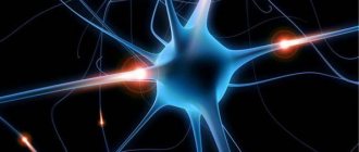
Neuroscience
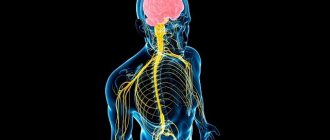
Modern science of the nervous system combines many scientific disciplines: along with classical neuroanatomy, neurology and neurophysiology, molecular biology and genetics, chemistry, cybernetics and a number of other sciences make an important contribution to the study of the nervous system. This interdisciplinary approach to the study of the nervous system is reflected in the term neuroscience. In Russian-language scientific literature, the term “neurobiology” is often used as a synonym. One of the main goals of neuroscience is to understand the processes occurring both at the level of individual neurons and neural networks, the result of which are various mental processes: thinking, emotions, consciousness. In accordance with this task, the study of the nervous system is carried out at different levels of organization, from the molecular to the study of consciousness, creativity and social behavior.
Professional societies and magazines
The Society for Neuroscience (SfN, the Society for Neuroscience)[4] is the largest non-profit international organization, uniting more than 38 thousand scientists and doctors involved in the study of the brain and nervous system. The society was founded in 1969 and is headquartered in Washington. Its main goal is the exchange of scientific information between scientists. To this end, an international conference is held annually in various cities in the United States and the Journal of Neuroscience is published[5]. The society carries out educational and educational work.
The Federation of European Neuroscience Societies (FENS, the Federation of European Neuroscience Societies)[6] unites a large number of professional societies from European countries, including Russia. The Federation was founded in 1998 and is a partner of the American Society for Neuroscience (SfN). The Federation holds an international conference in different European cities every 2 years and publishes the European Journal of Neuroscience[7].
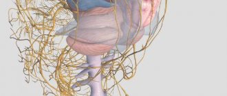
Features of the autonomic nervous system
Bishcha (1781) - divided the nervous system into:
- somatic (animal), i.e. formations that provide motor function and extrocentive sensitivity;
- vegetative - ensure the functioning of internal vessels, sweat glands and metabolic processes in skeletal muscles and the nervous system.
The autonomic nervous system is a set of central and peripheral formations that ensure work within the body.
The autonomic nervous system was studied in the 70s of the 19th century. The autonomic nervous system has a pronounced ability to function when the central nervous system is damaged. This is ensured by the autonomic ganglia (the body of postganglionic neurons of the central nervous system).
The autonomic nervous system includes three sections:
- sympathetic nervous system;
- parasympathetic nervous system;
- metasympathetic nervous system.
Anatomical features of the autonomic nervous system. The focal location of nerve centers is at various levels of the central nervous system, i.e., metasegmental structure. The centers of the sympathetic nervous system are in the spinal cord - the lateral horns of the Th and Z segments - (C (7)) Th (1) - Z (2-4) segments. The centers of the parasympathetic nervous system are in the brain and spinal cord. In the brain - the cranial section of the parasympathetic nervous system - at the level of the midbrain (nuclei VII, IX, X pairs of cranial nerves). The spinal region is at the level of the sacral segments - the nucleus of the pelvic nerve. The external cortical centers of the autonomic nervous system are in the hypothalamus (the anterior group of nuclei is the external center of the parasympathetic nervous system, the posterior group is the external center of the sympathetic nervous system).
The cerebral cortex - level 6-8 Brodmann fields (sensitive area) - is a point representation of the autonomic nervous system. There is the presence of autonomic ganglia (location of the efferent neuron). The sympathetic nervous system is the pre- and paravertebral location of the ganglia. Paravertebral ganglia - located on both sides of the spine - a chain of 20-22 nodes - the border sympathetic trunk (truncus sympaticus). Prevertebral ganglia - enter from the plexuses (solar, superior and inferior mesenteric). The sympathetic nervous system is characterized by a short preganglionic and long postganglionic pathway.
Parasympathetic nervous system - prevertebral and intramural location of ganglia. Prevertebral ganglia - in the plexuses around organs. Intramural - in the internal organs themselves. The parasympathetic nervous system is characterized by a long preganglionic and short postganglionic pathway.
The phenomenon of animation (multiplication) in the autonomic ganglion - in the autonomic ganglion the phenomena of convergence and divergence of impulses are simultaneously expressed: on the body of one postganglionic neuron, impulses from several preganglionic neurons converge and any preganglionic neuron innervates many postganglionic neurons. This ensures reliable excitation transmission.
Features of nerve fibers. The autonomic nervous system includes thin myelinated and unmyelinated fibers. Preganglionic fibers - group B. Postganglionic fibers - group C.
Physiological features of the autonomic nervous system:
Features of ganglia.
Autonomic ganglia have longer EPSP durations. There is a long period of trace hyperpolarization, so inhibition easily occurs after excitation. Very low speed of excitation - 5-10 times greater than in the central nervous system. Neurons of the autonomic ganglia are characterized by low lability, transmitting a small number of impulses from the central nervous system to the periphery. At a frequency of 100 impulses per second, a complete block occurs in the autonomic ganglia. Thus, the autonomic ganglia are an autonomous formation that regulates the conduction of impulses to the working organs.
Features of nerve fibers.
Low speed of excitation (group B is 13 - 18 m/s; group C is 0.5 - 3 m/s). Therefore, vegetative reactions arise slowly. Long-term chronoxia - low excitability of nerve fibers.
Most organs have double innervation.
The sympathetic nervous system innervates all organs and tissues. The parasympathetic nervous system does not innervate the smooth muscles of the walls of some blood vessels (skin, abdominal cavity), skeletal muscles, the central nervous system, receptors, the adrenal medulla, and the uterus.
Divisions of the autonomic nervous system, as a rule, have the opposite effect.
The autonomic nervous system provides 3 groups of reflexes:
- viscero-visceral,
- viscero-muscular,
- viscero-cutaneous (increased sensitivity in various areas of the skin with damage to the internal organs of the Zakharyin-Ged zone).
The sympathetic nervous system and parasympathetic nervous system provide different effects on organs and tissues. The sympathetic nervous system has a diffuse effect - when certain centers are excited, the work of a large number of organs is ensured. The parasympathetic nervous system is a more limited action.
Norepinephrine is a stable substance, easily absorbed into the blood - a diffuse effect of the sympathetic nervous system.
Acetylcholine - quickly destroyed - the action of the parasympathetic nervous system is limited, short-term.