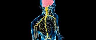
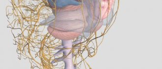
Diagram of the human nervous system
Nervous system
- an integral morphological and functional set of various interconnected nervous structures, which, together with the endocrine system, ensures the interconnected regulation of the activity of all body systems and the response to changes in internal and external environmental conditions. The nervous system acts as an integrative system, linking into one whole sensitivity, motor activity and the work of other regulatory systems (endocrine and immune).
Content
- 1 General characteristics of the nervous system 1.1 Neurons
- 1.2 Neuroglia
- 2.1 Types of nervous systems
- 2.2.6.1 Morphological division
- 2.3.1 Models 2.3.1.1 Regulatory mechanisms
- 3.1 Professional societies and journals
General development of the nervous system
Phylogeny of the nervous system
* in brief it boils down to the following. The simplest single-celled organisms (amoeba) do not yet have a nervous system, and communication with the environment is carried out using fluids located inside and outside the body - humoral (humor - liquid), a pre-nervous form of regulation.
* (Quote from the book: Sepp E.K. History of the development of the nervous system of vertebrates from skullless to humans. M., 1949.
)
Later, when the nervous system arises, another form of regulation appears - nervous. As the nervous system develops, nervous regulation increasingly subordinates humoral regulation, so that a single neuro-humoral regulation is formed with the leading role of the nervous system. The latter goes through a number of main stages in the process of phylogenesis (Fig. 265).
Rice. 265. Stages of development of the nervous system. 1 - diffuse nervous system; 2 - diffuse nervous system of hydra; 3 and 4 - nodal nervous system of the annelid worm
Stage I - reticular nervous system
. At this stage (coelenterates), the nervous system, such as hydra, consists of nerve cells, the numerous processes of which connect with each other in different directions, forming a network that diffusely permeates the entire body of the animal. When any point of the body is irritated, excitement spreads throughout the entire nervous network, and the animal reacts by moving its entire body. A reflection of this stage in humans is the network-like structure of the intramural nervous system.
Stage II - nodal nervous system
. At this stage (higher worms), nerve cells come together into separate clusters or groups, and from clusters of cell bodies, nerve nodes - centers are obtained, and from clusters of processes - nerve trunks - nerves. At the same time, in each cell the number of processes decreases, and they receive a certain direction. According to the segmental structure of the body of an animal, for example, an annelid, in each segment there are segmental nerve ganglia and nerve trunks. The latter connect nodes in two directions; transverse trunks connect nodes of a given segment, and longitudinal trunks connect nodes of different segments. Thanks to this, nerve impulses arising at any point in the body do not spread throughout the body, but spread along the transverse trunks within a given segment. Longitudinal trunks connect the nerve segments into one whole. At the head end of the animal, which, when moving forward, comes into contact with various objects of the surrounding world, sensory organs develop, and therefore the head nodes develop more strongly than others, being a prototype of the future brain. A reflection of this stage is the preservation of primitive features in humans (dispersion of nodes and microganglia on the periphery) in the structure of the autonomic nervous system.
Stage III - tubular nervous system
. At the initial stage of animal development, a particularly important role was played by the apparatus of movement, on the perfection of which depends the main condition for the existence of the animal - nutrition (movement in search of food, capturing and absorbing it).
In lower multicellular organisms, a peristaltic method of locomotion has developed, which is associated with smooth muscles and its local nervous apparatus. At a higher level, the peristaltic method is replaced by skeletal motility, i.e. movement using a system of rigid levers - over muscles (arthropods) and inside muscles (vertebrates). The consequence of this was the formation of striated muscles and a central nervous system that coordinates the movement of individual levers of the motor skeleton.
Such a central nervous system in chordates (lancelet) arose in the form of a metamerically constructed neural tube with segmental nerves extending from it to all segments of the body, including the movement apparatus - the trunk brain. In vertebrates and humans, the trunk cord becomes the spinal cord. Thus, the appearance of the trunk brain is associated with the improvement, first of all, of the animal’s motor weapons. Along with this, the lancelet also has receptors (olfactory, light). The further development of the nervous system and the emergence of the brain are mainly due to the improvement of receptor weapons.
Since most of the sense organs arise at that end of the animal’s body, which is facing the direction of movement, i.e. forward, then to perceive external stimuli coming through them, the anterior end of the trunk brain develops and the brain is formed, which coincides with the separation of the anterior end of the body into in the form of a head - cephalization (cephal - head).
E.K. Sepp in his manual on nervous diseases* gives a simplified but convenient for study diagram of the phylogeny of the brain, which we present here. According to this scheme, at the first stage of development, the brain consists of three sections: posterior, middle and anterior, and from these sections, the hind, or rhombencephalon, especially develops first (in lower fish). The development of the hindbrain occurs under the influence of acoustic and static receptors (receptors of the VIII pair of cephalic nerves), which are of key importance for orientation in the aquatic environment.
* (E. K. Sepp, M. B. Zucker, E. V. Schmidt, 1950.
)
In further evolution, the hindbrain differentiates into the medulla oblongata
, which is a transitional section from the spinal cord to the brain and is therefore called myelencephalon (myelos - spinal cord, encephalon - brain), and the
hindbrain itself
- metencephalon, from which the cerebellum and pons develop.
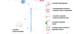
In the process of adapting the organism to the environment by changing metabolism, in the hindbrain, as the most developed part of the central nervous system at this stage, control centers for vital processes of plant life arise, associated, in particular, with the gill apparatus (breathing, blood circulation, digestion, etc. ). Therefore, the nuclei of the branchial nerves (group X pair - vagus) appear in the medulla oblongata. These vital centers of respiration and circulation remain in the human medulla oblongata, which explains the death that occurs when the medulla oblongata is damaged. At stage II (even in fish), the midbrain
, mesencephalon. At stage III, in connection with the final transition of animals from the aquatic environment to the air, the olfactory receptor intensively develops, perceiving chemical substances contained in the air, signaling with their smell about prey, danger and other vital phenomena of the surrounding nature.
Under the influence of the olfactory receptor, the forebrain—prosencephalon—develops, initially having the character of a purely olfactory brain. Subsequently, the forebrain grows and differentiates into the intermediate brain
- diencephalon and
final
- telencephalon.
In the telencephalon, as the highest part of the central nervous system, centers for all types of sensitivity appear. However, the underlying centers do not disappear, but remain, subordinate to the centers of the overlying floor. Consequently, with each new stage of brain development, new centers arise, subordinating the old ones. There seems to be a movement of functional centers to the head end and the simultaneous subordination of phylogenetically old rudiments to new ones. As a result, hearing centers that first arose in the hindbrain are also present in the middle and forebrain, vision centers that arose in the middle are also present in the forebrain, and olfactory centers are only in the forebrain. Under the influence of the olfactory receptor, a small part of the forebrain develops, therefore called the olfactory brain (rhinencephalon), which is covered with a gray matter cortex - the old cortex (paleocortex).
Improvement of receptors leads to the progressive development of the forebrain, which gradually becomes the organ that controls all animal behavior. There are two forms of animal behavior: instinctive, based on species reactions (unconditioned reflexes), and individual, based on the individual’s experience (conditioned reflexes). According to these two forms of behavior, two groups of gray matter centers develop in the telencephalon: subcortical nodes, which have the structure of nuclei (nuclear centers), and the gray matter cortex, which has the structure of a continuous screen (screen centers). In this case, the “subcortex” develops first, and then the cortex. The bark appears during the transition of an animal from an aquatic to a terrestrial lifestyle and is clearly found in amphibians and reptiles. The further evolution of the nervous system is characterized by the fact that the cerebral cortex increasingly subordinates the functions of all underlying centers to itself, and a gradual corticolization of functions
.
The necessary formation for the implementation of higher nervous activity is the new cortex, located on the surface of the hemispheres and acquiring a six-layer structure in the process of phylogenesis. Thanks to the enhanced development of the new cortex, the telencephalon in higher vertebrates surpasses all other parts of the brain, covering them like a cloak (pallium). The developing new brain (neencephalon) pushes into the depths the old brain (olfactory), which, as it were, curls up in the form of Ammon's horn (cornu Ammoni or pes hyppocampi), which still remains the olfactory center. The result is a cloak, i.e. a new brain
(neencephalon), sharply predominates over the rest of the brain - the old brain (paleencephalon).
So, the development of the brain takes place under the influence of the development of receptors, which explains that the highest part of the brain - the gray matter cortex - represents, as I. P. Pavlov teaches, a set of cortical ends of analyzers, i.e. a continuous perceiving (receptive) surface. Further development of the human brain is subject to other laws related to its social nature. In addition to the natural organs of the body, which are also found in animals, man began to use tools. Tools, which became artificial organs, complemented the natural organs of the body and constituted the technical equipment of man.
With the help of these weapons, man acquired the ability not only to adapt himself to nature, as animals do, but also to adapt nature to his needs. Labor, as mentioned above, was a decisive factor in the development of man, and in the process of social labor, a necessary means for people to communicate arose - speech. “First, work, and then, along with it, articulate speech, were the two most important stimuli, under the influence of which the monkey’s brain gradually turned into the human brain, which, for all its similarities with the monkey’s, far surpasses it in size and perfection” (K. Marx , F. Engels, Works, ed. 2, vol. 20, p. 490). This perfection is due to the maximum development of the telencephalon, especially its cortex - the new cortex (neocortex).
In addition to analyzers that perceive various stimuli from the external world and constitute the material substrate of concrete visual thinking characteristic of animals ( the first signal system
In reality, according to I.P. Pavlov), a person developed the ability of abstract, abstract thinking with the help of words, first heard (oral speech) and later visible (written speech).
This constituted the second signaling system
, according to I.P. Pavlov, which in the developing animal world was “an extraordinary addition to the mechanisms of nervous activity” (I.P. Pavlov). The material substrate of the second signal system was the surface layers of the neocortex. Therefore, the cerebral cortex reaches its highest development in humans. Thus, the entire evolution of the nervous system comes down to the progressive development of the telencephalon, which in higher vertebrates and especially in humans, due to the complication of nervous functions, reaches enormous sizes.
The stated patterns of phylogenesis determine the embryogenesis of the nervous system
person.
The nervous system originates from the outer germ layer, or ectoderm (see “Introduction”). This latter forms a longitudinal thickening called the medullary plate
(Fig. 266).
The medullary plate soon deepens into the medullary groove
, the edges of which (the medullary ridges) gradually become higher and then grow together, turning the groove into a tube (the
medullary tube
). The medullary tube is the rudiment of the central part of the nervous system. The posterior end of the tube forms the rudiment of the spinal cord, while its anterior extended end is divided by constrictions into three primary brain vesicles, from which the brain in all its complexity arises.
Rice. 266. Stages of embryogenesis of the nervous system in a cross-sectional schematic section. A - medullary plate; B and C - medullary groove; D and E - brain tube; 1 - horny leaf (epidermis); 2 - ganglion cushion
The medulla initially consists of only one layer of epithelial cells. During its closure into the brain tube, the number of cells in the walls of the latter increases, so that three layers appear: the inner one (facing the cavity of the tube), from which the epithelial lining of the brain cavities occurs (ependyma of the central canal of the spinal cord and ventricles of the brain); the middle one, from which the gray matter of the brain develops (nerve cells - neuroblasts), and finally, the outer one, almost free of cell nuclei, developing into the white matter (nerve cell processes - neurites). Bundles of neuroblast neurites spread either deep into the brain tube, forming the white matter of the brain, or extend into the mesoderm and then connect with young muscle cells (myoblasts). In this way motor nerves arise.
Sensory nerves arise from the rudiments of the spinal ganglia, which are visible already at the edges of the medullary groove at the place of its transition into the cutaneous ectoderm. When the groove closes into the brain tube, the rudiments are displaced to its dorsal side, located along the midline. Then the cells of these primordia move ventrally and are located again on the sides of the brain tube in the form of so-called ganglion ridges
. Both ganglionic ridges are laced clearly along the segments of the dorsal side of the embryo, as a result of which a number of spinal nodes, ganglia spinalia s, are obtained on each side. intervertebralia. In the head part of the brain tube they reach only the region of the posterior brain vesicle, where they form the rudiments of the nodes of the sensory head nerves. In the ganglion primordia, neuroblasts develop, taking the form of bipolar nerve cells, one of whose processes grows into the brain tube, the other goes to the periphery, forming a sensory nerve. Thanks to the fusion at some distance from the beginning of both processes, so-called false unipolar cells with one process dividing in the shape of the letter “T” are obtained from the bipolar ones, which are characteristic of the intervertebral nodes of an adult. The central processes of cells penetrating the spinal cord form the dorsal roots of the spinal nerves, and the peripheral processes, growing ventrally, form (together with the efferent fibers emerging from the spinal cord that make up the anterior root) a mixed spinal nerve. The rudiments of the autonomic nervous system also arise from the ganglion ridges.
General characteristics of the nervous system
All the variety of meanings of the nervous system follows from its properties.
- Excitability, irritability and conductivity are characterized as functions of time, that is, it is a process that occurs from irritation to the manifestation of the response activity of the organ. According to the electrical theory of the propagation of a nerve impulse in a nerve fiber, it spreads due to the transition of local foci of excitation to adjacent inactive areas of the nerve fiber or the process of spreading depolarization of the action potential, which is similar to an electric current. Another chemical process takes place in synapses, in which the development of an excitation-polarization wave belongs to the mediator acetylcholine, that is, a chemical reaction.
- The nervous system has the property of transforming and generating energies of the external and internal environment and converting them into a nervous process.
- A particularly important property of the nervous system is the ability of the brain to store information in the process of not only onto-, but also phylogenesis.
Descartes: “The irritation of the foot is transmitted along the nerves to the brain, interacts there with the spirit and thus gives rise to the sensation of pain.”
Neurons
Main article: Neuron
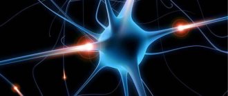
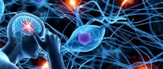
The nervous system consists of neurons, or nerve cells, and neuroglia, or neuroglial (or glial) cells. Neurons
- These are the main structural and functional elements in both the central and peripheral nervous systems.
Neurons are excitable cells, meaning they are capable of generating and transmitting electrical impulses (action potentials). Neurons have different shapes and sizes and form two types of processes: axons
and
dendrites
. There may be many dendrites, several, one, or none at all. Typically, a neuron has several short branched dendrites, along which impulses travel to the neuron’s body, and always one long axon, along which impulses travel from the neuron’s body to other cells (neurons, muscle or glandular cells). Neurons, according to the shape and nature of the processes from them, are: unipolar (single-process), biopolar (double-process), pseudounipolar (false-process) and multipolar (multi-process). The sizes of neurons are: small (up to 5 microns), medium (up to 30 microns) and large (up to 100 microns). The length of the processes of neurons is different: for example, in some the length of the processes is microscopic, while in others it is up to 1.5 m. For example, a neuron is located in the spinal cord, and its processes end in the fingers or toes. The transmission of a nerve impulse (excitation), as well as the regulation of its intensity, from one neuron to other cells occurs through specialized contacts - synapses.
Neuroglia
Main article: Neuroglia
Glial cells
are more numerous than neurons and make up at least half the volume of the central nervous system, but unlike neurons they cannot generate action potentials. Neuroglial cells are different in structure and origin; they perform auxiliary functions in the nervous system, providing support, trophic, secretory, delimitation and protective functions.
Structure and functions of the optic nerve
The optic nerve itself is a plexus of many thinnest nerve fibers, between which is the central arterial canal of the retina. Nerve fibers gather into the optic nerve at the posterior pole of the eye. In the place where they approach the optic nerve head (OND), the number of fibers increases, so a slight elevation above the retina is formed here. Next, the fibers gather into a disk, bend at an angle of 90˚ and limit the intraocular part of the optic nerve.
The optic nerve has three sheaths:
- Hard (brain) - connective tissue plate, no more than 0.35-0.50 mm thick, which thickens significantly at the point of its transition to the sclera
- Arachnoid - a thin layer (thickness only 10 microns) of collagen tissue covered with flat cells.
- Soft - loose connective tissue, which includes elastic, collagen, reticular fibers, as well as fibroblasts.
The space between them is filled with a liquid with a complex composition.
The optic nerve is divided into four sections: intraocular, intraorbital, intracanalicular and intracranial.
The optic nerve begins in the optic disc and ends in the chiasm, a kind of intersection of nerves. After this, part of the nerve fibers passes to the corresponding centers of the brain.
This is how the optic nerve performs its main function - delivering primary impulses to parts of the brain. From here, the impulses return the completed image to the visual center.
Comparative neuroanatomy
Types of nervous systems
There are several types of organization of the nervous system, represented in various systematic groups of animals.
- Diffuse nervous system - presented in coelenterates. Nerve cells form a diffuse nerve plexus in the ectoderm throughout the animal's body, and when one part of the plexus is strongly stimulated, a generalized response occurs - the whole body reacts.
- Stem nervous system (orthogon) - some nerve cells are collected into nerve trunks, along with which the diffuse subcutaneous plexus is preserved. This type of nervous system is present in flatworms and nematodes (in the latter the diffuse plexus is greatly reduced), as well as many other groups of protostomes - for example, gastrotrichs and cephalopods.
- The nodal nervous system, or complex ganglion system, is represented in annelids, arthropods, mollusks and other groups of invertebrates. Most of the cells of the central nervous system are collected in nerve nodes - ganglia. In many animals, the cells are specialized and serve individual organs. In some molluscs (for example, cephalopods) and arthropods, a complex association of specialized ganglia with developed connections between them arises - a single brain or cephalothoracic nerve mass (in spiders). In insects, some sections of the protocerebrum (“mushroom bodies”) have a particularly complex structure.
- A tubular nervous system (neural tube) is characteristic of chordates.
Nervous system of various animals
Nervous system of cnidarians and ctenophores
Cnidarians are considered the most primitive animals that have a nervous system. In polyps, it represents a primitive subepithelial nervous network ( nervous plexus
), entwining the entire body of the animal and consisting of neurons of different types (sensitive and ganglion cells), connected to each other by processes (
diffuse nervous system
), their especially dense plexuses are formed on the oral and aboral poles of the body.
Irritation causes rapid conduction of excitation through the body of the hydra and leads to contraction of the entire body, due to the contraction of epithelial-muscle cells of the ectoderm and at the same time their relaxation in the endoderm. Jellyfish are more complex than polyps; a central section begins to separate in their nervous system. In addition to the subcutaneous nerve plexus, they have ganglia along the edge of the umbrella, connected by processes of nerve cells into a nerve ring
, from which the muscle fibers of the velum and
rhopalia
- structures containing various sensory organs (
diffuse nodular nervous system
). Greater centralization is observed in scyphojellyfish and especially box jellyfish. Their 8 ganglia, corresponding to 8 rhopalia, reach quite large sizes.
The nervous system of ctenophores includes a subepithelial nerve plexus with condensations along rows of paddle plates that converge to the base of a complex aboral sensory organ. In some ctenophores, nearby nerve ganglia have been described.
Nervous system of protostomes
Flatworms
have a nervous system already divided into central and peripheral sections.
In general, the nervous system resembles a regular lattice - this type of structure was called orthogon
.
It consists of a cerebral ganglion, which in many groups surrounds the statocysts (endonal medulla), which is connected to the nerve trunks
running along the body and connected by annular transverse bridges (
commissures
).
Nerve trunks consist of nerve fibers extending from nerve cells scattered along their course. In some groups, the nervous system is quite primitive and close to diffuse. The following trends are observed among flatworms: ordering of the subcutaneous plexus with separation of trunks and commissures, an increase in the size of the cerebral ganglion, which turns into the central control apparatus, immersion of the nervous system into the thickness of the body; and, finally, a decrease in the number of nerve trunks (in some groups only two abdominal (lateral) trunks
).
In nemerteans, the central part of the nervous system is represented by a pair of connected double ganglia, located above and below the proboscis sheath, connected by commissures and reaching a significant size. Nerve trunks go back from the ganglia, usually in pairs, and they are located on the sides of the body. They are also connected by commissures; they are located in the skin-muscle sac or in the parenchyma. Numerous nerves depart from the head node, the most strongly developed are the spinal nerve (often double), abdominal and pharyngeal.
Gastrociliary worms have a suprapharyngeal ganglion, a peripharyngeal nerve ring, and two superficial lateral longitudinal trunks connected by commissures.
Nematodes have a peripharyngeal nerve ring, from which 6 nerve trunks extend forward and backward, the largest - the ventral and dorsal trunks - stretch along the corresponding hypodermal ridges. The nerve trunks are connected to each other by semicircular jumpers; they innervate the muscles of the abdominal and dorsal lateral bands, respectively. Nervous system of the nematode Caenorhabditis elegans
has been mapped at the cellular level[1]. Each neuron has been recorded, its origin has been traced, and most, if not all, neural connections are known[2]. In this species, the nervous system is sexually dimorphic: the male and hermaphroditic nervous systems have different numbers of neurons and groups of neurons to perform sex-specific functions.
In Kinorhynchus, the nervous system consists of a peripharyngeal nerve ring and a ventral (abdominal) trunk, on which, in accordance with their inherent body segmentation, ganglion cells are located in groups.
The nervous system of hairworms and priapulids has a similar structure, but their ventral nerve trunk is devoid of thickenings.
Rotifers have a large suprapharyngeal ganglion, from which nerves arise, especially large ones - two nerves that run through the entire body on the sides of the intestine. Smaller ganglia lie in the leg (pedal ganglion) and next to the masticatory stomach (mastax ganglion).
In acanthocephalans, the nervous system is very simple: inside the proboscis vagina there is an unpaired ganglion, from which thin branches extend forward to the proboscis and two thicker lateral trunks back; they emerge from the proboscis vagina, cross the body cavity, and then go back along its walls.
Annelids have a paired suprapharyngeal ganglion, with peripharyngeal connectives
(connectives, unlike commissures, connect opposite ganglia) connected to the ventral part of the nervous system.
In primitive polychaetes, it consists of two longitudinal nerve cords in which nerve cells are located. In more highly organized forms, they form paired ganglia in each segment of the body ( nervous scala
), and the nerve trunks come closer together.
In the majority of polychaetes, the paired ganglia merge ( ventral nerve cord
); in some, their connectives also merge.
Numerous nerves depart from the ganglia to the organs of their segment. In the series of polychaetes, the nervous system is immersed from under the epithelium into the thickness of the muscles or even under the skin-muscular sac. Ganglia of different segments can be concentrated if their segments merge. Similar trends are observed in oligochaetes. In leeches, the nerve chain lying in the abdominal lacunar canal consists of 20 or more ganglia, and the first 4 ganglia ( subpharyngeal ganglion
) and the last 7 are combined into one.
In echiurids, the nervous system is poorly developed - the peripharyngeal nerve ring is connected to the abdominal trunk, but the nerve cells are scattered evenly throughout them and do not form nodes anywhere.
Sipunculids have a suprapharyngeal nerve ganglion, a peripharyngeal nerve ring, and a nerveless ventral trunk lying on the inside of the body cavity.
Tardigrades have a suprapharyngeal ganglion, peripharyngeal connectives, and a ventral chain with 5 paired ganglia.
Onychophorans have a primitive nervous system. The brain consists of three sections: the protocerebrum innervates the eyes, the deutocerebrum innervates the antennae, and the tritocerebrum innervates the foregut. Nerves extend from the peripharyngeal connectives to the jaws and oral papillae, and the connectives themselves pass into distant abdominal trunks, evenly covered with nerve cells and connected by thin commissures.
Nervous system of arthropods
In arthropods, the nervous system is composed of a paired suprapharyngeal ganglion, consisting of several connected nerve ganglia (brain), peripharyngeal connectives and a ventral nerve cord, consisting of two parallel trunks. In most groups, the brain is divided into three sections - proto-
,
deutocerebrum
and
tritocerebrum
. Each body segment has a pair of nerve ganglia, but the fusion of ganglia to form large nerve centers is often observed; for example, the subpharyngeal ganglion consists of several pairs of fused ganglia - it controls the salivary glands and some muscles of the esophagus.
In a number of crustaceans, in general, the same trends are observed as in annelids: the convergence of a pair of abdominal nerve trunks, the fusion of paired nodes of one body segment (that is, the formation of the abdominal nerve chain), the fusion of its nodes in the longitudinal direction as the body segments unite. Thus, crabs have only two nerve masses - the brain and a nerve mass in the chest, while copepods and barnacles form a single compact formation, penetrated by the canal of the digestive system. The brain of crayfish consists of paired lobes - the protocerebrum, from which the optic nerves, which have ganglion clusters of nerve cells, depart, and the deutocerebrum, which innervates antennae I. Usually, a tritocerebrum is also added, formed by the fused nodes of the antennal segment II, the nerves to which usually arise from the peripharyngeal connectives. Crustaceans have a developed sympathetic nervous system
, consisting of a medulla and an unpaired
sympathetic nerve
, which has several ganglia and innervates the intestines.
An important role in the physiology of crayfish is played by neurosecretory cells
located in various parts of the nervous system and secreting
neurohormones
.
The brain of centipedes has a complex structure, most likely formed by many ganglia. The subpharyngeal ganglion innervates all oral limbs, from which a long paired longitudinal nerve trunk begins, on which there is one paired ganglion in each segment (in bipedal centipedes, in each segment, starting from the fifth, there are two pairs of ganglia located one after the other).
The nervous system of insects, also consisting of the brain and the ventral nerve cord, can achieve significant development and specialization of individual elements. The brain consists of three typical sections, each of which consists of several ganglia separated by layers of nerve fibers. Important associative
protocerebrum. Social insects (ants, bees, termites) have especially developed brains. The abdominal nerve chain consists of the subpharyngeal ganglion, which innervates the oral limbs, three large thoracic ganglia and abdominal ganglia (no more than 11). In most species, more than 8 ganglia are not found in adulthood; in many, these also merge, giving rise to large ganglion masses. It can go so far as to form only one ganglion mass in the thorax, innervating both the thorax and the abdomen of the insect (for example, in some flies). During ontogenesis, ganglia often unite. Sympathetic nerves arise from the brain. Almost all parts of the nervous system contain neurosecretory cells.
In horseshoe crabs, the brain is not externally divided, but has a complex histological structure. Thickened peripharyngeal connectives innervate the chelicerae, all limbs of the cephalothorax and gill covers. The abdominal nerve cord consists of 6 ganglia, the posterior one is formed by the fusion of several. The nerves of the abdominal limbs are connected by longitudinal lateral trunks.
The nervous system of arachnids has a clear tendency to concentrate. The brain consists only of protocerebrum and tritocerebrum due to the lack of structures innervated by deutocerebrum. The metamerism of the ventral nerve chain is most clearly preserved in scorpions - they have a large ganglion mass in the chest and 7 ganglia in the abdomen, in salpugs there is only 1, and in spiders all the ganglia have merged into the cephalothorax nerve mass; in harvestmen and ticks there is no distinction between it and the brain.
Sea spiders, like all chelicerates, do not have a deuterocerebrum. The ventral nerve cord in different species contains from 4-5 ganglia to one continuous ganglion mass.
Nervous system of mollusks
In primitive chiton mollusks, the nervous system consists of a peripharyngeal ring (innervates the head) and 4 longitudinal trunks - two pedal
(innervate the leg, which are connected in no particular order by numerous commissures, and two
pleurovisceral
, which are located outward and above the pedal ones (innervate the visceral sac, they are connected above the powder). The pedal and pleurovisceral trunks of one side are also connected by many jumpers.
The nervous system of monoplacophorans is structured similarly, but their pedal trunks are connected by only one bridge.
In more developed forms, as a result of the concentration of nerve cells, several pairs of ganglia are formed, which are shifted to the anterior end of the body, with the suprapharyngeal node (brain) receiving the greatest development.
Nervous system of deuterostomes
Vertebrate nervous system
The nervous system of vertebrates is often divided into the central nervous system (CNS) and the peripheral nervous system (PNS). The central nervous system consists of the brain and spinal cord. The PNS is made up of other nerves and neurons that do not lie within the CNS. The vast majority of nerves (which are actually the axons of neurons) belong to the PNS. The peripheral nervous system is divided into the somatic nervous system and the autonomic nervous system.
The somatic nervous system is responsible for coordinating body movement and receiving and transmitting external stimuli. This system regulates actions that are under conscious control.
The autonomic nervous system is divided into parasympathetic and sympathetic. The sympathetic nervous system responds to danger or stress, and, among many physiological changes, can cause an increase in heart rate and blood pressure and arousal of the senses due to an increase in adrenaline in the blood. The parasympathetic nervous system, on the other hand, is responsible for the state of rest, and ensures that the pupil contracts, slows the heart, dilates blood vessels and stimulates the digestive and genitourinary systems.
Mammalian nervous system
The nervous system functions as an integral unit with sensory organs, such as the eyes, and is controlled in mammals by the brain. The largest part of the latter is called the cerebral hemispheres (in the occipital region of the skull there are two smaller hemispheres of the cerebellum). The brain connects to the spinal cord. In all mammals, with the exception of monotremes and marsupials, unlike other vertebrates, the right and left cerebral hemispheres are connected to each other by a compact bundle of nerve fibers called the corpus callosum. In the brains of monotremes and marsupials there is no corpus callosum, but the corresponding areas of the hemispheres are also connected by nerve bundles; for example, the anterior commissure connects the right and left olfactory areas with each other. The spinal cord, the main nerve trunk of the body, passes through a canal formed by the foramina of the vertebrae and extends from the brain to the lumbar or sacral spine, depending on the species. On each side of the spinal cord, nerves extend symmetrically to different parts of the body. The sense of touch is, in general terms, provided by certain nerve fibers, countless endings of which are located in the skin. This system is usually supplemented by hairs that act as levers to press on areas riddled with nerves.
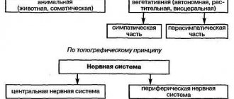
Morphological division
The nervous system of mammals and humans is divided according to morphological characteristics into central (brain and spinal cord) and peripheral (composed of nerves extending from the brain and spinal cord [3]).
The composition of the central nervous system can be represented as follows:
| |||||||||||||||||||
The peripheral nervous system includes cranial nerves, spinal nerves and nerve plexuses
Functional division
- Somatic (animal) nervous system
- Autonomic (autonomic) nervous system Sympathetic division of the autonomic nervous system
- Parasympathetic division of the autonomic nervous system
- Metasympathetic division of the autonomic nervous system (enteric nervous system)
Ontogenesis
Models
At the moment, there is no single position on the development of the nervous system in ontogenesis. The main problem is to assess the level of determinism (predestination) in the development of tissues from germ cells. The most promising models are the mosaic model
and
regulatory model
. Neither one nor the other can fully explain the development of the nervous system.
- The mosaic model assumes complete determination of the fate of an individual cell throughout ontogeny.
- The regulatory model assumes the random and variable development of individual cells, with only the neural direction being deterministic (that is, any cell of a certain group of cells can become anything within the scope of development for this group of cells).
For invertebrates, the mosaic model is almost flawless—the degree of determination of their blastomeres is very high. But for vertebrates everything is much more complicated. A certain role of determination here is undoubted. Already at the sixteen-cell stage of development of the vertebrate blastula, it is possible to say with a fair degree of certainty which blastomere is not
the predecessor of a certain organ.
Marcus Jacobson introduced a clonal model of brain development (close to regulatory) in 1985. He suggested that the fate of individual groups of cells representing the progeny of an individual blastomere, that is, “clones” of this blastomere, is determined. Moody and Takasaki (independently) developed this model in 1987. A map of the 32-cell blastula stage was constructed. For example, it has been established that descendants of the D2 blastomere (vegetative pole) are always found in the medulla oblongata. On the other hand, the descendants of almost all blastomeres of the animal pole do not have pronounced determination. In different organisms of the same species, they may or may not occur in certain parts of the brain.
Regulatory Mechanisms
It was found that the development of each blastomere depends on the presence and concentration of specific substances - paracrine factors, which are secreted by other blastomeres. For example, in an in vitro
with the apical part of the blastula, it turned out that in the absence of activin (paracrine factor of the vegetative pole), the cells develop into ordinary epidermis, and in its presence, depending on the concentration, in increasing order: mesenchymal cells, smooth muscle cells, notochord cells or cardiac muscle cells.
All substances that determine the behavior and fate of the cells that perceive them, depending on the dose (concentration) of the substance in a given area of the multicellular embryo, are called morphogens
.
Some cells secrete soluble active molecules (morphogens) into the extracellular space, decreasing from their source along a concentration gradient.
That group of cells whose location and purpose is specified within the same boundaries (with the help of morphogens) is called a morphogenetic field
. The fate of the morphogenetic field itself is strictly determined. Each specific morphogenetic field is responsible for the formation of a specific organ, even if this group of cells is transplanted into different parts of the embryo. The fates of individual cells within the field are not so rigidly fixed, so that they can, within certain limits, change their purpose, replenishing the functions of cells lost by the field. The concept of the morphogenetic field is a more general concept; in relation to the nervous system, it corresponds to the regulatory model.
The concept of embryonic induction is closely related to the concepts of morphogen and morphogenetic field. This phenomenon, also common to all body systems, was first shown in the development of the neural tube.
Development of the vertebrate nervous system
The nervous system is formed from the ectoderm, the outermost of the three germ layers. A paracrine interaction begins between the cells of the mesoderm and ectoderm, that is, a special substance is produced in the mesoderm - neuronal growth factor, which is transferred to the ectoderm. Under the influence of neuronal growth factor, part of the ectodermal cells turns into neuroepithelial cells, and the formation of neuroepithelial cells occurs very quickly - at a rate of 250,000 pieces per minute. This process is called neuronal induction (a special case of embryonic induction).
As a result, a neural plate is formed, which consists of identical cells. From it the neural folds are formed, and from them the neural tube, which is separated from the ectoderm (it is the change in the types of cadherins, cell adhesion molecules, that is responsible for the formation of the neural tube and neural crest), going under it. The mechanism of neurulation differs somewhat between lower and higher vertebrates. The neural tube does not close along its entire length simultaneously. First of all, the closure occurs in the middle part, then this process spreads to its rear and front ends. At the ends of the tube, two open sections remain - the anterior and posterior neuropores.
Then the process of differentiation of neuroepithelial cells into neuroblasts and glioblasts occurs. Glioblasts give rise to astrocytes, oligodendrocytes and epindymal cells. Neuroblasts become neurons. Next, the process of migration occurs - neurons move to where they will perform their function. Due to the growth cone, the neuron crawls like an amoeba, and the processes of glial cells indicate its path. The next stage is aggregation (sticking together of neurons of the same type, for example, those involved in the formation of the cerebellum, thalamus, etc.). Neurons recognize each other thanks to surface ligands - special molecules found on their membranes. Having united, the neurons are arranged in the order necessary for a given structure.
After this, the nervous system matures. An axon grows from the growth cone of the neuron, and dendrites grow from the body.
Then fasciculation occurs - the union of similar axons (formation of nerves).
The last stage is the programmed death of those nerve cells in which a malfunction occurred during the formation of the nervous system (about 8% of cells send their axon to the wrong place).
3.1. Nervous system of invertebrates
3. DEVELOPMENT OF THE NERVOUS SYSTEM IN PHYLOGENESIS
Invertebrate animals are characterized by the presence of several sources of origin of nerve cells. In the same type of animal, nerve cells can simultaneously and independently originate from three different germ layers. Polygenesis of invertebrate nerve cells is the basis for the diversity of mediator mechanisms in their nervous system.
The nervous system first appears in coelenterates.
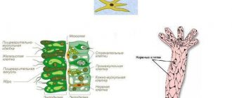
Coelenterates are two-layered animals.
Their body is a hollow sac, the internal cavity of which is the digestive cavity. The nervous system of coelenterates belongs to the diffuse type. Each nerve cell in it is connected by long processes to several neighboring ones, forming a nerve network. Nerve cells of coelenterates do not have specialized polarized processes. Their processes conduct excitation in any direction and do not form long pathways. Contacts between nerve cells of the diffuse nervous system are of several types. There are plasma contacts
that ensure continuity of the network (
anastomoses
).
Gap-like contacts
also appear between the processes of nerve cells, similar to synapses.
Moreover, among them there are contacts in which synaptic vesicles are located on both sides of the contact - the so-called symmetric synapses
, and there are also
asymmetric synapses:
in them the vesicles are located only on one side of the gap.
The nerve cells of a typical coelenterate animal, Hydra, are evenly distributed over the surface of the body, forming some clusters in the area of the mouth and sole (Fig. 8). The diffuse nervous network conducts excitation in all directions. In this case, the wave of spreading excitation is accompanied by a wave of muscle contraction.
Rice. 8. Scheme of the structure of the diffuse nervous system of a coelenterate animal:
1 - mouth opening; 2 - tentacle; 3 - sole
Rice. 9. Scheme of the structure of the diffuse-stem nervous system of turbellaria:
1 - nerve node; 2 - pharynx; 3 - abdominal longitudinal trunk; 4 - lateral nerve trunk
The next stage in the development of invertebrates is the appearance of three-layer animals - flatworms.
Like coelenterates, they have an intestinal cavity that communicates with the external environment through the mouth. However, they have a third germ layer - mesoderm and a bilateral type of symmetry. The nervous system of lower flatworms belongs to the diffuse type. However, several nerve trunks are already isolated from the diffuse network (Fig.
9
,
3
,
4
).
In free-living flatworms, the nervous apparatus acquires features of centralization. Nerve elements are collected in several longitudinal trunks (Fig. 10, 4
,
5
) (the most highly organized animals are characterized by the presence of two trunks), which are connected to each other by transverse fibers (commissures) (Fig. 10,
6
).
A nervous system ordered in this way is called an orthogon.
Orthogonal trunks are a collection of nerve cells and their processes (Fig. 10).
Along with bilateral symmetry, flatworms develop the anterior end of the body, on which sensory organs are concentrated (statocysts, “eyes,” olfactory pits, tentacles). Following this, an accumulation of nervous tissue appears at the anterior end of the body, from which the brain or cerebral ganglion is formed (Fig. 10, 3
). In cerebral cells
Rice. 10. Scheme of the structure of the orthogonal nervous system of the ciliated worm (anterior end):
1 - tentacular outgrowth; 2 - nerve innervating the outgrowth; 3 - cerebral ganglion; 4 - lateral longitudinal nerve trunk; 5 - abdominal longitudinal nerve trunk; 6 - commissure
ganglion, long processes appear that go into the longitudinal trunks of the orthogon (Fig. 10, 4
,
5
).
Thus, the orthogon represents the first step towards the centralization of the nervous apparatus and its cephalization (the appearance of the brain). Centralization and cephalization are the result of the development of sensory (sensitive) structures.
The next stage in the development of invertebrate animals is the appearance of segmented animals - annelids.
Their body is metameric, i.e.
consists of segments. The structural basis of the nervous system of annelids is the ganglion,
a paired collection of nerve cells located one in each segment.
Nerve cells in the ganglion are located along the periphery. Its central part is occupied by the neuropil -
an interlacing of nerve cell processes and glial cells. The ganglion is located on the ventral side of the segment under the intestinal tube. It sends its sensory and motor fibers to its segment and to two neighboring ones. Thus, each ganglion has three pairs of lateral nerves, each of which is mixed and innervates its own segment. Sensory fibers coming from the periphery enter the ganglion through the ventral nerve roots. Motor fibers exit the ganglion along the dorsal nerve roots. Accordingly, sensory neurons are located in the ventral part of the ganglion, and motor neurons are located in the dorsal part. In addition, the ganglion contains small cells that innervate internal organs (vegetative elements); they are located laterally - between sensory and motor neurons. Among the neurons of the sensitive, motor or associative zones of the ganglia of annelids, no grouping of elements was found; the neurons are distributed diffusely, i.e. do not form centers.
The ganglia of annelids are connected to each other in a chain. Each subsequent ganglion is connected to the previous one through
Rice. 11. Scheme of the structure of the nodal nervous system of an insect:
1 - suprapharyngeal nerve ganglion;
2 - subpharyngeal nerve ganglion;
3 - complex fused ganglion of the thoracic segment; 4 - abdominal ganglion; 5 - peripheral nerve; 6 - connective
nerve trunks, which are called connectives.
At the anterior end of the body of annelids, two fused ganglia form a large subpharyngeal ganglion. Connectives from the subpharyngeal ganglion, going around the pharynx, flow into the suprapharyngeal ganglion, which is the most rostral (anterior) part of the nervous system. The suprapharyngeal ganglion consists of only sensory and associative neurons. No motor elements were found there. Thus, the supra-pharyngeal ganglion of annelids is the highest association center; it exercises control over the sub-pharyngeal ganglion. The subpharyngeal ganglion controls the underlying nodes; it has connections with two or three subsequent ganglia, while the remaining ganglia of the ventral nerve chain do not form connections longer than to the neighboring ganglion.
In the phylogenetic series of annelids, there are groups with well-developed sensory organs (polychaetes). In these animals, three sections are separated in the suprapharyngeal ganglion. The anterior part innervates the tentacles, the middle part innervates the eyes and antennae. Finally, the back part develops in connection with the improvement of the chemical senses.
of arthropods has a similar structure.
, i.e. built according to the type of abdominal nerve chain, but can reach a high level of development (Fig. 11). It includes a significantly developed suprapharyngeal ganglion, which performs the function
Rice. 12. Diagram of the structure of the brain of an insect (bee). The left half is its cross-section:
1 - mushroom body; 2 - protocerebrum; 3 - visual blade; 4 - deutocerebrum; 5 - tritocerebrum
tion of the brain, the subpharyngeal ganglion, which controls the organs of the oral apparatus, and the segmental ganglia of the ventral nerve cord. The ganglia of the ventral nerve cord can fuse with each other, forming complex ganglion masses.
Brain
arthropods consists of three sections: anterior -
protocerebrum
, middle -
deuterocerebrum
and posterior -
tritocerebrum.
The insect brain has a complex structure. Particularly important associative centers of insects are the mushroom bodies located on the surface of the protocerebrum, and the more complex behavior the species is characterized by, the more developed its mushroom bodies are. Therefore, mushroom bodies reach their greatest development in social insects (Fig. 12).
exist in almost all parts of the nervous system of arthropods .
Neurosecrets play an important regulatory role in the hormonal processes of arthropods.
In the process of evolution, initially diffusely located bipolar neurosecretory cells perceived signals either by processes or by the entire surface of the cell, then neurosecretory centers, neurosecretory tracts and neurosecretory contact areas were formed. Subsequently, the specialization of nerve centers occurred, the degree of reliability in the relationship between the two main regulatory systems (nervous and humoral) increased, and a fundamentally new stage of regulation was formed - subordination of the neurosecretory centers of the peripheral endocrine glands.
Rice. 13. Scheme of the structure of the ganglion nervous system of the elasmobranch mollusk (toothless):
1 - cerebral commissure; 2 - cerebral ganglia; 3 - pedal ganglia; 4 - connective; 5 - visceral ganglia
Nervous system of mollusks
also has
a ganglion structure
(Fig. 13). In the simplest representatives of the type, it consists of several pairs of ganglia. Each pair of ganglia controls a specific group of organs: the leg, visceral organs, lungs, etc. - and is located next to the innervated organs or inside them. The ganglia of the same name are connected in pairs by commissures. In addition, each ganglion is connected by long connectives to the cerebral ganglion complex.
In more highly organized mollusks (cephalopods), the nervous system is transformed (Fig. 14). Its ganglia merge and form a common peripharyngeal mass - the brain.
Two large pallial nerves arise from the posterior part of the brain and form two large stellate ganglia. Thus, cephalopods exhibit a high degree of cephalization.
Return to contents
© 2000-NIV
Neuroscience
Modern science of the nervous system combines many scientific disciplines: along with classical neuroanatomy, neurology and neurophysiology, molecular biology and genetics, chemistry, cybernetics and a number of other sciences make an important contribution to the study of the nervous system. This interdisciplinary approach to the study of the nervous system is reflected in the term neuroscience. In Russian-language scientific literature, the term “neurobiology” is often used as a synonym. One of the main goals of neuroscience is to understand the processes occurring both at the level of individual neurons and neural networks, the result of which are various mental processes: thinking, emotions, consciousness. In accordance with this task, the study of the nervous system is carried out at different levels of organization, from the molecular to the study of consciousness, creativity and social behavior.
Professional societies and magazines
The Society for Neuroscience (SfN, the Society for Neuroscience)[4] is the largest non-profit international organization, uniting more than 38 thousand scientists and doctors involved in the study of the brain and nervous system. The society was founded in 1969 and is headquartered in Washington. Its main goal is the exchange of scientific information between scientists. To this end, an international conference is held annually in various cities in the United States and the Journal of Neuroscience is published[5]. The society carries out educational and educational work.
The Federation of European Neuroscience Societies (FENS, the Federation of European Neuroscience Societies)[6] unites a large number of professional societies from European countries, including Russia. The Federation was founded in 1998 and is a partner of the American Society for Neuroscience (SfN). The Federation holds an international conference in different European cities every 2 years and publishes the European Journal of Neuroscience[7].
Notes
- Due to the fact that the body of these animals always consists of the same set of cells.
- White JG, Southgate E., Thomson JN, Brenner S.
The structure of the nervous system of the nematode
Caenorhabditis elegans
//
Phil.
Trans. Royal Soc. London ,
314
, 1986. - P. 1-340. - Mednikov, 1994, p. 363.
- Society for Neuroscience -. www.sfn.org. Retrieved November 22, 2015.
- The Journal of Neuroscience. www.jneurosci.org. Retrieved November 22, 2015.
- Fens Home Page (English). FENS.org. Retrieved November 22, 2015.
- European Journal of Neuroscience.
- Harriet Cole: Drexel's Longest-Serving Employee.