What is the hippocampus?
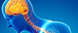
The hippocampus is located in the lower middle part of the brain, known as the temporal lobe, on the bilateral side. The hippocampus is 1/100 the size of the cerebral cortex and consists of three layers with characteristic pyramidal cells.
People have known about the hippocampus for 4 centuries, making it one of the most studied areas of the brain. Its main functions include learning and memory.
In the 1950s, a patient with epilepsy who failed treatment was given brain surgery. The part of the brain that seemed to cause the epileptic seizures was removed. These were the hippocampi.
The patient recovered from the operation, but developed serious memory problems. He remembered his early childhood, but could not remember how old he was. More importantly, he could not remember new events or words. The patient even forgot what he had recently said. Since his death in 2008, scientists have greatly expanded our understanding of memory and brain disease.
The hippocampus is part of the limbic system, which includes the area of the brain associated with feelings and reactions. Located on the periphery of the cortex, the limbic system includes the hypothalamus and amygdala. These structures help control various body functions such as the endocrine system.
Association with diseases
Dysfunction of the hippocampus is observed in the following diseases and syndromes:
- Alzheimer's disease, in the early stages manifests itself in short-term memory disorders, and later in long-term memory. In this case, the size of the hippocampus decreases. Pathology most often occurs in old age (over 65 years). Gradually, the patient experiences disturbances in speech, cognitive abilities, and orientation in a familiar environment. He becomes unable to take care of himself.
- Hypoxia (oxygen starvation).
- Inflammation of the brain (encephalitis).
- Temporal lobe epilepsy. In more than 30% of cases, this disease is associated with perinatal brain damage due to fetal hypoxia, infectious pathologies (measles, rubella, syphilis and others), and can also occur after birth injuries.
When the hippocampus is damaged, Korsakoff's syndrome is observed, in which a person cannot remember current events, but events from the distant past are retained in his memory. Patients are disoriented in time and space, do not know where they are, and cannot name the current date.
Functions of the hippocampus
The hippocampus is involved in two specific types of memory: declarative memory and spatial memory.
Declarative memory deals with facts and events. Learning how to remember a speech or line in a game is a good example of declarative memory in action.
Spatial memory involves remembering a route, such as when a taxi driver can remember the route of a city. Researchers can now say that spatial memory is stored in the right hippocampus.
The hippocampus also plays another important role in memory. This is where short-term memories are converted into long-term memories and then stored in another area of the brain. It was previously thought that new nerve cells developed only in embryos or young children, but new research has shown that nerve cells develop throughout adult life. The hippocampus is one of the few places in the brain where new nerve cells are formed.
When the hippocampus is damaged due to disease or injury, a person may experience memory problems. They cannot remember recent events, but they remember events that happened long ago.
Transient global amnesia
is a specific form of memory loss that develops suddenly, seemingly on its own. Most patients with transient global amnesia regain their memory, but researchers are not entirely clear why this happens.
(c) Wikimedia/Life Sciences Database
Memory
Many scientists agree that the hippocampus plays an important role in the formation of new memories. This area of the brain is also involved in explicit memory, which is associated with conscious memories, such as events from the distant past. However, as research shows, when the hippocampus is damaged, the functions of acquiring new skills (playing a musical instrument and others) are not completely lost.
There is an opinion that this part of the brain helps a person retain information while awake, and during sleep transfers it to the cerebral cortex. The hippocampus is also involved every time when it is necessary to remember and reproduce spatial landmarks, that is, it contributes to the formation of cognitive maps or an image of a familiar object environment.
Diseases affecting the hippocampus
The hippocampus is a sensitive area of the brain and can be negatively affected by many different conditions, including long-term exposure to severe stress.
Three diseases that affect the ability of the hippocampus to perform its function:
- Alzheimer's disease;
- Epilepsy;
- Depression.
Alzheimer's disease
is a leading cause of dementia and memory loss. As the disease progresses, the affected areas of the brain begin to shrink. The hippocampus loses volume and is unable to function normally.
There is a strong connection between the hippocampus and epilepsy
. In 50 - 75% of patients suffering from epilepsy, damage to the hippocampus was found after autopsy. As the researchers note, it is not yet clear whether epilepsy is a cause or a consequence of damage to the hippocampus.
The hippocampus also loses volume in cases of severe depression.
There is considerable evidence that stress has a negative effect on the hippocampus. Thus, Russian scientists have discovered the mechanism of how stress affects the hippocampus, and people with Cushing's disease have a number of symptoms associated with high levels of cortisol. This hormone is produced when people are under stress. One of the symptoms is a decrease in the size of the hippocampus. The hippocampus is currently the subject of new research. Scientists believe that exercise in old age can strengthen the ability of this structure to generate new nerve cells. This would preserve and potentially improve memory.
How the hippocampus is connected to our episodic memory
Scientists from Chicago tested an experiment in which they stimulated neural connections between the cerebral cortex and the hippocampus. This stimulation caused hippocampal activity to decrease, causing participants to better recall details in memory tests. It turns out that the hippocampus is not only the conductor of our episodic memory, but also an optimizer of our memories, making them more monotonous and less vivid. The article was published in the journal Current Biology.
Hippocampus
What is episodic memory from the brain's perspective? Neuroscience says that our memories of certain events are the re-activation of the same neurons that were involved at the moment of remembering. Firing the right set of neurons requires control, which is exerted by a structure in the brain called the hippocampus.
The hippocampus is a kind of human memory center, a structure that is responsible for the consolidation of memories. The absence of the hippocampus (as in the famous case of Henry Molaison) or malfunctions in its functioning lead to the fact that a person does not form new long-term memories. Experiments with functional magnetic resonance imaging (fMRI) have shown that the hippocampus seems to orchestrate the brain at the moment when we remember an event: the activity of the hippocampus correlates well with the activity of other areas of the cortex. In this way, the hippocampus seems to integrate details of memories that are stored in very different areas (for example, a beautiful landscape that we remember is partly encoded in the visual cortex, and the birdsong we heard while observing the landscape is partly encoded in the auditory cortex).
The hippocampus controls episodic memory, connecting brain areas such as the medial and lateral temporal lobes, the parietal cortex, the medial frontal cortex, and a number of subcortical areas.
Knowing the areas of the brain responsible for episodic memories, scientists can use various technologies to influence this type of memory. One of these technologies is transcranial magnetic stimulation, or rather its specific type: targeted stimulation of the hippocampal network. TMS is applied to those areas of the brain that are functionally connected to the hippocampus. In this experiment, stimulation was provided by short-term biphasic triple theta bursts with a frequency of 50 Hz, produced every 200 ms. This frequency is believed to successfully replicate natural brain activity. Data from previous experiments have shown that such stimulus parameters can improve performance on tests of word or location memory.
Credit: public domain
One of the areas most closely associated with the hippocampus is the lateral parietal area. At the beginning of the experiment, the scientists stimulated 20 participants in this area, and then placed them in an fMRI scanner. Next, the participants were shown several video clips that they had to remember as best as possible.
Experimental design
Credit: Hebscher, M., et al. Current Biology, 2021
The video clips, filled with a lot of detail, told about various scenes from everyday life. After watching the clips, the researchers tested how participants remembered specific details of the video using two questionnaires: one set of questions tested memory for the general meaning of the video, the other focused on its context. Participants were also asked to answer a question about how vivid the memories were in their subjective opinion.
Credit: Hebscher, M., et al. Current Biology, 2021
The next day, the scientists invited participants again and repeated the procedure (session 2). However, this time they stimulated a different area, which is in no way connected with the hippocampus. Thus, scientists were able to collect data on how the brain works when it remembers and remembers some material in the presence and absence of magnetic stimulation.
The researchers, having a pattern of neural activity at the time of memorization and reproduction, then compared them. The results of the experiment showed that after applying stimulation, participants answered better (more accurately) questions of the second category, which were related to the context of the video fragments. However, in the control group, where stimulation occurred in those areas of the brain that were not related to episodic memory, the situation was different, as can be seen from the graph below.
Credit: Hebscher, M., et al. Current Biology, 2021
The fact that memory accuracy increased suggests that stimulation of the parietal cortex affected the functions of the hippocampus, and not the parietal cortex itself. As previous studies have shown, targeting the parietal cortex would affect the vividness of memories, but this did not change.
By comparing the activity of different hemispheres, scientists were convinced that stimulation affected mainly the left half of the brain.
It is also worth noting that neural patterns when remembering the same information are similar to each other. Thus, it can be assumed that the stimulation affected both relevant neurons (which were activated in response to the shown video) and irrelevant neurons (which were activated in response to other videos already in memory). Thus, perhaps stimulation did not affect specific memorization, but only changed the properties of the neurons responsible for the process itself? Scientists have answered this question as well. It turned out that stimulation contributed to better recovery of activity patterns for the relevant video fragment, and reduced memory for details of irrelevant material.
How to explain the results obtained?
Scientists hypothesized that stimulation leads to decreased activity in the hippocampus in the left hemisphere. This in turn affects other areas of the brain (in the left hemisphere).
It is possible that the more activity in the hippocampus at the moment of memorization, the more our brain generalizes what we saw. As a result, when answering questions, all videos (both relevant and irrelevant at a particular point in time) seem the same to us. The hippocampus, as it were, generalizes all the details, and also suppresses other areas that try to capture the unique features of a video fragment. The cortex, in turn, on the contrary, helps us remember specifics. As a result, without stimulation, the accuracy of our episodic memory decreases.
Credit: Hebscher, M., et al. Current Biology, 2021
By comparing neural patterns of activity, the scientists were convinced that the functioning of the hippocampus led to more frequent overlap of different neural regions. That is, the specificity of the information was indeed lost.
At the same time, less activity in the hippocampus was associated with participants being better at recalling various details for specific video segments. This was the positive effect of stimulation, which seemed to suppress the activity of the hippocampus at the time of memorization. Suppressed generalization.
It turns out that stimulating the parietal cortex, which is closely connected to the hippocampus (functionally), increased memory accuracy and also caused the pattern of activity (in the parietal and occipital cortex) during retrieval to more accurately replicate the pattern during memorization. Even one session of theta stimulation led to this result. What then can happen if such stimulation is carried out constantly? In the future, scientists plan to answer this question.
“That’s why sometimes it seems to us that remembering some events from life is like mental time travel,”
says lead researcher, Melissa Hebscher.
“Our results show that stimulation improves this mental journey and improves its accuracy. This, in turn, opens the way for the development of safe and effective ways to improve memory in people.”
Text: Nikita Otstavnov
Hebscher, M., Kragel, J. E., Kahnt, T., & Voss, J. L. (2021). Enhanced reinstatement of naturalistic event memories due to hippocampal-network-targeted stimulation. Current Biology
. doi:10.1016/j.cub.2021.01.027
Treatment
To reduce the symptoms of the disease, neurologists at the Yusupov Hospital prescribe antiepileptic drugs. The drug of first choice is Carbamazepine. Second-choice drugs include Valproate, Diphenin and Hexamidine. After treatment, some patients stop having attacks and go into long-term remission.
In cases of resistance to therapy and progression of hippocampal sclerosis, surgical treatment is performed in partner clinics. It involves removing the temporal lobe of the brain (lobectomy). After surgery, the number of attacks decreases in 70-95% of cases. If you are faced with the problem of hippocampal sclerosis and would like to receive qualified specialized medical care, call us. You will be scheduled for a consultation with a neurologist at the Yusupov Hospital.
Almond nuclei
In a series of brain autopsies of patients with schizophrenia, no significant increase in the volume of the amygdala nuclei was detected (Steven A. et al., 2002). However, data obtained using MRI currently refers to rather crude imaging methods, not to mention CT, and may be less reliable compared to autopsy.
In a comparative review by Wright et al (2000), who studied the size of 44 brain regions in schizophrenia, it was noted that the amygdala of the left and right hemispheres was reduced in volume by 10%, and this was noticeable to a greater extent than the reduction in volume of other brain regions. Loss of gray matter in the amygdala and changes in the shape of the amygdala have also been demonstrated through studies that looked at ventricular-brain index scores. At the same time, many authors note methodological difficulties in studying the amygdala of the limbic system of patients with schizophrenia.
Return to Contents