What is it about?
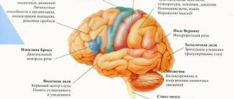
We all know very well that the human brain is a very complex unique structure in which absolutely all elements are inextricably and firmly connected through millions of neural connections. The brain contains gray and white matter. The first is a common accumulation of many nerve cells, and the second is responsible for the speed of impulse transmission between neurons. In addition to the cortex, of course, there are other structures. They are nuclei or basal ganglia, consisting of gray matter and found in white matter. In many ways, they are responsible for the normal functioning of the nervous system.
Definition and structure
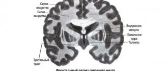
The subcortical nuclei, also known as the basal ganglia, are a collection of gray matter located beneath the cortical structures. The nuclei are located deep in the white matter within the cerebral hemispheres within the brain near the lateral ventricles and include the striatum and amygdala. Between the nuclei lie capsules formed by projection paths. The striatum is located under the cortex and consists of structures:
- Caudate nucleus. Located below the lateral ventricle, slightly above and to the side of the thalamus. The caudate nucleus consists of a head (lateral wall of the anterior ventricular horn), body (lies at the bottom of the ventricle) and tail (rises to the upper wall of the occipital ventricular horn). On the medial (closer to the median plane) side, the tail is close to the thalamus, from which it is separated by a thin strip consisting of white matter.
- Lenticular nucleus. The anatomical structure suggests the presence of a globus pallidus and putamen within this brain structure. Located next to the thalamus and lateral to the caudate nucleus. On the side of the lenticular nucleus, closer to the edge of the brain relative to the median plane, there is an external capsule; on the other edge, closer to the median plane, there is an internal capsule. The internal capsule serves as the boundary between the lenticular and caudate nuclei. A horizontal section of the lenticular nucleus is presented in the form of a wedge.
- Fence. It is represented by a plate consisting of gray matter, the thickness of which does not exceed 2 mm. It is located outward from the lenticular region.
The striped brain got its name because of the appearance on a section of the brain, which is represented by alternating white and gray stripes. The striatum consists of a group of nuclei that perform the tasks of motor centers. Layers consisting of white matter divide the lenticular structure of the brain into two globus pallidus (medial, lateral) and the putamen.
The amygdala lies in the region of the temporal lobe in the thickness of the white matter, is part of the striatum, interacts with the olfactory brain, forming a single structure, and complements the limbic system, responsible for the functions of emotions and memory. The tasks of the limbic system include the regulation of eating behavior and the emergence of a sense of danger. Human behavioral reactions are formed under the influence of the limbic system and hormones produced by the hypothalamus.
Emotional activity caused by the limbic system is difficult to consciously control on the part of a person. The basal structures within the brain interact with each other, representing part of the functional system - the extrapyramidal. The nuclei that make up the striatum and their pathways (afferent, efferent) form the striopallidal system within the extrapyramidal system.
The tasks of the extrapyramidal system include maintaining uncontrolled, involuntary motor activity, which occurs automatically and changes under the influence of external conditions. The extrapyramidal system ensures the readiness of skeletal muscles to perform voluntary, purposeful movements. Under its control, motor automatisms occur, motor components of emotions appear (contraction of facial muscles when crying, laughing).
Motor automatisms are formed through repeated repetitions of voluntary movements. Movement parameters are recorded in the brain's motor centers - the basal ganglia, and are reproduced with the participation of the cerebellum and the substantia nigra. The more the cerebellum is involved in the motor process, the less voluntary control is required when performing a movement - it becomes completely automatic.
Basal ganglia: physiology
These nuclei are located near the cerebral hemispheres. They have many long processes called axons. Thanks to them, information, that is, nerve impulses, is transmitted to different structures of the brain.
The basal nuclei can be considered the red and caudate nuclei, globus pallidus, putamen, substantia nigra and reticular formation.
Symptoms of basal ganglia damage
Damage to the basal ganglia is accompanied by a wide variety of movement disorders. Of all these disorders, Parkinson's syndrome is the most famous.
The gait is cautious, in small steps, slow, reminiscent of an old man’s gait. The initiation of movement is impaired: it is not possible to move forward immediately. But in the future, the patient cannot stop immediately: he still continues to be pulled forward.
Facial expressions are extremely poor, the face takes on a frozen mask-like expression. A smile, a grimace of crying with emotions appear belatedly and disappear just as slowly.
The usual posture is the back bent, the head tilted to the chest, the arms bent at the elbows and wrists, the legs at the knee joints (supplicant pose).
Speech is quiet, monotonous, dull, without sufficient modulation and sonority.
Akinesia - (hypokinesia) - great difficulties in the manifestation and motor initiation: difficulty in starting and completing movements.
Muscle stiffness is a constant increase in muscle tone, independent of joint position and movement. The patient, having adopted a certain position, maintains it for a long time, even if it is not comfortable. “Frozen” in the accepted position - plastic or waxy rigidity. During passive movements, the muscles do not relax gradually, but intermittently, as if in steps.
Rest tremor is a trembling that is observed at rest, expressed in the distal parts of the limbs, sometimes in the lower jaw, and is characterized by low amplitude, frequency and rhythm. The tremor disappears during purposeful movements and resumes after their completion (difference from cerebellar tremor, which appears during movement and disappears at rest).
Parkinson's syndrome is associated with the destruction of the pathway (inhibitory) running from the substantia nigra to the striatum. In the region of the striatum, the neurotransmitter dopamine is released from the fibers of this pathway. The manifestations of parkinsonism and, in particular, akinesia are successfully treated by introducing the dopamine precursor - dopa. On the contrary, destruction of the areas of the globus pallidus and thalamus (ventrolateral nucleus), in which the paths to the motor cortex are interrupted, leads to the suppression of involuntary movements, but does not relieve akinesia.
When the caudate nucleus is damaged, athetosis develops - slow, worm-like, writhing movements are observed in the distal parts of the limbs at certain intervals, during which the limb takes on unnatural positions. Athetosis can be limited or widespread.
When the shell is damaged, chorea develops - it differs from athetosis in the rapidity of twitching and is observed in the proximal parts of the limbs and on the face. Characterized by rapid changes in the localization of convulsions, then the facial muscles twitch, then the muscles of the leg, at the same time the eye muscles and the arm, etc. In severe cases, the patient becomes like a clown. Grimacing, smacking, and speech disturbances are often observed. Movements become sweeping, excessive, and the gait becomes dancing.
Structure
The structure of the basal ganglia is varied. Basically, according to this classification, they are divided into those that belong to the extrapyramidal and limbic system. Both of these systems have a huge impact on the functioning of the brain and are in close interaction with it. They affect the thalamus, parietal and frontal lobes. The extrapyramidal network consists of the basal ganglia. It completely permeates the subcortical parts of the brain, and it has a major influence on the functioning of all functions of the human body. These modest formations very often remain underestimated, and yet their work has not yet been fully studied.
What are they involved in?
There are a number of processes in which nuclei are directly involved. The basal ganglia, the structure, development and functions of which we are considering, are involved in the following actions:
- affect a person’s dexterity when using scissors;
- accuracy of driving nails;
- reaction speed, dribbling the ball, accuracy of hitting the basket and dexterity of hitting the ball when playing basketball, football, volleyball;
- control of the voice while singing;
- coordination of actions while digging the earth.
These nuclei also influence complex motor processes, such as fine motor skills. This is expressed in the way the hand moves when writing or drawing. If the functioning of these brain structures is disrupted, then the handwriting will be illegible, rough, and “uncertain.” In other words, it will seem as if the person has only recently picked up a pen.
New research has shown that the basal ganglia can also influence the type of movement:
- controllable or sudden;
- repeated many times or new, completely unknown;
- simple monosyllabic or sequential and even simultaneous.
Many researchers, not unreasonably, believe that the functions of the basal ganglia are that a person can act automatically. This suggests that many actions that a person performs on the go, without paying special attention to them, are possible precisely thanks to the cores. The physiology of the basal ganglia is such that they control and regulate human automatic activities without taking resources away from the central nervous system. That is, we must understand that it is these structures that largely control how a person acts under stress or in an incomprehensible dangerous situation.
In ordinary life, the basal ganglia simply transmit impulses that come from the frontal lobes to other brain structures. The goal is to purposefully perform known actions without stressing the central nervous system. However, in dangerous situations, the ganglia “switch” and allow a person to automatically make the most optimal decision.
Login to the site
Hello, friends!Today we continue the conversation about the basal ganglia.
First, there are symptoms that may indicate that something is wrong with them. But don’t try to fall into despair (to yourself) based on one symptom or to “diagnose” something in others! If you have read previous issues, you may well have noticed that the same symptoms (say, panic and anxiety) can be “common” for problems in a wide variety of systems!
So, if the basal ganglia are working unbalanced, then anxiety, hypervigilance and panic are likely manifestations. Such a person will avoid conflicts, but at the same time his muscles can be tightly squeezed, overstrained, forming a hard “shell.” Such a person will predict the worst outcome, while constantly fearing judgment from other people. In communication he will be timid and shy, and in alarming situations he will first of all freeze. The same fear of other people can lead to the fact that having started work, such a person will not be able to stop it, even if it is obvious that it would be high time. And incorrect functioning of the basal ganglia can manifest itself, oddly enough, in nail biting or skin picking. Hm...
If you let all this go, then you can “easily” end up with anxiety disorders, physical stress, and even unhealthy workaholism! He is unhealthy for the reason that he lacks enthusiasm, “but” he has enough fatigue and fear...
How to “treat” the basal ganglia?
If you have such an opportunity, talk to a good doctor. He may well be able to help you select dietary supplements (or recommend a diet) so that more GABA (gamma-aminobutyric acid) enters the body. By the way, tincture of valerian or kava-kava may be useful here - but, I repeat, I would only undertake such things after consulting a doctor.
If there is no doctor, I would recommend starting not with supplements, but with restrictions. First of all, by sharply reducing your caffeine and alcohol . Yes, yes, and caffeine too! Caffeine can be stimulating, and excessively so. Plus, it draws water from all tissues, including nervous tissue, drying them out. So – less coffee, more clean water !
Relaxation exercises, such as auto-training, as well as relaxing music will also be very useful. By the way, Timur Shaov with his “personally, auto training helps me get rid of all kinds of crap” is also quite suitable!
If you know how to meditate, meditate more. True, I have a suspicion that if you really know how to meditate, then there won’t be any particular problems with the basal ganglia...
And finally, about psychotherapy. If the problem is localized in the basal ganglia, then you should work first on increasing self-confidence and neutralizing negative thoughts (I talked about this in “Rational Psychohygiene”).
That's all for now. See you later!
With respect and sympathy,
Pavel Grigal.
Transforming problems into resources: How to work with feelings of excess and lack
The price for “Transforming problems into resources” is another 1,500 rubles . The materials include recordings of two webinars and a meditation session “Self Acceptance.”
On May 13 I attended a webinar. I remember 2 important points that I always avoided before (I didn’t have enough time), but today Pavel presented the information in such a way that it was impossible not to follow through... Elena
I attended a webinar on May 13. Before him, I thought that negative thoughts were not about me at all. But during these two days I realized that they were happening. I began to track them and work through them. At the end of the day I returned home with a smile . Dariga
Thank you for the information received in the webinar “Excess and Deficiency”. Previously, I divided everything into positive-negative, like-dislike, pleasant-irritating, etc., but presenting the problem as an excess or deficiency already removes the charge from it and makes a smaller gap between you and the problem. Thanks again. Elena
I attended the webinar “Transformation of problems into resources”, the effect of today’s meditation was somewhat shocking ... there seems to be no end to the work... it’s difficult to talk about the effect yet, we need to repeat it again and again... thank you, Pavel, your classes give a great effect and good results. Alyona
Original reviews and descriptions of webinars are here: https://pavelgrigal.ru/wppage/racionalnaya-psihogigiena/
Come, read, take it!
https://pavelgrigal.ru/wppage/racionalnaya-psihogigiena/
Pathologies
Lesions of the basal ganglia can be very different. Let's look at some of them. These are degenerative lesions of the human brain (for example, Parkinson's disease or Huntington's chorea). These may be hereditary genetic diseases that are associated with metabolic disorders. Pathologies characterized by malfunctions of enzyme systems. Diseases of the thyroid gland can also occur due to disturbances in the functioning of the nuclei. Possible pathologies resulting from manganese poisoning. Brain tumors can affect the functioning of the basal ganglia, and this is perhaps the most unpleasant situation.
Forms of pathologies
Researchers conventionally identify two main forms of pathology that can occur in humans:
- Functional problems. This often occurs in children. The cause in most cases is genetics. May occur in adults after a stroke, severe trauma or hemorrhage. By the way, in old age it is disruptions in the human extrapyramidal system that cause Parkinson’s disease.
- Tumors and cysts. This pathology is very dangerous and requires immediate medical intervention. A characteristic symptom is the presence of serious and protracted neurological diseases.
It is also worth noting that the basal ganglia of the brain can influence the flexibility of human behavior. This means that a person begins to get lost in various situations, cannot quickly react, adapt to difficulties, or simply act according to his usual algorithm. It is also difficult to understand how to logically act in a situation that is simple for a normal person.
Damage to the basal ganglia is dangerous because a person becomes practically unteachable. This is logical, because learning is similar to an automated task, and as we know, these cores are responsible for such tasks. However, it is treatable, albeit very slowly. In this case, the results will be insignificant. Against this background, a person ceases to control his coordination of movements. From the outside it seems that he is moving sharply and impetuously, as if he is twitching. In this case, tremors of the limbs or some involuntary actions over which the patient has no control may indeed occur.
The role of the basal ganglia in human movement
It is believed that the afferent input of the basal ganglia is the neostriatum, i.e., the caudate body and putamen. There are three main incoming afferentation streams. The first carries sensory information from the thalamus. The second stream of afferentation comes from the midbrain (mainly from the substantia nigra), and the third - from the cerebral cortex, including from the sensory zones of the cortex, from the motor zones (pyramidal and extrapyramidal cortex), from the anterior associative area, as well as from the cingulate gyrus .
It has been established that the globus pallidus is the main structure of the basal ganglia, from which efferent pathways extend to almost all parts of the central nervous system. Numerous pathways also extend from the neostriatum, i.e., the caudate nucleus, and the putamen. The most powerful of them is the path to the globus pallidus, through which the neostriatum is also connected to almost the entire central nervous system. In addition, from the neostriatum there are direct pathways to the substantia nigra, to the red nucleus, to the vestibular nuclei, to the fence, to the medial group of thalamic nuclei, to the cerebellum, and also to the spinal cord. It is possible that there are direct pathways to the cerebral cortex, although the main part of the information from the caudate nucleus and putamen to the cortex passes sequentially through the globus pallidus and thalamus. In general, there is a closed circle of connections between the neostriatum and the cerebral cortex: neostriatum - globus pallidus - thalamus - cerebral cortex - neostriatum.
Of particular interest are the connections between the neostriatum and the substantia nigra, since disruption of these connections leads to the development of pathology (for example, Parkinson's disease). The interaction of the substantia nigra and the caudate nucleus is based on direct and feedback connections between them. Thus, it has been established that stimulation of the caudate nucleus increases the activity of neurons in the substantia nigra. On the other hand, it was found that stimulation of the substantia nigra increases the content of dopamine in the caudate nucleus, and the destruction of the substantia nigra reduces it.
Regarding the fence, it is known that it forms bilateral connections with various lobes of the cerebral cortex, with the olfactory bulbs, with the substantia nigra of the midbrain, with the putamen, caudate nucleus, amygdala complex, thalamus and globus pallidus. This indicates that the fence has some important function, including in the regulation of movements.
It is believed that the basal ganglia, together with the substantia nigra, regulate motor automatisms, ensure normal tone distribution and adequate movement dynamics. One of the main functions of the basal ganglia, according to many researchers, is their participation in the formation of motor programs. In particular, it is assumed that the basal ganglia, like the cerebellum, are used as a system in which the program for performing complex movements (automatisms and voluntary movements) is specified. In other words, the basal ganglia, like the cerebellum, are considered an integral part of the extrapyramidal system. At the same time, it is believed that in order to form a motor program, information from the associative areas of the cortex, i.e., from the places where the “plan” of movement originates (parallel to the flow of information to the cerebellum), comes to the neostriatum, i.e., to the caudate nucleus and putamen. Information from the neostriatum goes through two channels:
1)
to the substantia nigra, from which it returns to the neo-striatum (dopaminergic pathway) and at the same time goes through the thalamus to the motor cortex of the cerebral hemispheres;
2)
information from the neostriatum enters the globus pallidus, and from it, through the thalamus, reaches the motor cortex.
Thus, all information coming to the motor cortex from the neostriatum, globus pallidus and substantia nigra goes through the thalamus. Having entered the motor cortex, information (i.e., a refined action program) is used to control movement. To this end, the motor cortex sends motor commands to the muscles along the pyramidal and extrapyramidal pathways to the alpha motor neurons of the spinal cord. When all the connections described above are disrupted, changes occur in the human motor sphere.
There is evidence that from the substantia nigra there are also descending pathways to the gamma motor neurons of the spinal cord and (or) to the Renshaw cells, due to which the substantia nigra is able to directly control the activity of the alpha motor neurons of the spinal cord. It is possible that the globus pallidus also has direct access to the structures of the brain stem, in particular to the reticular formation, and from it to the reticulospinal tract. But these influences are probably not decisive in motion control processes. So, the path “associative cortex – neostriatum – globus pallidus (and in parallel – substantia nigra) – thalamus – motor cortex” plays an important role in the processes of movement control. It has now been concluded that the caudate nucleus and putamen exercise inhibitory control over the behavioral reactions of the body. In particular, the neostriatum exercises inhibitory control over the activity of the globus pallidus, as a result of which maximum accuracy and efficiency of motor acts are achieved.
Correction
Treatment for the disorder depends entirely on what caused it. The treatment is carried out by a neurologist. Very often, the problem can be solved only with the help of constant medication. These systems are not capable of recovering on their own, and traditional methods are extremely rarely effective. The main thing that is required of a person is to consult a doctor in a timely manner, since only this will improve the situation and even avoid very unpleasant symptoms. The doctor makes a diagnosis by observing the patient. Modern diagnostic methods such as MRI and CT scan of the brain are also used.
Summing up the article, I would like to say that for the normal functioning of the human body, and in particular the brain, the correct functioning of all its structures and even those that at first glance may seem completely insignificant.