Olfactory brain, rhinencephalon
(Fig. 291, 292), is phylogenetically the most ancient part of the forebrain, which arose in connection with the olfactory analyzer, when the forebrain had not yet become an organ of animal behavior. Therefore, all its components are different parts of the olfactory analyzer (for the concept of the analyzer, see “Morphological bases of localization of functions”).
In fish, almost the entire forebrain is the organ of smell. With the development of the new cortex, which is observed in mammals and humans, a new part of the forebrain (neencéphalon) develops - the cloak, pallium. But the cloak also goes through a long path of development and contains three parts of different phylogenetic dates. Older parts.
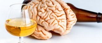
Rice. 291 . Development of new bark (neopallium).
a - snakes; b - marsupial mammal; 1 , 3 - neopallium: 2 - archipallium; 4 - hippocampus.
Rice. 292 . Olfactory brain (diagram).
1 - gyrus dentatus; 2 - gyrus parahippocampalis; 3 - uncus; 4 - substantia perforata anterior; 5 , 6 - striae olfactoriae; 7 - tr. olfactorius; 8 - bulbus olfactorius; 9 - commissura anterior; 10 - fornix; 11 - septum pellucidum; 12 - corpus callosum; 13 - gyrus for meatus.
1. Paleopallium, part of the temporal lobe. Initially, this section was located on the lateral surface of the hemisphere, but later, under the influence of the greatly enlarging neopallium, it curled up into a sausage-shaped formation - the hippocampus and shifted medially into the cavity of the lateral ventricle of the telencephalon in the form of a protrusion of its lower horn. The hippocampus is covered with ancient cortex, paleocortex.
2. Archipallium - a small area of the cortex on the ventral surface of the frontal lobe, lying near the bulbus olfactórius and covered with old cortex, archicórtex.
3. Neopallium, a new cloak, in the cortex of which, neocórtex, the highest centers of smell appeared - the cortical ends of the analyzer. This is the úncus, which is part of the vaulted gyrus.
As a result, the human olfactory brain contains a number of formations of different origins, which can be topographically divided into two sections. The peripheral section is the olfactory lobe, lóbus olfactórius, which refers to a number of formations lying at the base of the brain: 1) bulbus olfactórius; 2) tráctus olfactórius; 3) trigónum olfactórium; 4) substántia perforáta antérior. The central section is the convolutions of the brain: 1) parahippocampal gyrus, gýrus parahippocampalis; 2) dentate gyrus, gyrus dentatus; 3) vaulted gyrus, gyrus fornicatus, with its anterior part located near the temporal pole - the hook, úncus.
Lateral ventricles
In the hemispheres of the telencephalon, two lateral ventricles, ventriculi laterales, lie below the level of the corpus callosum,
(Fig. 293, 294, 295[no figure]), separated from the superolateral surface of the hemispheres by the entire thickness of the medulla.
Rice. 293. Lateral ventricles, opened from above by removing part of the hemispheres along with the corpus callosum.
1 - cornu anterius; 2 - nucl. caudatus (caput); 3 - for. interventriculare; 4 - nucl. lentiformis (in section); 5 - stria terminalis; 6 - upper surface of thalamus; 7 - hippocampus; 8 - eminentia collateralis; 9 - fimbria hippocampi; 10 - crus fornicis; 11 - cornu posterius ventriculi lateralis; 12 - medial wall of the posterior horn; 13 - calcar avis; 14 , 15 - cornu posterius; 16 - splenium corporis callosi; 17 , 19 - plexus choroideus in the central part of the lateral ventricle and its continuation into the lower horn; 18 - commissure fornicis; 20 - columnae fornicis; 21 - septum pellucidum; 22 - cavum septi pellucidi; 23 - corpus callosum.
The cavity of each lateral ventricle (see Fig. 294) corresponds to the shape of the hemisphere: it begins in the frontal lobe in the form of an anterior horn , córnu antérius , from here it stretches through the region of the parietal lobe called the central part , pars centralis , which at the level of the posterior edge of the corpus callosum it is divided into the inferior horn , córni inférius , (in the thickness of the temporal lobe) and the posterior horn , córnu postérius (in the occipital lobe).
Rice. 294. Sagittal section of the left hemisphere (done slightly lateral to the midplane to demonstrate the sections of the lateral ventricle).
Olfactory brain: its central and peripheral parts.
The olfactory brain is located on the lower and medial surfaces of the cerebral hemispheres and is conventionally divided into peripheral and central sections.
The peripheral part of the olfactory brain includes the olfactory bulb and tract, located in the olfactory sulcus. The olfactory tract ends with the olfactory triangle, which diverges in front of the anterior perforated substance into two olfactory stripes. The lateral stripe goes around the bottom of the lateral sulcus and ends in the uncinate cortex of the temporal lobe.
The medial strip is directed to the medial longitudinal fissure into the subcallosal gyrus and periolfactory field, which are located under the beak of the corpus callosum.
The central part of the olfactory brain includes: the vaulted gyrus, the hippocampus, the dentate gyrus, the uncinus, the intramarginal gyrus, the fasciculate gyrus and the gray layer above the corpus callosum.
The vaulted gyrus goes around the corpus callosum and is located on the medial surface of the hemispheres. The vaulted gyrus consists of three parts: the cingulate gyrus and the parahippocampal gyrus, connected by an isthmus.
The cingulate gyrus lies above the corpus callosum on the medial surface of the cerebral hemisphere and is not only the center of smell, but also the regulation of the function of internal organs. It is bounded above by the cingulate groove and below by the groove of the corpus callosum. Posteriorly, at the level of the parieto-occipital sulcus, it passes into the isthmus of the fornix, which below the posterior edge of the corpus callosum connects with the gyrus of the hippocampus.
The hippocampus is an invagination of the gray matter due to the medial wall of the inferior horn of the lateral ventricle. It is bounded on the lateral side and in the posterior part by the colateral groove, and in front by the nasal groove. The hippocampus at the anterior perforated substance is bent in the form of a hook.
The dentate gyrus represents the convoluted part of the cortex of the medial margin. The gray matter of the dentate gyrus extends to the inner edge of the hippocampus, as well as to the dorsal surface of the corpus callosum, forming a gray vestment that ends in the supracallosal gyrus.
The hook represents the anterior end of the hippocampal sulcus, which is divided by a cord into two parts: anterior and posterior. The anterior part belongs to the hook, and the posterior part forms the intramarginal gyrus.
Lateral ventricles of the brain, their walls. Choroid plexuses. Pathways for the outflow of cerebrospinal fluid.
The lateral ventricle is located deep within the cerebral hemisphere. There are two lateral ventricles: left and right. The parietal lobe of the cerebral hemisphere corresponds to the central part of the lateral ventricle, the frontal lobe - the anterior horn, the occipital lobe - the posterior horn, the temporal lobe - the inferior horn.
The central part is a horizontally located slit-like space, bounded above by the corpus callosum. The bottom of the central part is represented by the body of the caudate nucleus, part of the dorsal surface of the thalamus and the terminal strip, which separates these two formations from each other.
The medial wall of the central part of the lateral ventricle is the body of the fornix. Between the body of the fornix above and the thalamus below there is a vascular fissure, to which the choroid plexus of the lateral ventricle is adjacent from the central part. Laterally, the roof and the bottom of the central part of the lateral ventricle are connected at an acute angle. The side wall of the central part seems to be missing.
The anterior horn looks like a wide slit, curved downwards and laterally. The medial wall of the anterior horn is the transparent septum. The lateral and partly lower walls of the anterior horn are formed by the head of the caudate nucleus. The anterior, superior and inferior walls of the anterior horn are limited by the corpus callosum.
The inferior horn is the cavity of the temporal lobe. The lateral wall and roof of the lower horn are formed by the white matter of the hemisphere. The roof includes the tail of the caudate nucleus. In the area of the bottom of the lower horn, a collateral elevation is noticeable. The medial wall is formed by the hippocampus. On the medial side, the hippocampal fimbria is fused with the hippocampus, to which is attached the choroid plexus of the lateral ventricle, descending here from the central part.
The posterior horn projects into the occipital lobe of the hemisphere. Its upper and lateral walls are formed by the corpus callosum, the lower and medial walls are formed by the protrusion of the white matter of the occipital lobe into the cavity of the posterior horn. Two protrusions are noticeable on the medial wall of the posterior horn. The upper one - the bulb of the posterior horn - is represented by fibers of the corpus callosum. The lower protrusion - the bird's spur - is formed due to the depression of the medulla, located in the depths of the calcarine groove, into the cavity of the posterior horn. On the lower wall of the posterior horn there is a collateral triangle - a trace of the substance of the cerebral hemisphere being pressed into the ventricular cavity.
In the central part and lower horn of the lateral ventricle there is the choroid plexus of the lateral ventricle. This plexus is attached to the vascular band below and to the band of the fornix above. The choroid plexus continues into the inferior horn, where it also attaches to the fimbria of the hippocampus.
The choroid plexus of the lateral ventricle includes the vessels of the pia mater, penetrating through the vascular fissure in the central part of the ventricles. It is covered with an epithelial plate - part of the inner lining of the ventricles - ependyma. The plexus is present only in the central part and the lower horn. The circulatory plexus is attached to the lower wall using a vascular band made of an epithelial plate; on the medial wall - by the band of the fornix, in the lower horn - by the fimbria of the hippocampus.
The cerebrospinal fluid circulation pathways include the subarachnoid space with cisterns, the ventricles of the brain and the central canal of the spinal cord.
The arachnoid membrane is a thin, transparent, connective tissue film without blood and lymphatic vessels. It covers the brain with a mesh that is located between the hard and soft membranes. Under it there is a subarachnoid space filled with cerebrospinal fluid. In the convex area
parts of the convolutions and protruding structures of the brain stem, the arachnoid membrane fuses with the pia mater, and in the grooves, depressions, and pits it forms expansions called subarachnoid cisterns.
These include:
- the cerebellocerebral cistern is the largest, arising during the transition of the membrane from the cerebellum to the medulla oblongata;
- cistern of the lateral fossa and groove - in the same pit and groove;
- optic chiasm cistern - around the chiasm;
- interpeduncular cistern - between the cerebral peduncles;
- cistern of the corpus callosum - under the corpus callosum;
- lateral pontine or cerebellopontine cistern and other smaller containers.
Arachnoid (Pachionian) granulations are outgrowths of the membrane that penetrate the lumen of the meningeal sinuses, which is necessary for the exchange of cerebrospinal fluid.
Liquor is formed in the choroid plexuses of the ventricles. From the lateral ventricles, through the interventricular foramina, the fluid enters the third ventricle, and from it through the aqueduct into the fourth. From this ventricle, the cerebrospinal fluid leaves into the subarachnoid space (cerebellocerebral cistern) through the paired lateral and unpaired median foramina. From here, the fluid disperses throughout the subarachnoid space and is excreted into the venous blood of the meningeal sinuses through Pachionian granulations. From the 4th ventricle, the cerebrospinal fluid passes into the central canal of the spinal cord.
Microglia of the olfactory bulb in protecting the brain from infection
One of the specific symptoms in people with Covid-19 is the development of anosmia (hyposmia), which occurs as a result of viral damage to the olfactory zone of the nasal cavity. The proximity of this zone to brain structures and the possibility of their infection deserve special attention. Contrary to these assumptions, clinical practice demonstrates the absence of direct viral damage to the central nervous system. Possible mechanisms of antiviral protection of the nervous system were presented on June 5, 2021 in the Press release: Microglia in the olfactory bulb have a nose for protecting the brain from infection by the National Institute of Neurological Disorders and Stroke (NINDS), part of the National Institutes of Health (NIH). National Institutes of Health) USA, on the NIH website (https://www.nih.gov).
Researchers from the National Institute of Neurological Disorders and Stroke (NINDS), part of the National Institutes of Health (NIH), have identified a specific line of defense that limits olfactory bulb infection and protects olfactory bulb neurons from viral damage. Sensory neurons in the nasal cavity respond to inhaled odors and send this information to an area of the brain called the olfactory bulb. Although the location of nasal neurons and their environmental exposure make them easy targets, viral respiratory infections rarely spread from the olfactory bulb to the rest of the brain, where they can cause potentially fatal encephalitis. The study was published in the journal Science Immunology.
Using special viruses that can be tracked using fluorescence microscopy, researchers led by Dr Dorian McGavern, a senior researcher at NINDS, found that a viral infection that started in the nose was stopped just before it could spread from the olfactory bulb to the rest of the central nervous system.
“Airborne viruses constantly challenge our immune systems, but we rarely see viral infections leading to neurological disease,” said Dr. McGavern. "This means that the immune system in this area must be surprisingly good at protecting the brain."
Additional experiments showed that microglia (a specialized class of glial cells of the central nervous system, which are phagocytes that destroy infectious agents and destroy nerve cells, they originate from blood monocytes and are activated during the development of the inflammatory process, undergoing significant changes - in the activated state they release numerous processes, resembling amoebas - author's note) plays an underappreciated role in helping the immune system recognize the virus, and does so in a way that limits damage to the neurons themselves. And this protection is critical because neurons in most other tissues do not regenerate.
The evolution of the central nervous system has led to the development of a number of protective mechanisms that prevent the entry of pathogens. Airborne viruses, when inhaled, pass through the nasal passages and interact with tissue called the olfactory epithelium, which is responsible for our sense of smell. The peripheral neurons of the olfactory system pass through small projections through the bone that forms the nasal cavity, this provides access to odors present in the air. Olfactory epithelial neurons may provide an easy way to bypass traditional CNS barriers and provide a direct pathway to the brain.
"If a virus infects the dangling neurons in the airways, that virus has a chance to enter the brain and eventually cause encephalitis or meningitis," Dr. McGavern said. "We are interested in understanding the immune responses that develop at the interface between the nasal olfactory neurons terminating in the olfactory bulb and the rest of the brain."
Dr. McGavern's team was able to demonstrate that CD8 T cells, which are part of the immune system responsible for controlling viruses, are very important in protecting the brain during nasal infections. Using advanced microscopy, his team observed in real time how CD8 T cells protected the brain from nasal viral infection.
Interestingly, CD8 T cells did not appear to interact directly with infected neurons. Instead, they recruited microglia, which are immune cells in the central nervous system that act as scavengers of cellular debris and dead cells. When a viral infection occurs, microglia appear to absorb viral material from the environment and present it to the immune system.
Thus, infected olfactory neurons transmit viral particles to microglia, which are then detected by T cells, which cause the development of an antiviral response that clears the neuron of viruses, ensuring the safety of the nerve cells. Since microglia are a renewable cell type, this mode of interaction makes sense from an evolutionary perspective.
“The immune system has developed strategies to preserve neurons at all costs,” Dr. McGavern said. “Here we show that microglia are able to ‘take the hit’ from neurons by recruiting T cells, allowing the development of an antiviral program.”
Respiratory viral infections have received significant attention recently due to the current COVID-19 pandemic. Dr. McGavern noted that although this virus was not studied in these experiments, some of the symptoms it causes suggest that the described mechanism may be involved.
“One of the interesting symptoms associated with infection with the new coronavirus is that many people are losing their sense of smell and taste. This suggests that the virus is not only a respiratory pathogen, but most likely affects or destroys olfactory sensory neurons."
It's important to note that widespread infection of olfactory sensory neurons, whether by the novel coronavirus, the virus used in this study, or any other similar virus, will likely impair our sense of smell. However, unlike other CNS neurons, these sensory neurons, which begin in the nose and end in the brain, are capable of regenerating after the infection is complete.
“The immune response we describe does not protect olfactory sensory neurons and the sense of smell,” explains Dr. McGavern. “…. It is essential to protect the brain and central nervous system from encephalitis or meningitis - our sense of smell can often be restored over time.”
Dr. McGovern continued by saying that given the importance of microglia in stimulating the antiviral response, factors that can lead to their depletion or loss of function may increase susceptibility to CNS infection.