Cerebellar ataxia
Treatment of the causative disease is fundamental. If cerebellar ataxia has an infectious-inflammatory origin, it is necessary to prescribe antibacterial or antiviral therapy. If the cause lies in vascular disorders, then measures are taken to normalize blood circulation or stop cerebral bleeding. For this purpose, angioprotectors, thrombolytics, antiplatelet agents, vasodilators, and anticoagulants are used in accordance with indications. For ataxia of toxic origin, detoxification is performed: intensive infusion therapy in combination with the prescription of diuretics; in severe cases - hemosorption.
Ataxia of a hereditary nature does not yet have a radical treatment. Metabolic therapy is mainly carried out: vitamins B12, B6 and B1, ATP, meldonium, ginko biloba preparations, piracetam, etc. To improve metabolism in skeletal muscles, increase its tone and strength, massage is recommended for patients.
Tumors of the cerebellum and posterior cranial fossa often require surgical treatment. Removal of the tumor should be as radical as possible. If the malignant nature of the tumor is established, a course of chemotherapy or radiotherapy treatment is additionally prescribed. For cerebellar ataxia caused by cerebrospinal fluid duct occlusion and hydrocephalus, shunt operations are used.
Prognosis and prevention
The prognosis depends entirely on the cause of cerebellar ataxia. Acute and subacute ataxia caused by vascular disorders, intoxication, inflammatory processes, with timely elimination of the causative factor (vascular occlusion, toxic effects, infection) and adequate treatment, can completely regress or partially persist as residual effects. Chronically progressive, hereditary ataxias are characterized by an increasing aggravation of symptoms, leading to disability of the patient. The most unfavorable prognosis is for ataxia associated with tumor processes.
The prevention of injuries, the development of vascular disorders (atherosclerosis, hypertension) and infection is of a preventive nature; compensation of endocrine and metabolic disorders; genetic counseling when planning pregnancy; timely treatment of pathology of the cerebrospinal fluid system, chronic cerebral ischemia, Chiari syndrome, processes of the posterior cranial fossa.
3.3. GROUP OF CRANIAL NERVES OF THE PONTOCEREBELLAR ANGLE – Pain Clinic – Pain Clinic
3.3.3.
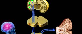
VIII PAIR: VESTIBULAR-COCHLEARIAN NERVE
Consists of two functionally different parts: the vestibular (pars vestibularis) and the cochlear (pars cochlearis). The vestibular part provides the function of body balance, orientation of the head and body in space. The cochlear part provides hearing (sound perception, sound conduction).
A) vestibular part
The receptors of the vestibular part of the vestibulocochlear nerve are located inside the ampullae of the semicircular canals in two membranous sacs (succulus and utriculus) of the vestibule. Vestibular fibers begin in the Scarpa spiral ganglion (ganglion spirale), located in the inner ear (1st neuron).
The central processes of this node go into the cranial cavity through the porus acusticus internus, enter the brain stem in the cerebellopontine angle, and end in the superior (Bechterew's nucleus), inferior (Roller's nucleus), medial (Schwalbe's nucleus) and lateral (Deiters' nucleus) vestibular nuclei
(2 th neuron).
From the lateral nucleus of Deiters, axons form the vestibulospinal tract (vestibulospinal fasciculus of Leventhal),
which on its side, as part of the lateral cord of the spinal cord, approaches the anterior horns of the spinal cord. Axons extending from the lateral nucleus of Deiters.
From the vestibular medial nucleus of Schwalbe and the inferior nucleus of Roller, axons approach the nuclei of the oculomotor nerves on the opposite side, and from the superior nucleus of Bechterew - on their side. Along these vestibuloculomotor tracts
impulses are transmitted to the muscles of the eye.
The vestibulocerebellar fascicle also extends from the ankylosing spondylitis nucleus
to the nucleus of the tent (n. fastigii) of the cerebellum and the cerebellar vermis.
To carry out the function of body balance, the vestibular nuclei have connections with proprioceptive conductors (Gaull and Burdach bundles) from the spinal cord.
Axons of neurons transmit impulses to the contralateral thalamus, globus pallidus ( 3rd neuron)
and the cortex of the temporal, partially parietal and frontal lobes
(4th neuron).
FUNCTION STUDY
1. Study of the oculocephalic reflex - the patient fixes his gaze, the doctor quickly (2 cycles per second) turns the patient’s head to the right or left. Normally, the movements of the eyeballs are smooth, proportional to the speed of head movement and directed in the opposite direction. The patient continues to fix his gaze on the object.
2. Study of nystagmus.
3.
Study of the Romberg pose.
SYMPTOMS OF DEFEAT
1. Spontaneous nystagmus.
2. Oscillopsia (illusion of surrounding objects shaking)
3. Vestibular ataxia (see page).
4. Violation of the oculocephalic reflex. This reflex is absent when the brain stem is damaged. The eyeballs turn along with the head, the gaze is not fixed.
5. Dizziness, accompanied by nausea and vomiting.
B) Cochlear part
Sound waves are perceived by the receptors of the spiral (corti) organ. The auditory fibers begin together with the vestibular fibers in the spiral ganglion (1st neuron).
The axons of the cells of this node run in the internal auditory canal along with the vestibular part.
Coming out of the pyramid of the temporal bone, the nerve is located in the cerebellopontine angle and penetrates the cerebral pons lateral to the olive. The fibers of the cochlear part end in two auditory nuclei - ventral and dorsal (2nd neuron).
Most of the axons of the 2nd neuron move to the opposite side of the brain pons and end in
the olive nucleus and trapezoid body (3rd neuron)
, a smaller part of the fibers approaches the olive and trapezoid body nuclei on their side.
The axons of the 3rd neuron form the lateral loop (lemniscus lateralis), which rises upward and ends in the inferior colliculi of the midbrain roof and the medial geniculate body
(corpus geniculatum mediale)
(4th neuron)
.
From the cells of the medial geniculate body, axons pass as part of the posterior limb of the internal capsule and end in the transverse temporal gyrus (
Heschl's gyrus)
FUNCTION STUDY
When examining auditory functions,
the following are determined: hearing acuity, air and bone conduction of sound.
1. Hearing acuity
determined as follows. The subject stands at a distance of 5 m from the doctor, turning the ear being examined towards him, the second ear is tightly closed by pressing the finger on the tragus. The doctor whispers the words and offers to repeat them.
2. Air conductivity of sound is determined using a sounding tuning fork located at the ear canal.
3.
Bone conduction is tested by placing a tuning fork on the mastoid process and crown. In a healthy person, air conduction lasts longer than bone conduction; a sounding tuning fork placed on the head in the midline is heard equally on both sides.
Rinne test
used to compare bone and air conduction. The physician places the base of a vibrating tuning fork (516 Hz) on the mastoid process. After the patient stops hearing it, the tuning fork is moved to the ear (without touching it). Normally, the sound of the tuning fork continues to be perceived (positive Rinne test). A disease of the sound-conducting apparatus causes the opposite results: the tremor of a tuning fork, indistinguishable by the ear, is again detected when the tuning fork is placed on the mastoid process (negative Rinne test).
Weber test:
In a healthy person, a sounding tuning fork (516 Hz), placed on the head in the midline, is heard evenly on both sides. In diseases of the middle ear, air conduction is disrupted, and bone conduction is greater. Therefore, when the middle ear is damaged, the sound of a tuning fork placed on the crown of the head is perceived more strongly on the affected side. When the pathological process is localized in the inner ear, sound is better perceived on the healthy side. Consequently, an increase in the duration of bone conduction indicates damage to the sound-conducting apparatus (tympanic membrane, auditory ossicles), and its shortening indicates damage to the sound-receiving apparatus (cochlea, auditory tract).
Other reasons for the development of the disorder
In some cases, cerebellar damage is not a consequence, but a symptom. This applies to oncological diseases of the following organs and systems:
- lungs;
- brain;
- ovaries;
- lymph nodes.
The “first bell” of brain cancer can be cerebellopontine angle syndrome. As a result of the growth of a malignant tumor, parts of the brain are compressed and nutrition and neural communication in their cells are disrupted.
With prolonged alcohol dependence, substance abuse and drug addiction, irreversible damage to the cerebellum occurs. Ataxia can also become a hereditary disease. In this case, therapy must be selected in a special way.
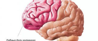
What is the cerebellum, its functions and structure:
Clinical picture of cerebral neuroma
Symptoms depend on the location and volume of the tumor. Patients complain of whistling or tinnitus. Gradually the noise gives way to partial deafness. The patient may have trouble hearing high pitched tones.
If a neuroma of the right cerebellopontine angle develops, the patient complains of hearing impairment on the right side. Accordingly, with a tumor on the left, hearing loss occurs on the left side. After partial deafness in one ear, complete deafness develops.
Patients with neuroma experience periodic dizziness and involuntary movements of the eyeballs (nystagmus). Other symptoms include:
- occipital pain on the side of the tumor;
- loss of sensation in the facial nerve.
If a tumor develops in the area of the internal auditory canal, the patient experiences impaired salivation, partial loss of taste and sensitivity in the nasal cavity on the side of the tumor. If the tumor grows and affects the vagus nerve, the following symptoms appear:
- weakening of the vocal cords;
- changing the sound modification during a conversation;
- swallowing disorder.
When the cerebellum is compressed, the patient experiences characteristic symptoms:
- weakening of the muscle tone of the arms and legs;
- slow movements;
- inability to perform rapidly alternating movements;
- tremor during purposeful movements;
- miss;
- spontaneous movement of the eyeball on the affected side.
With large neuromas, intracranial hypertension may develop. Patients complain of severe headaches in the morning, which are accompanied by vomiting. Typically, this symptom appears several years after the onset of neuroma formation.
Features of surgery to remove a tumor of the cerebellopontine angle
Removal of the tumor of the cerebellopontine angle is carried out surgically. Before surgery, patients of this profile undergo general clinical and neurootological examination, CT and MRI. According to indications, myo- and angiography and neuropsychological testing are prescribed.
Removal of a tumor of the cerebellopontine angle is performed using modern methods of endoscopic microsurgery under conditions of constant neurophysiological monitoring (including stimulation and EMG of the facial nerve). After neuroimaging, the doctor develops a surgical plan, determines its volume and the point of best access. The intervention is carried out using high-speed drills and pneumatic drills, which ensure minimal invasiveness and reduce the extent of damage to nearby tissues.
The final result of surgical intervention is determined by the characteristics of tumor growth, the degree of damage to the base of the skull, and its fusion with neurovascular structures. In most cases, I, together with a team of assistants at the N. N. Burdenko Research Institute of Neurosurgery, manage to solve all the problems facing us.
We will call you back within 1 minute
Moscow, Balaklavsky prospect, building 5
Numerous types of skin tumors are either completely safe for health or can cause harm to surrounding tissues and even pose a threat to human life.
The method, which is called shock wave therapy, is used to treat musculoskeletal diseases and any disease of the musculoskeletal system.
the process of removing cells or tissue from the body for further microscopic examination to check for cancer
- News
- Tumor
- Tumors of the cerebellopontine angle
Tumors can affect any organ in the human body. Some of them are well-known, others are rarer and more complex. Tumors of the cerebellopontine angle touch the vestibulocochlear nerve, which occupies an intermediate position between the cerebellum and the so-called pons.