Introduction
PONTOCEREBELLAR ANGLE
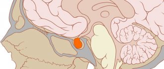
- the space where the pons (pons), medulla oblongata and cerebellum meet. M. u. open in front, to the base of the skull, in the region of the posterior cranial fossa. On the ventral side of M. u. covered by an arachnoid membrane, which does not go deep into it, but is located superficially, as a result of which a container for cerebrospinal fluid is formed in this area - the lateral pontine cistern, often identified in the literature with M. at. in the broad sense of the word. In this case, under M. u. understand a narrow space resembling a flattened irregular pyramid in shape, bounded in front and on the side by the posterior surface of the pyramid of the temporal bone, from the inside by the junction of the pons, medulla oblongata and cerebellum, constituting the apex of the cerebellopontine region, behind by the surface of the cerebellar hemisphere, and above by the tentorium of the cerebellum. In the area of M. u. the roots of the V-XI pairs of cranial nerves, the anterior inferior cerebellar and labyrinthine arteries and numerous cerebellar veins flowing into the superior petrosal sinus are located, among which the flocculus vein is distinguished by its constancy.
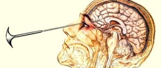
MRI is an expensive method of hardware research, so the cerebellopontine angles are most often examined in conjunction with brain diagnostics. For a targeted study of this department, it is necessary to have symptoms indicating degenerative changes in the structure.
| Layout of the cerebellopontine angle | MRI image of the cerebellopontine angle |
Traditional methods of treating neuroma
Folk remedies for treating neuroma include the use of tinctures and decoctions and diet.
Important! Any traditional medicine recipe, even a harmless one, should be discussed with a doctor.
Traditional medicine recipes:
- Horse chestnut tincture. 50 g of flowers are poured into 0.5 liters of vodka, left for 10 days, and squeezed out. Apply 10 drops 3 times a day. The tincture is diluted with water. The course of treatment is 2 weeks. After a break of 7 days, the course is repeated.
- Tincture of Sophora japonica. 50 g of raw material is poured with 0.5 alcohol, left for 40 days, filtered, and squeezed. Take 10 ml tincture every day. The tincture is diluted with water. The course of admission is 40 days. If necessary, the course is repeated after a two-week break.
Patients are recommended to include in their diet foods that contain chlorophyll (dandelion leaves, nettles, cabbage, greens), orange and red vegetables, and fruits (tomatoes, carrots, oranges, apricots).
The following fruits and vegetables have antioxidant effects:
Dangerous foods include fatty meat and dairy foods, smoked foods, sugar, flour products, and canned food. Proper nutrition helps cells recover, improves the patient’s well-being, protects against inflammatory processes, and improves metabolism.
In conclusion, it is worth noting that if you experience tinnitus or deafness, you should consult a doctor and undergo a comprehensive examination. Thanks to timely detection and removal of the neuroma, the patient has an increased chance of maintaining hearing and cranial nerve function.
The X-ray method of examination is especially important for diagnosing tumors of the cerebellopontine angle, i.e., the space limited by the posterior surface of the pyramids of the temporal bones, the bottom of the posterior cranial fossa, the cerebellar tentorium and the pons. In the area of the cerebellopontine angle, pathological processes of two types can occur: tumors (most often acoustic neuromas) and inflammatory changes in the membranes (arachnoiditis).
Arachnoiditis in the area of the cerebellopontine angle is not detected by X-ray examination methods. When diagnosing tumors of this localization, the x-ray method is the leading one.
Acoustic neuroma is a benign, slow-growing tumor that develops in the vast majority of cases from that part of the vestibular portion of the VIII nerve, which is located in the internal auditory canal (V. G. Egorov, 1949).
Initially, the tumor causes irritation of the auditory nerve; patients note noise in the ear, squeaking, and ringing. Then the hearing in that ear gradually decreases. As the tumor grows in the internal auditory canal, it also compresses the facial nerve, which leads to an asymmetry in the innervation of the facial muscles. Subsequently, the tumor emerges from the pyramid at the base of the skull and compresses the cerebellum and brain stem.
Depending on the direction of tumor growth, compression and displacement of the brain stem, one or another neurological symptoms develop. Wedged into the lateral cistern of the bridge, the tumor displaces and stretches the cranial nerves passing here and compresses the vessels. In the late stage, due to compression of the cerebrospinal fluid ducts, general cerebral symptoms occur.
The clinical picture of eurinoma of the VIII pair is sometimes very similar to arachnoiditis of the cerebellopontine angle and multiple sclerosis. In such cases, x-ray examination of the temporal bone pyramids is crucial. Since tumor growth begins from the internal auditory canal, one of the early symptoms of the tumor is the expansion of the internal auditory canal on the side of the tumor, sometimes with destruction of its walls. The internal auditory canal is most often evenly expanded, its walls remain parallel to each other, but there may be a spindle-shaped and flask-shaped expansion. Changes in the internal auditory canal can be detected on direct radiographs of the skull with a projection of the pyramids into the orbits, but they appear especially clearly on photographs of the pyramids of the temporal bones according to Stenvers.
Ch., 24 years old. Complaints of deafness in the left ear, slight asymmetry of the face, awkwardness when moving in the left hand. Ill for 3 years. In photographs with the projection of the pyramids into the orbits and in photographs of the pyramids according to Stenvers, an expansion of the internal auditory canal on the left is revealed (Fig. 56). During surgery, an acoustic neuroma was discovered.
R., 41* year. Complaints of unsteadiness of gait, dizziness, feeling of awkwardness in the left arm and left leg, deafness in the left ear. Ill for 12 years. The disease was regarded as arachnoiditis of the cerebellopontine angle. Stenvers' photographs of the pyramids of the temporal bones reveal an expansion of the internal auditory canal on the left (Fig. 57); tomograms show destruction of the edges of the jugular foramen. During surgery, an acoustic neuroma was discovered.
Upon exiting the internal auditory canal, the tumor can put pressure on the pyramid of the temporal bone, which is manifested by osteoporosis and destruction of its apex. The spread of the tumor posteriorly along the bottom of the posterior cranial fossa can lead to destruction of the edges of the corresponding jugular foramen. Growing forward along the clivus of Bloomsnbach can cause destruction of the dorsum of the sella turcica and its inclination anteriorly.
In the later stages, changes in the opposite pyramid may be observed: osteoporosis of its apex and expansion of the internal auditory canal due to the pressure of the pons pushed to the opposite side, as well as general signs of increased intracranial pressure.
In addition to images with the projection of the pyramids into the orbits and Stenvers images, a posterior semi-axial Altschul image and an image of the base of the skull can be used. But changes are especially clearly visible on tomograms. Tomograms are performed in a posterior viewing position at depths of 7, 8 and 9 cm. The tomograms clearly show changes in the petrous part of the pyramid, the bottom of the posterior cranial fossa and the edges of the jugular foramen.
Acoustic neuromas can be bilateral. Bilateral damage is observed in Recklinghausen's neurofibromatosis. Clinical diagnosis of bilateral tumors is difficult. In addition to damage to the auditory nerves, Recklinghausen's disease reveals small tumor-like nodules under the skin, and other cranial nerves and spinal roots are often affected. X-rays reveal bilateral destruction of the pyramids of the temporal bones.
In addition to acoustic neuromas, other tumors can occur in the area of the cerebellar-pontine angle - sarcomas of the base of the cerebellum (see Fig. 14 on page 44), gastrointestinal node tumors, cholesteatomas, arachnoidal lotelioma of the Blumenbach clivus and others. However, they are much less common. All these tumors can cause destructive changes in the pyramids of the temporal bones and adjacent parts of the bones of the base of the skull.
N., 44 years old. Complaints of pain in the left half of the face, deafness in the left ear, asymmetry of facial muscles, unsteadiness of gait, dizziness and difficulty moving in the left limbs. Ill for 5 years. The photographs reveal destruction of the apex of the left pyramid with a smooth sclerosed edge (Fig. 58). During the operation, a tumor was discovered - a neuroma, emanating from the Gasserian ganglion.
Summarizing the section on craniographic changes in brain tumors, it should be emphasized that this method can detect tumors of not every location and any histological structure. The leading methods of X-ray diagnosis of brain tumors are contrast studies of the cerebrospinal fluid tract and blood vessels. These methods are described in the next section of the book.
Study of the cerebrospinal fluid spaces of the brain
refers to benign neurogenic tumors of the cerebellopontine angle with an unfavorable clinical course. It comes from the cells of the Schwann membrane of the vestibular portion of the VIII nerve from the bottom of the internal auditory canal to the entrance to the medulla oblongata.
There are three stages of the disease.
The first stage of development is otiatric (tumor up to 1.5 cm) is characterized by cochleo-vestibular symptoms: constant noise in the ear, sensorineural hearing loss, tone-speech dissociation (speech intelligibility is impaired with relative preservation of tonal hearing), occasionally ear pain or headache, slight disturbances in static balance, some uncertainty in gait, dizziness.
The tuning experiments of Rinne and Federici are positive. The pure-tone audiogram has a horizontal and then descending character, predominantly in the high-frequency region, with the absence of an air-bone interval. There is an increase in the level of auditory discomfort and a lack of lateralization of sounds in the audible range in Weber’s experiment in the presence of lateralization of ultrasounds into the healthy ear. FUNG is not detected, the reverse adaptation time increases to 15 minutes, its threshold is shifted to 30-40 dB (normally 0-15 dB). Impedance measurements show a decay of the acoustic reflex of the stapes. Normally, within 10 s the amplitude of the reflex remains constant or decreases to 50%. A reflex half-life of 1.5 s is considered pathognomonic for neuroma of the VIII nerve. The stapes reflex (ipsilateral and contralateral) may not be evoked when the affected side is stimulated. Otoacoustic emissions (OAE) are not recorded on the affected side. During audiometry using auditory evoked potentials, the interpeak interval of I and V ACEP is prolonged. In large tumors, KSVP is not caused.
Patients have difficulty understanding words during a telephone conversation, and severe hearing fatigue is noted. 75% of patients have a chronic violation of static balance with instability when walking, horizontal spontaneous nystagmus in the healthy direction. With caloric and rotational tests, pronounced asymmetry of nystagmus is often observed.
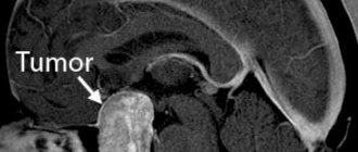
X-rays of the temporal bones according to Stenvers may show expansion of the internal auditory canal. Computer and magnetic resonance imaging scans reveal a tumor of the YIII nerve. Magnetic resonance imaging has greater resolution in the diagnosis of neuroma, especially in combination with the introduction of contrast agents that increase the information content of the image (Fig. 1.13.1).
The second stage - otoneurological (tumor from 1.5 to 4 cm) is characterized by headache, increased hearing loss, statokinetic disorders, unilateral cerebellar symptoms, absence of caloric nystagmus on the affected side, dysfunction of the trigeminal nerve (paresthesia, decreased or absent corneal reflex), paresis abducens nerve (convergent strabismus and diplopia).
The third stage - neurosurgical (tumor from 4 to 6 cm or more) is manifested by severe hearing loss, even deafness, loss of vestibular function. Symptoms of damage to the cerebellum, pyramidal system and severe intracranial hypertension (congestive optic nipples, severe headache, nausea, vomiting, etc.) are added. Along with damage to the facial, intermediate and abducens nerves, dysfunction of the trigeminal and abducens nerves in the cerebellopontine angle is more often observed. Subsequently, bulbar disorders develop, damage to many cranial nerves, including visual impairment up to blindness, gaze paralysis, swallowing disorder, phonation, and decreased sense of smell. Severe hydrocephalus develops. The statokinetic function is disturbed according to the central type with dissociation and disharmonization of reactions.
For otiatrists, the first two stages of neuroma are of particular interest, when with timely diagnosis and surgical treatment, further spread of the tumor can be prevented. During the initial diagnosis of unilateral sensorineural hearing loss, it is necessary to exclude neuroma using the most modern diagnostic methods.
Neuromas are differentiated from Meniere's disease, arachnoiditis of the cerebellopontine triangle and hearing loss of various origins with an intact tympanic membrane.
Treatment is surgical. The most favorable outcomes are for tumor stages I and II. Neurosurgical approaches to neuroma are carried out through the posterior and middle cranial fossae, and otiatric access is transpyramidal through the mastoid process, temporal bone to the internal auditory canal. The otiatric method is more gentle (Gorokhov A.A., 1989).
Patients with benign and malignant ear tumors are immediately sent to the hospital. After treatment, they are under the dynamic supervision of a unit doctor. A follow-up examination by an otolaryngologist is carried out at least once every 6 months. When testifying, military personnel are examined under articles 8,9,10 of Order No. 315 of the 1995 Ministry of Defense of the Russian Federation.
Indications
The reason for conducting targeted MRI diagnostics of MMU is considered to be the presence of sensorineural hearing loss if ENT disorders are excluded from the causes of the pathology.
Symptoms that determine the need for magnetic resonance imaging:
- Dizziness and lack of coordination.
- Paralysis of facial muscles.
- Impaired taste perception.
- Copious, causeless lacrimation.
- Partial or complete loss of facial skin sensitivity.
These are typical signs of damage to 5-8 pairs of cranial nerves passing through the MMU.
The following factors are also indications for prescribing MR tomography of the cerebellopontine angles:
- Anomalies of the structure and malformations of the brain.
- Infectious lesions of the central nervous system.
- Abscess of the cerebellopontine zone.
- Suspicion of tumor processes.
- Assessing the need for surgical intervention.
- Analysis of the dynamics of the disease.
- Postoperative control.
- Studying the effectiveness of conservative treatment of benign and malignant tumors. Search for metastases.
Diagnosis and treatment methods for neuroma
When diagnosing, the patient is excluded from cholesteotoma, Meniere's disease, auditory neuritis, arachnoiditis, and vascular pathologies. In addition, aneurysm of the vertebral arteries, tuberculous or syphilitic meningitis are excluded.
For diagnostics use:
- computer diagnostics;
- X-ray examination;
- MRI;
- angiography.
Since the tumor grows slowly and may in some cases regress, patients are offered conservative treatment. To eliminate cerebral edema, brain shunt surgery is indicated.
If the tumor is small, microsurgical removal is indicated. In this case, patients may retain hearing and nerve function. Rehabilitation after removal of small neuromas up to 2 cm is much faster. With total removal of a large neuroma, postoperative complications may occur - paresis and paralysis of the facial nerve. If the neuroma is partially removed, the issue of radiation therapy is considered.
Possible complications after surgery:
- temperature increase;
- cramps, nausea;
- loss of sensation in certain areas of the body;
- dyspnea;
- headache;
- tachycardia.
If pathological symptoms appear, re-diagnosis is carried out in order to correct treatment and further observation.
What does it show
The decoding protocol describes the state of the structures of the pontocerebellar region and their symmetry. In the presence of pathologies, MRI allows you to identify a number of diseases with an accuracy of up to 90%:
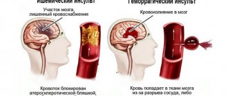
- Circulatory disorders: thrombosis of venous vessels, rupture of the vessel membrane and hemorrhage into the brain cavity, aneurysm.
- Infectious and inflammatory nerve lesions.
- Tumors: neuroma, meningioma, cholesteatoma, lipoma, arachnoid cysts.
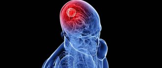
Types of tumors of the cerebellopontine angle
Medical statistics note an important fact. It lies in the fact that in ten percent of a hundred formations in the brain are located in a place called the cerebellopontine angle.
Types of tumors associated with the affected area:
- neuroma of the vestibulocochlear nerve;
- meningioma;
- cholesteatoma.
The first disease accounts for 95 percent of all formations of the cerebellopontine angle. The detected tumor is benign and does not become a source of damage to other organs. Patients of working age are at risk. Neuromas are often found in women. Today, doctors prefer to remove the tumor surgically, performing unilateral or bilateral removal.
Doctors often diagnose “cerebellopontine angle syndrome.” It should be noted that it is a consequence of another disease called neuroma.
Ultrasound or MRI, which is better?
Ultrasound and MRI of the cerebellopontine angles are based on different physical processes. To compare them correctly, it is necessary to evaluate the following factors:
Method of obtaining an image.
A magnetic resonance imaging machine produces images of the internal structures of the brain by processing vibrational pulses of hydrogen atoms under the influence of an electromagnetic field. Ultrasound equipment processes reflected ultrasound signals and builds an image based on the data received.
Device capabilities.
MRI provides a more complete clinical picture with a low reporting error rate. Ultrasound of the cerebellopontine angle is not informative enough, since dense bone tissue impedes the passage of the ultrasound signal. The method is more effective in examining children.
Image quality.
An ultrasound image has a large number of artifacts and noise, and low image resolution. MRI projection is performed in high resolution with great detail.
Research speed.
To perform an ultrasound diagnosis, 3-5 minutes are enough, while an MRI can take from 15 minutes to an hour.
Cost and availability of the study.
Ultrasound diagnostics is included in the list of free services provided by the general health insurance program. The procedure can be done free of charge at your clinic. MRI on an outpatient basis is provided for a fee. To undergo the study, you must take a referral to a regional diagnostic center or go to a commercial clinic. The cost of an MRI is several times higher than the cost of an ultrasound.
| Ultrasound of the cerebellopontine angles in a child | MRI of the cerebellopontine angles |
General characteristics of the disease
Neuroma (or schwannoma) is a benign tumor that forms in Schwann cells of the spinal, cranial and peripheral nerves. Thus, a neuroma is a growth in the cells that cover the nerves. They are round, sometimes lobular, capsule-shaped formations. They appear most often in the root of the auditory nerve (vestibular portion), progress in the cerebellopontine angle (facial and auditory nerves), less often in the root of the V nerve (maxillary and mandibular, orbital nerves). And extremely rarely, neuromas arise in the roots of the glossopharyngeal and vagus nerves.
The percentage of neuroma disease is relatively small and accounts for about 8% of all intracranial neoplasms and about 20% of all primary spinal formations. Middle-aged and older people are susceptible to this disease, with women getting sick more often. Because Since neuroma is a benign tumor with slow growing growth, it very rarely develops into a malignant tumor.
The most common types of neuroma are:
1. Morton's neuroma. This benign tumor forms in the area of the plantar nerve of the foot. In most cases, it occurs at the site of the nerve of the third and fourth toes, sometimes between the second and third toes. It mainly affects one foot, very rarely both.
2. Acoustic neuroma (also called vestibular schwannoma). In most cases, such a tumor arises in the vestibular branch of the auditory nerve, and its growth continues in the cerebelloptine angle. The neuroma increases in volume, resulting in pressure on the cerebellum and brain stem. Direct contact of the mass with the facial nerves sometimes even affects the lower cranial and trigeminal nerves.
There are unilateral acoustic neuromas (about 95%) and bilateral (5%). The latter is usually caused by neurofibromatosis, and its first manifestations are possible already in the second decade of life, in contrast to unilateral, which most often affects people aged 40-50 years.
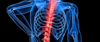
3. Spinal neuroma. It is caused by a tumor of the spinal nerve roots, and in most cases occurs in the thoracic and cervical region, less often in the lumbar region. It is considered the most common disease of primary neoplasms on the spinal cord. Develops as an extramedullary-intradural tumor in Schwann cells.
Neuromas of the spinal roots can spread through the intervertebral foramen extradurally. This form of neuroma is of the “hourglass” type and is characteristic of the cervical spine. The consequences of spinal neuroma are bone changes, which are diagnosed by conventional spondylography.
Neuroma of the trigeminal and other nerves is a consequence of a tumor of the auditory nerve, and such a type as mediastinal neuroma is a subtype of tumor on the spine.
Photo
| Tumor of the cerebellopontine angle |
| Arachnoid cyst of the cerebellopontine angle |
| Lipoma of the cerebellopontine angle |
| Cerebellopontine meningioma.jpeg |
| neuroma of the cerebellopontine angle |
Pontine cerebellar ganglion neuroma: symptoms, treatment, rehabilitation
In women, neuroma of the cerebellar angle pons is detected more often. There is also a relationship between tumor growth from hormones and exposure to radiation. The growth of cerebellar neuroma leads to its compression, compression of the 5th and 7th cranial nerves, the bridge, a group of nerves of the medulla oblongata and spinal cord.
The rate of tumor growth varies in intensity among patients. Most often, cerebral neuroma grows slowly at a rate of 2 to 10 mm per year.
In some patients, pathology may not appear until the tumor has grown to a significant size.
Neuroma of the cranial nerves is surrounded by a capsule, is not able to grow into adjacent tissues, and can form cysts.
Clinical picture of cerebral neuroma
Symptoms depend on the location and volume of the tumor. Patients complain of whistling or tinnitus. Gradually the noise gives way to partial deafness. The patient may have trouble hearing high pitched tones.
If a neuroma of the right cerebellopontine angle develops, the patient complains of hearing impairment on the right side. Accordingly, with a tumor on the left, hearing loss occurs on the left side. After partial deafness in one ear, complete deafness develops.
Patients with neuroma experience periodic dizziness and involuntary movements of the eyeballs (nystagmus). Other symptoms include:
- occipital pain on the side of the tumor;
- loss of sensation in the facial nerve.
If a tumor develops in the area of the internal auditory canal, the patient experiences impaired salivation, partial loss of taste and sensitivity in the nasal cavity on the side of the tumor. If the tumor grows and affects the vagus nerve, the following symptoms appear:
- weakening of the ligaments;
- changing the sound modification during a conversation;
- swallowing disorder.
When the cerebellum is compressed, the patient experiences characteristic symptoms:
- weakening of the muscle tone of the arms and legs;
- slow movements;
- inability to perform rapidly alternating movements;
- tremor during purposeful movements;
- miss;
- spontaneous movement of the eyeball on the affected side.
With large neuromas, intracranial hypertension may develop. Patients complain of severe headaches in the morning, which are accompanied by vomiting. Typically, this symptom appears several years after the onset of neuroma formation.
Causes of brain tumors
Modern medicine does not know the exact reasons for the development of space-occupying brain tumors. Experts suggest that such changes occur under the influence of a combination of factors. In pediatric patients, genetic factors for the occurrence of neoplasms prevail.
They are associated with mutations in certain regions of chromosomes that control cell growth and division. Such changes most often provoke the formation of a tumor in newborns and young children. In adult patients, cancer can also be genetic.
Scientists identify a number of factors that can provoke a failure of the cell cycle and the development of a space-occupying tumor in the brain (ICD code D33):
- Ultraviolet and infrared radiation can change the structure of DNA, as a result of which the likelihood of the formation of a tumor, including melanoma, increases.
- Some viruses have the ability to potentiate uncontrolled cell growth, which then degenerates into oncology. These viruses include, for example, papillomavirus, which causes warts.
- Eating foods that contain GMOs. They can have a teratogenic effect - cause cancer and the formation of deformities.
Classification of brain tumors
The prognosis for recovery from a brain tumor (according to ICD-10 D33) depends on the stage of the pathological process, as well as the individual characteristics of the patient’s body and the histology of the tumor formation.
Cerebral lesions are divided into two large groups: gliomas, the formation of which occurs directly from nervous tissue; non-gliomas formed from the meninges, lymphoid elements. Secondary formations are also identified, which are the result of metastasis of the primary lesion along the lymphatic tract and blood vessels.
As a rule, a similar process is found in organs where there is active blood flow - the spleen, lungs, liver.
Gliomas
The group of gliomas is represented by various pathologies; they are the most common space-occupying formations of the corpus callosum of the brain. They are diagnosed in 80% of cases. Gliomas are classified according to their level of malignancy, and therefore the prognosis and clinical picture may vary. The most common type of glioma is glioblastoma. Also distinguished:
- Oligodendrogliomas. This pathology is very rare; in this case, a neoplasm is formed from protective elements that also support the process of hemostasis in the brain. The peculiarity of such space-occupying formations of the brain stem is that most often they form in middle-aged patients. Such neoplasms are moderately malignant.
- Astrocytomas. This neoplasm is diagnosed in 3/5 cases of brain cancer. Astrocytomas are formed from cells that separate neurons from vessels. These cells (astrocytes) are involved in the nutrition and natural development of nervous tissue. Astrocytomas are differentiated based on the degree of malignancy.
- Ependymomas. They are formations formed by the cells of the inner layer of the cerebral ventricles. These cells take part in the process of cerebrospinal fluid production, and therefore the symptoms of ependymoma are associated with a violation of this particular function. Ependymomas are classified into malignant and high-grade.
- Mixed gliomas. This type of tumor is formed from several types of tissue that are normally present in the brain.
Source: https://yazdorov.win/pechen-i-zhelchnyj-puzyr/nevrinoma-mosta-mozzhechkovogo-uzla-simptomy-lechenie-reabilitatsiya.html
Price
On our website, many clinics list prices for MRI, including MRI of the cerebellopontine angles.
To see prices, you need to go to the list of clinics in your city. To do this, select your city on the “Addresses” page.
On the page for the list of city clinics, in the form for selecting a clinic based on parameters, in the “Research area” field, select “MRI of the cerebellopontine angles.”
The list will be filtered and the clinics will be sorted by ascending price, which will allow you to find out where to get an MRI of the cerebellopontine angles cheaper.
| List of clinics filtered by MRI of the brain using the example of the city of Moscow |
If prices are not indicated on the website, then you need to call all the clinics and find the best price for you yourself.
When talking with the operator, be sure to ask if there are any promotions or discounts. For example, some 24-hour clinics offer discounts on MRIs at night.
You can get an MRI with compulsory medical insurance or VHI insurance. To do this, you need to find out which clinic does MRI under insurance, using the information on the website or calling the clinics yourself if there is no information on the website. In the form for selecting a clinic by parameters, there is a field “MRI under insurance”. Next, you need to find out the list of documents that the clinic operator will tell you, collect them and you will be able to undergo an MRI with insurance, which will allow you to save a lot.
Symptoms of neuroma
The first noticeable symptoms of acoustic neuroma are ringing in the ears; about 60% of all patients complain about it, as well as hearing loss, especially noticeable during a telephone conversation. In most cases, the functioning of the auditory function changes gradually, but in 10-20% there is an immediate decrease in hearing. Lesions of the vestibular nerve can occur when the head or body turns sharply, causing an unpleasant feeling of instability or loss of balance. In cases where the formation is quite large, hydrocephalus (fluid accumulation in the brain) may occur. At the same time, as a concomitant symptom, barely noticeable facial paralysis is possible.
The main symptom of spinal neuroma is radicular pain caused by the development of the disease in the posterior sensory roots. Spinal neuromas develop slowly. Therefore, the correct diagnosis is established only after several years of pain, if localization is noticed in the wide lumbosacral region. A narrow cervical spine allows the disease to be detected at an early stage because there are signs of pressure on the spinal cord. If there is a tumor in the lateral triangle of the neck and in the supraclavicular zone (which, as a rule, arise in the roots of the cervical or brachial plexus), pain in the neck or shoulder girdle is possible.
Because peripheral neuromas are long-lasting and asymptomatic, the tumor is detected as a subcutaneous mass formation without painful signs.
Reviews
On our website, people leave reviews for clinics about their MRI experience.
To read patient reviews, you need to go to the list of clinics in your city. To do this, select your city on the “Addresses” page.
The list in the block of each clinic will show a button to go to the list of reviews, if there are reviews, or a button to go to write a review, if there are no reviews yet.
| This is what review buttons look like, using the example of the city of Moscow |
You can also leave a review about your MRI experience so that other people can choose the most suitable clinic.
To leave a review, you need to click the “Your review” button and you will be taken to the review writing form. Or if there are already reviews, then you need to go to the end of the list of reviews and there will be a form for writing a review.
To open the review writing form, click the “Leave a review” button.
Fill out the form and send it for moderation.