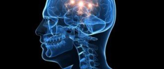
The brain is a powerful control center that sends commands throughout the body and controls the progress of their implementation. It is thanks to him that we perceive the world and are able to interact with it. The kind of brain a modern person has, his intelligence, thinking, are the result of millions of years of continuous evolution of mankind, his structure is unique.
The brain is characterized by division into zones, each of which specializes in performing its own specific functions. It is important to have information about what functions each zone performs. Then you can easily understand why specific symptoms appear in such common diseases as Parkinson’s disease, Alzheimer’s disease, stroke, etc. Disorders can be regulated with medication, as well as with the help of special exercises and physical procedures.
The brain is structurally divided into:
- rear;
- average;
- front.
Each of them has their own role.
In an embryo, the head develops faster than other parts of the body. In a one-month-old embryo, all three parts of the brain can be easily seen. During this period they look like “brain bubbles”. The brain of a newborn is the most developed system in his body.
Scientists attribute the hindbrain and midbrain to more ancient structures. It is this part that is entrusted with the most important functions - maintaining breathing and blood circulation. The boundaries of their functions are clearly separated. Each gyrus does its job. The more pronounced the groove became during development, the more functions it could perform. But the anterior section provides everything that connects us with the external environment (speech, hearing, memory, ability to think, emotions).
There is an opinion that a woman's brain is smaller than a man's brain. Data from modern hardware studies, in particular on a tomograph, have not confirmed this. This definition can easily be called erroneous. The brain of different people may differ in size and weight, but this does not depend on gender.
Knowing the structure of the brain, you can understand why certain diseases appear and what their symptoms depend on.
Structurally, the brain consists of two hemispheres: right and left. Outwardly, they are very similar and are interconnected by a huge number of nerve fibers. Each person has one side that is dominant, right-handers have the left side, and left-handers have the right side.
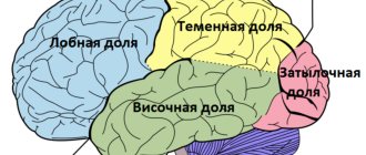
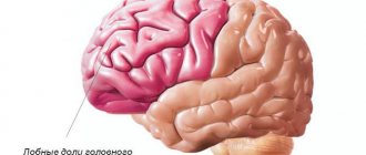
There are also four lobes of the brain. You can clearly see how the functions of the shares are differentiated.
Frontal
These lobes have a frontal location, they occupy the forehead area. Let's figure out what the frontal lobe is responsible for. The frontal lobes of the brain are responsible for sending commands to all organs and systems. They can be figuratively called a “command post.” It would take a long time to list all their functions. These centers are responsible for all actions and provide the most important human qualities (initiative, independence, critical self-esteem, etc.). When they are defeated, a person becomes carefree, changeable, his aspirations have no meaning, he is prone to inappropriate jokes. Such symptoms may indicate atrophy of the frontal lobes, leading to passivity, which is easily mistaken for laziness.
Each lobe has a dominant and auxiliary part. For right-handed people, the left area will be dominant and vice versa. If you separate them, it is easier to understand which functions are assigned to a specific area.
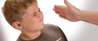
It is the frontal lobes that control human behavior. This part of the brain sends commands that prevent a specific antisocial action from being performed. It is easy to notice how this area is affected in dementia patients. The internal limiter is turned off, and the person can tirelessly use obscene language, indulge in obscenities, etc.
The frontal lobes of the brain are also responsible for planning, organizing voluntary actions, and mastering the necessary skills. Thanks to them, those actions that seem very difficult at first become automatic over time. But when these areas are damaged, the person performs the actions as if anew each time, and automaticity is not developed. Such patients forget how to go to the store, how to cook, etc.
When the frontal lobes are damaged, perseveration can occur, in which patients literally become fixated on performing the same action. A person may repeat the same word, phrase, or constantly move objects around aimlessly.
The frontal lobes have a main, dominant, most often left, lobe. Thanks to her work, speech, attention, and abstract thinking are organized.
It is the frontal lobes that are responsible for maintaining the human body in an upright position. Patients with their lesions are distinguished by a hunched posture and a mincing gait.
Functions of the cortex covering the brain
To understand the significance of the cortex, you need to understand what it is, where it is located in the brain and what it is responsible for. With the participation of cortical brain structures, new movements are mastered and habitual physical skills are improved, as well as any meaningful and unconscious activity. The main function of the cortex located in the brain is to maintain the process of homeostasis.
Homeostasis is the body’s ability to self-regulate, the ability to maintain a constant internal state and overcome negative influences directed from the external environment. Sections of the cortex, covering the deep layers of the brain, coordinate all physiological processes occurring in the body. Thanks to its multi-layered, finely organized structure, the cortex, located in the brain, performs the following functions:
- Maintains balance of internal state when interacting with the external environment.
- Reacts to the slightest impulses, signaling changes within the body due to the penetration of toxic, foreign substances.
- Regulates all physiological processes, including the functioning of the circulatory and respiratory systems.
Control of organs, systems and processes occurs through excitation and inhibition of neurons. At the same time, a balance of states is maintained. If excitation occurs in one of the functional areas of the cortex, inhibition occurs in another area of the brain.
The interaction of the cortex with the subcortical and deep centers located in the brain is also carried out according to the principle of balanced inhibition and excitation. The higher parts of the central nervous system are interconnected with all reflex reactions. Signals entering the brain centers along afferent pathways are perceived in a complex manner, which allows you to accurately and objectively perceive the surrounding reality.
Pulse processing area
Perception of information occurs through sensory systems. The impulse processing zones are located mainly in the posterior parts of the cortical structures of the hemispheres. As we move towards the cortical sections, information is processed at least at three levels - receptor-effector (receptors, muscles), segmental (spinal cord, stem complexes), subcortical (brain sections).
The sequence reflects the process of movement of an impulse to the cortical sections and the order of making a chosen decision with the subsequent commission of a purposeful action. Data enters the cortical zones in a compressed form - as it moves from the receptors to the brain, unimportant, unimportant details are eliminated.
Sensory zone
The sensory zones constantly receive auditory, visual, olfactory, gustatory, and somatosensory signals from peripheral receptors. Processing of the received data occurs in associative zones, where information about models and images of information coming from outside is stored. During the analysis, processing, comparison of existing and new information, the images are adjusted - updated, specified, detailed.
Association zone
Information from the outside enters the brain, in particular to the centers of the cortex, along afferent pathways. The pathways of conscious sensitivity continue to cortical structures. The pathways of unconscious sensitivity end in the subcortical layers. During the perception of information, it is compared with the data in memory and signals sent by other receptors. Afferent pathways of general sensitivity carry impulses coming from pain, temperature, and tactile receptors.
The structural organization of the cortex includes association zones, which are also called functional zones. The comparative analysis takes place in the associative zones of the cortex covering the cerebral hemispheres, which is of greatest importance in the development of intellectual (cognitive) abilities. Sensory signals entering the associative zones are interpreted, differentiated, and comprehended. Based on the results of the analysis, an adequate response is selected, and the corresponding information is sent to the motor zone.
The work of associative zones is interconnected with the processes of memorizing data, learning, and mental activity, and therefore play a decisive role in increasing intelligence. In the occipital region there is an associative zone that interacts with the organs of vision, which works in concert with the sensory zone and is responsible for the interpretation of visual sensations. Among the main associative zones:
- Sound. Sound analysis.
- Speech. Perception and comprehension of words, phrases, expressions.
- Motor. Planning and reproduction of complex motor activity.
The division of zones in the cortical region is carried out according to the somatotopic principle. Information coming from the face area is projected into the central posterior gyrus, into its lower sections, hands - into the middle part of the same gyrus, legs - into the upper part. The more complex the functional tasks of body parts, the larger the area of projection of impulses in the cortex.
Temporal
They are responsible for hearing, turning sounds into images. They provide speech perception and communication in general. The dominant temporal lobe of the brain allows you to fill the words you hear with meaning and select the necessary lexemes in order to express your thoughts. The non-dominant helps to recognize intonation and determine the expression of a human face.
The anterior and middle temporal regions are responsible for the sense of smell. If it is lost in old age, it may signal incipient Alzheimer's disease.
The hippocampus is responsible for long-term memory. It is he who stores all our memories.
If both temporal lobes are affected, a person cannot assimilate visual images, becomes serene, and his sexuality goes through the roof.
Parietal
In order to understand the functions of the parietal lobes, it is important to understand that the dominant and non-dominant side will do different jobs.
The dominant parietal lobe of the brain helps to understand the structure of the whole through its parts, their structure, order. Thanks to her, we know how to put individual parts into a whole. The ability to read is very indicative of this. To read a word, you need to put the letters together, and you need to create a phrase from the words. Manipulations with numbers are also carried out.
The parietal lobe helps to link individual movements into a complete action. When this function is disrupted, apraxia is observed. Patients cannot perform basic actions, for example, they are not able to get dressed. This happens with Alzheimer's disease. A person simply forgets how to make the necessary movements.
The dominant area helps you feel your body, distinguish between the right and left sides, and relate parts and the whole. This regulation is involved in spatial orientation.
The non-dominant side (in right-handed people it is the right side) combines information that comes from the occipital lobes and allows you to perceive the world around you in three-dimensional mode. If the non-dominant parietal lobe is disrupted, visual agnosia may occur, in which a person is unable to recognize objects, landscapes, or even faces.
The parietal lobes are involved in the perception of pain, cold, and heat. Their functioning also ensures orientation in space.
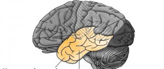
Structure and functions of the hindbrain
The metencephalon (posterior part) develops at the stage of embryonic ontogenesis, originating from the anterior part of the rhomboid region. During development, the structure of the hindbrain is supplemented and gives rise to the pons and cerebellum. Thus, the rhomboid section becomes the myelencephalon (secondary medullary vesicle) from which the medulla oblongata of the CNS organ originates.
The anatomy of this structure is quite well studied and in evolutionary development it belongs to the most ancient area. The physiology of the hindbrain was studied using the ablative method (removing part of the organ and then observing changes in the body), which helped to deeply study the functioning.
Having studied the back of the brain in section, scientists were able to describe which cavity communicates it with the medulla oblongata of the central nervous system - the fourth ventricle. The cranial nerves pass through it; the medullary striae serve as the border.
The posterior (diamond-shaped) section of the central nervous system consists of:
- Oblong section;
- The hindbrain itself.
The second structure, in turn, is divided into the pons and the cerebellum. The rhomboid part of the central nervous system is involved in reflex activity, since it contains nerve bundles and cranial nerves that perform various functions.
Hindbrain - structure and functions of reflex activity:
- Gaulle's bundle - represented by axons, regulates muscle and joint sensitivity (from the lower extremities);
- Burdach's bundle - includes axons, regulates muscle and joint sensitivity of the entire upper body (from the neck).
These bundles form the path of proprioceptive sensitivity and allow you to recognize the position of body parts in space, perceive postures and feel both active and passive movements. If there are disturbances in the cortical direction, the coordination of movements is lost and they become disproportionate.
Reflex activity is also carried out with the help of innervation by cranial nerves (nuclei from 5 to 12 pairs), which form the classification of various reflexes in the corresponding structures of the central nervous system.
The hindbrain is involved in the implementation of the following reflexes:
- Responsible for tactile sensitivity - the function is provided by the trigeminal nerve (5th pair), located between the pons and the middle cerebellar peduncle. Supports unconscious reflexes in response to pain or touching a hot object. At the exit from the bridge it connects with the mandibular nerve and innervates the muscles of mastication. Damage to the fibers is accompanied by sharp pain and hyperemia of the skin on the face. If disturbances affect the motor nuclei, then atony of the masticatory and temporal muscles occurs.
- Responsible for the work of the oculomotor muscle (rectus) - the function is supported by the abducens nerve (6th pair), lies in the thickness of the bridge, exiting in the oblong part of the central nervous system and penetrates into the orbital area. Damage to the fibers leads to blurred vision (double vision) and the inability to direct gaze in any direction.
- Provides facial expressions - innervation of the facial nerve (7th pair). The roots extend from the bridge tire. After which a loop is formed and then the fibers run through the thickness of the bridge, exiting through the gap between the oblong structure of the central nervous system organ. Damage to the fiber (its branches) leads to a complete absence of facial expressions, the face becomes like a mask (all folds are smoothed out, blinking movements are impossible, eyelids do not droop).
- Responsible for hearing and vestibular regulation - the vestibular nerve (8th pair), is divided into two parts. The first (cochlea) conducts auditory impulses to the central nervous system, the second (vestibular) is located at the bottom of the auditory canal and regulates balance. From the first part, the nerve fibers end in the tegmentum of the bridge, from the second, in the cavity of the diamond-shaped structure - the diamond-shaped fossa. Damage to the nuclei leads to hearing loss and impaired balance (staggering gait).
- Responsible for swallowing movements - muscle contraction is provided by the glossopharyngeal nerve (9th pair), and in addition to the pharynx, it innervates the middle ear and tongue (posterior third). It comes out of the hole in the skull and runs to the cavity of the rhomboid region (4th ventricle). Damage to the fibers causes difficulty swallowing (or pain) and impaired taste sensitivity of the tongue.
- Responsible for the functioning of organs in the abdominal and thoracic cavities - the function is provided by the vagus nerve (10 pair), starting from the oblong part of the central nervous system organ, extending down through the neck to the cavities. Damage to the nuclei causes a malfunction of the internal organs, and paresis of the pharynx or larynx is possible.
- Controls contractions of the trapezius and large superficial muscles of the neck - the innervation comes from the accessory nerve (11th pair), the upper part of the fibers starts from the oblong structure, the lower part - from the anterior horns of the spinal part of the central nervous system (upper segments). At the exit from the cranium it intertwines with the vagus nerve. Damage to these fibers causes paresis or paralysis of the innervated muscles.
- Responsible for movements of the tongue - innervation is provided by the hypoglossal nerve (12 pair), located in the rhomboid fossa and has multiple branches, the final branch ends in the tongue. Damage to the fibers leads to impaired motor abilities in the tongue (muscle atrophy).
All parts of the diamond-shaped structure are connected to each other through neural connections that act as conductors.
Occipital
The occipital lobes process visual information. It is with these lobes of the brain that we actually “see.” They read signals that come from the eyes. The occipital lobe is responsible for processing information about shape, color, and movement. The parietal lobe then turns this information into a three-dimensional image.
If a person stops recognizing familiar objects or loved ones, this may indicate a dysfunction in the occipital or temporal lobe of the brain. In a number of diseases, the brain loses the ability to process received signals.
Brain disorders
Unlike epileptic conditions, which are caused by dysfunction of the right temporal lobe of the brain, the feelings of an ordinary person manifest themselves systematically, and not in fits and starts.
As a result of voluntary subjects, it was revealed that forced activation of the temporal lobes of the brain is felt by a person as supernatural experiences, sensations of the presence of a non-existent object, angels, aliens, and a feeling of transition beyond the limits of life and approaching death was also recorded.
Awareness of a double or “other self” arises due to a mismatch between the hemispheres of the brain, according to experts. If emotional perception is stimulated, extraordinary, so-called spiritual experiences arise.
The passive temporal lobe hides intuition; it is activated when you have the feeling that someone you know is not feeling well, although you cannot see them.
Among patients suffering from an illness of the medial areas of the temporal lobe, there were cases of extreme emotionality, as a result of which highly ethical behavioral manifestations developed. The behavior of patients with hyperactive gyri of the temporal lobe was monitored by rapid and coherent speaking, and at the same time, a relative decrease in sexual activity was noticeable.
Conclusion
So, each department has its own functional load. If a separate lobe suffers due to injury or disease, another zone may take over some of its functions. Psychiatry has accumulated a lot of evidence of such redistribution.
It is important to remember that the brain cannot function fully without nutrients. The diet should have a variety of foods from which the nerve cells will receive the necessary substances. It is also important to improve blood supply to the brain. It is promoted by sports, walks in the fresh air, and a moderate amount of spices in the diet.
If you want to maintain full brain function until old age, you should develop your intellectual abilities. Scientists note an interesting pattern - people with intellectual work are less susceptible to Alzheimer's and Parkinson's diseases. The secret, in their opinion, lies in the fact that with increased brain activity in the hemispheres, new connections are constantly created between neurons. This ensures constant tissue development. If a disease affects one part of the brain, its functions are easily taken over by the neighboring zone.
Telencephalon structure
In the hemispheres of the telencephalon, there are three surfaces (superior lateral, medial, inferior), three edges (superior, inferolateral, inferomedial) and three poles (frontal, occipital and temporal).
The outer surfaces of the hemispheres have a peculiar shape, due to the presence of convolutions and grooves passing between them.
On the superolateral surface of the hemisphere, the frontal and temporal lobes are separated by a deep lateral, or Sylvian fissure, covering the insula.
The frontal lobe is separated from the parietal lobe by a central sulcus, which continues on a small part of the medial surface of the hemisphere and ends before reaching the Sylvian fissure. Closer to the frontal pole, the precentral sulcus runs along the central one. It projects forward the superior and inferior frontal sulci, which determine the presence of the superior, middle and inferior frontal gyri.
The precentral and central sulci define the precentral sulcus. The inferior frontal sulcus of the telencephalon is cut through by the ascending and anterior branches of the lateral sulcus, dividing it into the tegmental, triangular and orbital parts.
From the central to the conventional border of the parieto-occipital sulcus, located on the medial surface of the hemispheres and dissecting the upper edge, the parietal lobe continues. Within its boundaries lies the postcentral gyrus, separating the postcentral gyrus. On the medial surface, the pre- and postcentral sulcus merge to form the paracentral lobule. From the postcentral gyrus towards the occipital lobe there is an intraparietal sulcus separating the superior and inferior parietal lobes.
The occipital lobe is located from the continuation of the parieto-occipital sulcus to the occipital pole.
Within its boundaries, the transverse occipital groove is well defined.
The temporal lobe is horizontally crossed by two parallel grooves - the superior and inferior temporal, separating the superior, middle and inferior temporal gyri.
The insula is hidden deep in the lateral sulcus.
It also has several small convolutions and grooves, the deepest of which is the circular groove.
What is the telencephalon? On the medial surface of the hemisphere, directly above the corpus callosum, there is a fissure of the corpus callosum, which continues below into the hippocampal fissure.
Parallel to it above lies the cingulate groove, which follows to the upper edge of the hemisphere and separates the cingulate gyrus. Posterior to the corpus callosum, there is an isthmus on the cingulate sulcus, which continues into the parahippocampal gyrus. These three formations are combined into the vaulted gyrus. The posterior part of the medial surface contains the parieto-occipital and calcarine grooves, closer to the occipital pole.
The triangular-shaped space between these grooves is called a wedge.
On the lower surface of the hemisphere, the grooves are less pronounced. Within the frontal lobe there are several orbital gyri and sulci. Closer to the occipital pole, the collateral and occipitotemporal grooves are defined.
The telencephalon cortex, covering the hemispheres, is an important functional element of the brain.
Its thickness in different sections reaches from 1.5 to 4.5 mm. The morphology and arrangement of neurons in different parts of the cortex varies. The specificity of cell distribution in the cerebral cortex V.A. Betz defined the term “cytoarchitecture”. The term “myeloarchitecture” refers to the heterogeneity of the arrangement of fibers. The new cortex is divided into layers; In total, six layers are distinguished in it.
Old and ancient bark may only contain two or three layers. In the neocortex, the molecular, outer granular, outer pyramidal, inner granular, inner pyramidal and granular layers are distinguished from the outside to the inside. Studying the features of the distribution of fibers and neurons in the cortex, K. Brodman substantiated the concept of cytoarchitectonic fields. He proposed the presence of 52 fields responsible for specific functions.
For example, the motor area of the cortex is determined in the fourth and sixth fields, the nucleus of the visual analyzer is in the 17th, 18th and 19th Brodmann fields.