Craniotomy is a surgical procedure that can be performed in a hospital at any level as an emergency medical aid for patients with intracranial hypertension.
Craniotomy has been known since ancient times. Even ancient people used trepanation to treat almost all diseases, believing that the evil spirit of the disease escaped through the hole in the skull. Now this medical manipulation is carried out exclusively for health reasons or to improve the prognosis of brain disease.
Types of trepanations and indications for them
Indications for craniotomy are gradually narrowing due to the emergence of new, more gentle treatment methods, but in many cases it is still the only way to quickly eliminate the pathological process and save the patient’s life.
decompressive trepanation is performed without intervention on the brain
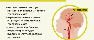
The reason for decompressive trephination (resection) are diseases that lead to a rapid and threatening increase in intracranial pressure, as well as causing a displacement of the brain relative to its normal position, which can result in infringement of its structures with a high risk of death:
- Intracranial hemorrhages;
- Injuries (crushed nerve tissue, bruises combined with hematomas, etc.);
- Brain abscesses;
- Large inoperable neoplasms.
Trepanation for such patients is a palliative procedure that does not eliminate the disease, but eliminates the most dangerous complication (dislocation).
Osteoplastic trepanation serves as the initial stage of surgical treatment of intracranial pathology, providing access to the brain, vessels, and membranes. It is shown when:
- Malformations of the skull and brain;
- Tumors that can be removed surgically;
- Intracerebral hematomas;
- Vascular aneurysm and malformations;
- Abscesses, parasitic lesions of the brain and membranes.
osteoplastic trepanation for brain surgery
In modern medicine, craniotomy is also called craniotomy (but not brain trepanation). Another name does not change the fact that this is a very complex surgical procedure. The emergence of new methods of combating many brain diseases makes it possible to resort to it less often than before. However, two types of such operations on the skull still take place in neurosurgical practice - osteoplastic, resection trepanation.
Trepanation is done when you need to gain access directly to the contents of the skull for surgical treatment:
- malformations of the skull and brain;
- tumors;
- intracerebral hematomas (for example, with hemorrhagic stroke);
- vascular aneurysm;
- abscesses;
- brain damage by parasites.
The operation begins with choosing a location for the burr hole: it should be as close as possible to the affected area. First of all, the surgeon cuts the soft tissue in a horseshoe shape so that the base of the flap is located in the lower part, since the blood vessels pass from bottom to top, and it is very important not to violate their integrity.
Next, using special instruments, the periosteum and bone are dissected at an angle of 45°. Such a cutting angle is necessary so that the outer surface of the bone flap exceeds the inner one, and when restoring the integrity of the skull, the removed fragment does not fall inward. Having reached the meninges, the surgeon performs manipulations directly in the cranial cavity (removes the tumor, eliminates hemorrhage).
The craniotomy ends with suturing:
- the dura mater of the brain is sutured with absorbable threads;
- the flap is fixed with special threads or wire;
- the skin and muscles are sutured with catgut.
The pretexts for performing resection craniotomy are pathologies that provoke a rapid increase in intracranial pressure, which threatens life, or contribute to the displacement of brain structures, which is fraught with their infringement and death. These conditions include:
- cerebral hemorrhages;
- cerebral edema;
- injuries (bruises, hematomas, crushing tissue as a result of an impact);
- inoperable large tumors.
Trepanation in such cases is a palliative procedure, that is, it does not eliminate the disease, but only eliminates a dangerous complication.
The best place for surgery is the temporal area. Here, after removal of the bone flap, the membrane of the brain will be protected by the powerful temporalis muscle.
How is resection craniotomy performed? As with osteoplastic craniotomy, soft tissue and bone are cut. The bone fragment is removed so that the diameter of the hole is 5 - 10 cm. Having discovered swelling of the brain membrane, the surgeon is in no hurry to dissect it so that there is no displacement of the brain structures.
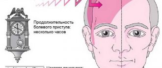
Craniotomy of any kind can last several hours, and is used only for serious indications that threaten the patient's life. No one will perform such an operation, for example, in case of a mini-stroke - to eliminate its consequences, there are more gentle methods of therapy.
Scope of operation
Surgical intervention affecting the brain is so serious that it is carried out in the only case - if not just a person’s health is at risk, but his life. Trepanation is prescribed:
- if a tumor is maturing in the patient’s brain, even if it has nothing to do with oncology, as it grows it will compress parts of the brain, cause monstrous migraines and hallucinations, making normal life almost impossible;
- if cancer develops in the patient’s brain, as the tumor develops, it will not only begin to compress neighboring sections, but also affect them with metastases, which can lead to disability, and subsequently death;
- if an inflammatory infectious process occurs in the patient’s brain, the further it goes, the greater the likelihood of irreversible damage that will lead to the failure of certain parts and, accordingly, body functions;
- if the patient’s skull was damaged due to a traumatic brain injury, trephination can be performed to remove bone fragments, assess the damage caused and, if possible, compensate for it;
- if the patient has experienced a stroke caused by thrombosis, trepanation is performed to remove the blood clot that has plugged the vessel;
- if the patient suffers from thrombosis and the risk of stroke is very high, trepanation is performed to remove blood clots;
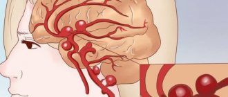
- if the patient suffers from brain bleeding caused by a sudden rupture of a vessel, trephination is intended to give the doctor access to the brain and the ability to cope with the bleeding;
- If there is a suspicion of brain cancer and a biopsy is needed, trepanation opens the brain so that tissue samples can be taken from it.
Historical reference
In neurosurgery, trephination is the making of a hole in some part of the skull to gain direct access to brain tissue. However, such surgery should not be considered an invention of modern medicine. Archaeological finds indicate that our ancestors could have drilled holes in the skull for medicinal purposes several thousand years ago.
Hippocrates proposed trepanation as a way to treat head wounds, including removing bone fragments from the brain after injury. For this procedure, his followers came up with a special drill. Prehistoric trepanations in the culture of ancient civilizations of Peru were performed with a ceremonial knife called a tumi.
The purpose of trephination was not always to open access to the brain for further manipulation. In ancient times, a hole in the skull often served as an outlet for evil spirits, which were considered the cause of disease. Also, the hole in the skull was seen as a kind of channel for obtaining special knowledge and spiritual experience. In Egypt, pharaohs underwent such an operation, presumably to make it easier for the soul to leave the body after death.
Preparing for surgery
If penetration into the cranial cavity is necessary, good preparation of the patient for surgery is important. If there is enough time, the doctor prescribes a comprehensive examination, including not only laboratory tests, CT and MRI, but also consultations with specialists and examinations of internal organs. An examination by a therapist is required to decide whether the intervention is safe for the patient.
However, it happens that the opening of the skull is performed urgently, and then the surgeon has very little time, and the patient undergoes the necessary minimum of studies, including general and biochemical blood tests, a coagulogram, MRI and/or CT scan to determine the state of the brain and the localization of the pathological process.
During a planned operation, after six o'clock in the evening the day before, it is forbidden to eat and drink, the patient once again talks with the surgeon and anesthesiologist, and takes a shower. It is advisable to rest and calm down, and in case of severe anxiety, sedatives may be prescribed.
Before the intervention, the hair on the head is carefully shaved, the surgical field is treated with antiseptic solutions, and the head is fixed in the desired position. The anesthesiologist puts the patient under anesthesia, and the surgeon begins the manipulations.
Opening the cranial cavity can be done in different ways, therefore the following types of trepanation are distinguished:
- Osteoplastic.
- Resection.
Regardless of the type of surgery planned, the patient must be placed under general anesthesia (usually nitrous oxide). In some cases, trepanation is performed under local anesthesia with a solution of novocaine. To enable artificial ventilation of the lungs, muscle relaxants are administered. The surgical area is carefully shaved and treated with antiseptic solutions.
Postoperative period
Rehabilitation and prognosis on the first day after surgery
The first day the patient is in the intensive care unit, unconscious. The functions of vital systems are provided by a ventilator and parenteral nutrition. At this time, it is important to monitor the patient’s condition, as there is a risk of missing the onset of a serious complication. In terms of rehabilitation, it is important to ensure complete not only physical, but also emotional peace for the patient. The prognosis on the first day is questionable, since it is impossible to predict the brain’s reaction to this type of intervention.
Osteoplastic trepanation
In this type of operation, a burr hole is made where the path to the affected area of the brain will be the shortest. The first step is a horseshoe-shaped incision into the soft tissues of the head. It is important that the base of this flap is at the bottom, since the vessels supplying the skin and underlying tissue run radially from bottom to top, and their integrity must not be compromised to ensure normal blood flow and healing. The width of the base of the flap is about 6-7 cm.
After the musculocutaneous flap with the aponeurosis is separated from the surface of the bone, it is turned down, fixed on napkins soaked in saline solution or hydrogen peroxide, and the surgeon proceeds to the next stage - the formation of the osteoperiosteal flap.
stages of osteoplastic trepanation according to Wagner-Wolf
The periosteum is cut and peeled off according to the diameter of the cutter, which the surgeon uses to make several holes. The sections of bone preserved between the holes are cut out using a Gigli saw, but one “lintel” remains intact, and the bone in this place is broken. The bone flap will be connected to the skull through the periosteum in the area of the fractured area.
To ensure that the fragment of the skull bone does not fall inward after being placed in its original place, the cut is made at an angle of 45°. The area of the outer surface of the bone flap turns out to be larger than the inner one, and after this fragment is returned to its place, it is firmly fixed in it.
Having reached the dura mater, the surgeon dissects it and enters the cranial cavity, where he can perform all the necessary manipulations. After the intended goal is achieved, the tissues are sutured in the reverse order. Sutures of absorbable threads are placed on the dura mater of the brain, the bone flap is returned to its place and fixed with wire or thick threads, and the musculocutaneous area is sutured with catgut. It is possible to leave a drainage in the wound for the outflow of discharge. The sutures are removed by the end of the first week after surgery.
Operation process
After the patient is put under anesthesia, the scalp is carefully treated with an antiseptic. An incision is made to expose the required area of the skull. The trephinated skull bone is dissected, removed, and brain surgery is performed.
Once the intervention is complete, the exposed area of the brain is closed. The removed portion of the skull bone is returned to its original location, and surgical sutures are placed on the scalp. To ensure the outflow of fluid and remove blood, drainage tubes are inserted into the operated area, and a bandage is applied to the head. After a few days, the drainage can be removed. The operation itself lasts several hours.
The patient is then sent to the recovery room, where his vital signs are carefully monitored. Pulse, body temperature, breathing and blood pressure are checked regularly. After some time, the operated patient will be transferred to the intensive care ward, and then to the hospital ward.
Complications after surgery
The operation is performed under general anesthesia; in some cases, part of the intervention takes place under local anesthesia, that is, the person remains conscious. After trephination, the patient is admitted to the intensive care unit or recovery room. When the patient comes to his senses without incident, he is transferred to the neurosurgery department, the duration of his stay there is about 2 weeks.
The consequences of trephination (anesthesia) may be thirst, headache, but this is not critical. But swollen facial tissues and bruises around the eyes may indicate the progression of cerebral edema. There are a number of other possible complications after this surgery.
- The occurrence of inflammatory processes in the wound, encephalitis, meningitis is a consequence of infections.
- Neurological disorders (convulsions, problems with coordination of movements, intellectual disorders) are associated with damage to the meninges and tissues.
- Deformation of the skull after removal of part of the bone, formation of a keloid scar.
One of the main uncontrollable factors that aggravate the course of the postoperative period is the age of the patient. Craniotomy is most easily tolerated by young people without serious concomitant diseases. The situation is somewhat worse with children. This is due to the insufficient development of compensatory mechanisms of the child’s body and anatomical features.
The most severe consequences occur in older people. Due to natural disturbances in the regulation of blood circulation, metabolism and recovery processes, the postoperative period is very difficult. The recovery period after craniotomy rarely goes smoothly, absolutely without complications.
The individual characteristics of each organism are no less significant. This is determined by numerous genetic characteristics. Each person has unique deviations in metabolic processes, the structure of certain anatomical formations and the severity of reactions to surgical intervention.
The effects of craniotomy are influenced by surgery performed in the past. Sometimes, during repeated surgical interventions on the brain part of the skull, adhesions (adhesions) between the membranes of the brain and its substance can be detected, which occupy the trepanned area of the bones of the cranial vault. In this case, the duration of surgical intervention and the risk of complications increases significantly.
Premorbid background is also important in terms of prognosis. This concept means the entire spectrum of diseases that arose before the operation and have persisted to the present day. Some diseases significantly complicate the course of the postoperative period. For example, diabetes mellitus, which causes significant damage to the capillary beds of all organs, including the brain with all its membranes.
Rehabilitation after trepanation
Rehabilitation after such a serious intervention is aimed at restoring the functions of the damaged area and improving the general condition of the patient.
This part is final, and, one might say, the most important. Without necessary measures after surgery
full recovery is impossible. Moreover, the affected person may return to the condition that caused the problem.
Rehabilitation after trephination is complex and is aimed at consolidating the results of the operation and neutralizing all kinds of negative consequences.
Main tasks of the rehabilitation period:
- Neutralization of the cause that caused brain diseases after surgery;
- Smoothing out the consequences of surgery;
- Early identification of risk factors that can lead to complications;
- Maximum restoration of impaired brain functions.
The recovery process after trepanation is the most complex, which is why it consists of many successive stages, each of which is equally important. The duration of treatment and technique may vary in each specific case.
The duration and outcome of the operation are influenced by many factors, including:
- The patient's initial health status;
- Doctor's experience;
- Patient's age;
- The presence of complications and concomitant diseases.
The main thing to remember for those who have undergone such an operation or have a relative who underwent trepanation is that stress and noise are an absolute contraindication.
The patient should not be overloaded in the first ten days, until the sutures are removed.
After this stage, it is necessary to gradually introduce more active measures along with drug treatment.
In addition to ensuring complete rest, it is necessary to take a number of the following sequential measures:
- Select painkillers . Pain causes additional stress, which brings the patient back into the risk zone;
- Antiemetic drugs are part of the treatment, since due to impairment of certain functions and increased sensitivity and susceptibility, the patient may suffer from attacks of vomiting and headaches;
- Ongoing physical therapy and brain function testing are necessary;
- Weekly consultations with a psychologist and neurologist . This stage is important because it allows you to detect the slightest changes in consciousness or behavior, which is a signal of disturbances;
- Testing neural connections of the brain;
- Constantly maintaining the cleanliness of the wound , monitoring the healing and disinfection processes;
- Preventive measures to prevent the development of complications.
After 14-20 days of stay in the hospital ward under strict supervision, the patient is discharged and sent to secondary rehabilitation on an outpatient basis.
The full range of restoration procedures consists of:
- monitoring the condition of the wound;
- a complex of various physiotherapeutic procedures;
- restoration of lost or damaged skills;
- occupational therapy and other approaches;
- Exercise therapy and massages;
- walking outside the hospital buildings;
- control of diet and lifestyle;
- psychotherapy.
In addition, the patient is prescribed medications that help cope with the disease and its consequences from the inside.
It is imperative for patients to constantly maintain contact with a doctor, who must be contacted at the slightest deviation from the norm, which may be:
- physical and mental (failures of thinking, logic, memory, motor processes and reactions, sensations);
- inflammation and swelling of scars;
- the appearance of regular headaches;
- nausea and vomiting;
- difficulty breathing;
- convulsions and fainting;
- numbness of the face;
- general weakness, chills, fever;
- blurred vision;
- chest pain.
When starting rehabilitation, you need to remember that even the right approach may not lead to a complete recovery, but it will teach you how to live well with the problem and gradually improve your condition.
Resection trepanation
Resection trephination is performed for intracranial tumors that can no longer be removed, with a rapid increase in cerebral edema due to hematomas with a risk of dislocation of nerve structures. Its location is usually the temporal region. In this area, the skull bone is located under the powerful temporal muscle, so the trepanation window will be covered with it, and the brain will be reliably protected from possible damage. In addition, temporal decompressive trephination provides better cosmetic results compared to other possible trepanation sites.
resection (decompressive) trephination according to Cushing
At the beginning of the operation, the doctor cuts out a musculoskeletal flap linearly or in the shape of a horseshoe, turns it outward, dissects the temporalis muscle along the fibers and incises the periosteum. Then a hole is made in the bone with a milling cutter, which is expanded using special Luer bone cutters. This results in a round trepanation hole, the diameter of which varies from 5-6 to 10 cm.
After removing the bone fragment, the surgeon examines the dura mater of the brain, which, with severe intracranial hypertension, can be tense and bulge significantly. In this case, it is dangerous to dissect it immediately, since the brain can quickly shift towards the trepanation window, which will cause damage and wedging of the trunk into the foramen magnum.
The operation is completed by sequential suturing of tissues with the exception of the dura mater. The bone section is not put into place, as in the case of osteoplastic surgery, but subsequently, if necessary, this defect can be eliminated using synthetic materials.
Operation technique
During craniotomy, the cranial cavity – the bones of the skull – is opened. This is needed for two purposes:
- Relieve intracranial hypertension (edema fluid or blood will flow out through the artificial opening, which will prevent a life-threatening complication - wedging of the brain).
- Carry out medical manipulations on a living brain. For example, removing a brain tumor.
Opening the bones is done using special instruments. If you just need to relieve hypertension, you usually make one small hole in the parietal bone with a milling cutter. This is less traumatic, and therefore more favorable in terms of rehabilitation and health consequences. If wide access to the brain is necessary, extensive trephination is performed with the removal of part of the bone.
Early postoperative consequences
Frequent complications after craniotomy include bleeding. They can occur both during the surgical intervention itself and immediately after its completion. Due to the abundant blood supply to the tissues of the head, the patient can lose a significant amount of blood in a short period of time.
In this case, emergency blood transfusion (transfusion of someone else's blood) may be required. Therefore, in the preoperative period, if the patient’s condition allows, a full laboratory and instrumental examination is performed. This includes determining the blood group and Rh factor, since when massive bleeding develops, every second counts.
At the present stage of development of neurosurgical technology, unintentional damage to the brain is extremely rare. However, in some situations it is quite possible. Depending on the degree of damage (size and depth) of the brain matter, further consequences are formed.
The brain reacts to damage (concussion, bruise or penetrating wounds) in a very similar way. Edema and swelling of its substance develops. At the histological level, this is manifested by the release of a significant amount of liquid blood from the capillary bed into the interstitial space and the “leakage” of nerve fibers by it.
This leads to a significant increase in the volume of the medulla. The brain seems to be pressing on the skull from the inside. When trephination is carried out carelessly or inadequate infusion therapy, the brain substance is displaced into the trepanation hole with the development of damage, ruptures and other irreparable changes in the structure.
Considering the complexity of any intervention on the brain and the seriousness of the reasons that may be the reason for this intervention, there remains the risk of death right on the operating table. In this case, a number of circumstances that are beyond the control of medical personnel are decisive.
The duration of some operations for craniotomy is associated with the risk of complications that are not a direct consequence of the intervention itself. Firstly, these may be the consequences of a long stay in a narcotic sleep. Which is associated with many respiratory and cardiac disorders.
The patient's limbs may remain in an unnatural position for a long time. This is associated with an increase in pressure on individual neurovascular bundles and can lead to damage to these structures and the occurrence of flaccid paralysis and paresis in the postoperative period.
Staying in one position for several hours in the absence of spontaneous breathing (since such surgical interventions are performed under inhalation anesthesia) can cause the development of pneumonia.
After the intervention, the patient is taken to the intensive care unit or recovery room, where doctors carefully monitor the function of vital organs. On the second day, if the postoperative period is successful, the patient is transferred to the neurosurgery department and spends there for up to two weeks.
It is very important to control the discharge through the drainage, as well as the hole during resection trepanation. Bulging of the bandage, swelling of the facial tissues, bruising around the eyes may indicate an increase in cerebral edema and the appearance of a postoperative hematoma.
Trephination is accompanied by a high risk of various complications, including infectious and inflammatory processes in the wound, meningitis and encephalitis, secondary hematomas with inadequate hemostasis, suture failure, etc.
The consequences of craniotomy can be various neurological disorders when the meninges, vascular system and brain tissue are damaged: disorders of the motor and sensory sphere, intelligence, convulsive syndrome. A very dangerous complication of the early postoperative period is the leakage of cerebrospinal fluid from the wound, which is fraught with the addition of infection with the development of meningoencephalitis.
The long-term result of trephination is deformation of the skull after resection of a section of bone, the formation of a keloid scar when regeneration processes are disrupted. These processes require surgical correction. To protect brain tissue and for cosmetic purposes, the hole after resection trepanation is closed with synthetic plates.
Some patients after craniotomy complain of frequent headaches, dizziness, decreased memory and performance, feeling tired and psycho-emotional discomfort. There may be pain in the area of the postoperative scar. Many symptoms following the operation are associated not with the intervention itself, but with the pathology of the brain, which was the root cause of trephination (hematoma, bruise, etc.).
Recovery after craniotomy includes both drug therapy and the elimination of neurological disorders, social and work adaptation of the patient. Before removing the sutures, wound care is required, including daily monitoring and changing dressings. You can wash your hair no earlier than two weeks after the operation.
For intense pain, analgesics are indicated; in case of seizures, anticonvulsants are indicated; the doctor may also prescribe sedatives for severe anxiety or agitation. Conservative treatment after surgery is determined by the nature of the pathology that brought the patient to the operating table.
If various parts of the brain are damaged, the patient may have to learn to walk, speak, restore memory and other impaired functions. Complete psycho-emotional rest is indicated; it is better to avoid physical activity. An important role at the rehabilitation stage is played by the patient’s relatives, who, already at home, can help cope with some inconveniences in everyday life (taking a shower or cooking, for example).
In case of severe brain damage with paralysis and paresis, disorders of speech, thinking, memory, etc., the patient needs additional care and cannot not only go to work, but also take care of himself independently. Of course, such cases require the establishment of disability. After craniotomy, the disability group is determined by a special medical commission of different specialists and depends on the severity of the patient’s condition and the degree of disability.
The rehabilitation period after craniotomy lasts different times, depending on the pathology of the brain. At first, you need careful care of the wound and monitoring the condition of the sutures. Intense pain is relieved with analgesics, severe anxiety is relieved with sedatives. The patient is advised to have physical and emotional rest - the doctor will tell you how long you can’t go to work.
To quickly restore strength, your doctor may recommend a diet. Most likely, you will have to switch to proper nutrition, giving up foods that increase blood pressure and clog blood vessels with cholesterol. We are talking about caffeinated drinks, alcohol, fatty, fried foods.
If the disease has led to damage to certain areas of the brain and functional impairment, trepanation is not able to instantly put everything in order. In such situations, rehabilitation may include the need to relearn walking, talking, etc.
Possible complications
There are two options for complications that can develop in a patient who is scheduled for surgery:
- Early. Their occurrence occurs directly during the intervention and often does not even depend on the skill of the surgeon. Among them:
- Bleeding. Since the brain is richly supplied with blood, the loss will be rapid and heavy - which is why surgeons always have blood ready for transfusion.
- Brain damage. At the current level of medical development, they are rare, but can lead to complete failure of the affected area of the brain.
- Edema. This is how the brain reacts to any emergency situation. In case of inaccurate trepanation, the brain matter may be displaced towards the area of intervention, often with pathologies and ruptures.
- Death. It can develop for a variety of reasons, up to simple heart failure due to anesthesia and the excessive stress it causes.
- Late. Their occurrence should be expected after trepanation, during the recovery period. They can be provoked by non-compliance with the doctor’s recommendations, an inaccurate operation and weakness of the body after the intervention. Among them:
- Wound infection. If hygienic standards have not been maintained strictly enough, there is a chance that the edges of the wound will become inflamed and swollen, causing pain to the patient.
- Brain infections. They are very rare, but have dire consequences, causing forgetfulness, irreversible personality changes, convulsions, and failure of certain departments.
- Blood clots and blood stagnation. After surgery, a person usually moves little, so there is a high probability of developing thrombosis, which can lead to complications, including strokes and heart attacks.
- Neurological disorders. The brain tissue may swell, which will disrupt the functioning of its parts. A person may experience problems with everything from speech to coordination—either permanent or temporary, depending on the extent of the damage.
The patient may also feel depressed, have problems sleeping and eating, suffer from speech and coordination problems, and may become irritable or tearful. The main thing is to carefully monitor any suspicious symptoms and, while rehabilitation after craniotomy lasts, carefully report them to your doctor.
There are no unimportant symptoms - if something causes concern in the patient, it needs to be talked about.