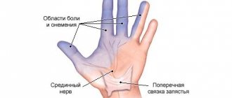
Pinched nerves can occur in more than just the neck, lower back, or extremities. In some cases, neurological pain occurs in the groin. This is a fairly rare symptom, but it occurs in some patients. In the groin area there are branches of nerves located in the lumbar and sacrum areas. The endings of the ilioinguinal nerve are located here. It is rarely subject to compression. However, with some pathologies and injuries, infringement of this part of the peripheral nervous system is observed. This is accompanied by chronic pain and sometimes a feeling of numbness.
Anatomical structure
The ilioinguinal nerve begins in the lumbar region. In the groin area it branches into 2 parts. One of them innervates the thigh. The second is called the inguinal branch. The further continuation of this nerve ending depends on the gender of the person:
- In men, the inguinal branch passes through the spermatic cord and ends in the scrotum.
- In women, the inguinal branch exits in the area of the uterine ligament and ends in the area of the labia majora.
The inguinal branch is responsible for the innervation of the following organs:
- muscles and skin of the anus;
- anal sphincter;
- external genitalia;
- perineal muscles;
- bladder sphincter.
The ilioinguinal nerve affects sensations during sexual intercourse, the process of defecation and urination. Its work is regulated by the autonomic nervous system.
In some pathologies, compression of the nerve occurs, which is accompanied by chronic aching pain.
Treatment
Drug therapy
Medicines are used to relieve pain . Usually a painful attack lasts 2-5 minutes. But because of its intensity, it seems to the patient that its duration is much longer.
If the pain is caused by the pressure of muscle tissue on the nerve endings, then it is advisable to take antispasmodics. NSAIDs - this group of drugs is used to relieve swelling, inflammation and pain. The course of treatment is short - 3-5 days.
Medicines with a diuretic effect are prescribed to relieve swelling. Reparants - prescribed to improve and accelerate the recovery of nerve fibers . Even after pain relief, you need to consult a doctor and find out its cause.
Physiotherapy
Physiotherapeutic procedures are necessary to warm up the joints and tendons. The most commonly used procedures are:
- electro- and phonophoresis;
- Magnetotherapy;
- UHF;
- hot baths;
- wraps;
- splinting;
- applications.
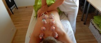
Massage is a separate item of physiotherapy. It is most often prescribed to treat neuralgia. The procedure is aimed at relieving pinching and reducing the inflammatory process.
However, please note that all procedures should be carried out only under the supervision of the attending doctor.
Gymnastics for inflammation of the pudendal (genital) nerve
Gymnastics is aimed at improving blood flow in the inflamed area. Start with a regular walk. Often in the first days of walking you may feel pain, but you need to overcome it and walk for at least 10-15 minutes at a slow pace.
As soon as the patient's condition improves, you need to move on to gymnastics. Basic exercises:
- Lie on the floor (or other hard surface), exhale, bend your knees and clasp them with your hands, while inhaling, rise slightly and touch your forehead to your knees.
Stay in this position for a few minutes, then relax, straighten up completely and rest for a minute. - Slowly sit on your heels, straighten your arms forward and stretch towards the floor, keeping your back straight.
- Lie on the floor, put your legs on the wall and bend them at a right angle, slightly lift your buttocks off the floor (literally a few centimeters) and stay in this position for 10 minutes.
- Stand straight, but at the same time bend your legs slightly at the knees, straighten your back and tilt forward a few degrees, rest your palms on your thighs and begin to press - this way the lumbar vertebrae are stretched.
- Sit on the floor, straighten your legs completely, put your hands behind your head and fasten them with a lock, slowly lie on your back and begin to raise your legs as high as possible and at the same time spread them to the sides, stay in this position for 2 minutes.
Radical methods
Doctors try to treat the disease using conservative methods. However, in some cases it is necessary to resort to surgery, which most often involves destroying the affected nerve or neutralizing the pressure on the nerve.
Radical methods are used in the following cases:
- lack of positive results after conservative treatment;
- rapid progression of the disease;
- the presence of serious motor and sensory contractures;
- the appearance of tumors.
Folk recipes
Effective recipes:
- Take a medium-sized onion, peel, finely chop and boil in a glass of milk for 5 minutes. Strain the finished broth and drink a glass of milk three times. The course of treatment is seven days.
- Take 50 g of thyme and boil it in a glass of water. Cool the broth and soak a gauze bandage in it. Apply to the affected area and insulate. Leave the compress overnight.
- Grate 100 g of horseradish on a fine grater, squeeze out the juice, soak gauze in the juice and apply again to the inflamed area. Insulate and leave for at least 5-6 hours, preferably overnight.
This method of treatment cannot be independent; it goes in combination with traditional therapy. Before using any folk remedy for neuralgia, you should consult a doctor.
Causes of a pinched nerve
What causes a pinched nerve? Most often, the inguinal branch is compressed after operations for a hernia. During surgery, the doctor closes the hernial opening. In this case, the nerve branches are often damaged. In addition, after surgery, scar tissue forms, which can put pressure on the nerve.
There are other factors that can cause neuropathy (pinched nerve):
- pelvic fractures;
- difficult childbirth;
- tumors of the groin area;
- anal sphincter spasms;
- pathology of the round ligament of the uterus;
- varicocele;
- hypertonicity of the pelvic muscles;
- herpes zoster;
- horseback riding;
- long bike rides.
Inguinal region anatomy. MCPC Rusakov V.I. 1997
The groin area has the shape of a right triangle. Its boundaries are: below - the Poupart ligament, above - the line connecting the right and left anterior superior iliac spines, and on the inside - the outer edge of the rectus abdominis muscle (Fig. 39).
Inguinal region anatomy. MCPC Rusakov V.I. 1997
The groin region contains the following layers: skin, subcutaneous tissue, superficial fascia, deep fascia, aponeurosis of the external oblique muscle, internal oblique muscle, transversus abdominis muscle, transversalis fascia, preperitoneal fatty tissue and peritoneum.
The fibers of the aponeurosis of the external oblique muscle of the abdomen go from top to bottom and inside. The lower thickened edge of the aponeurosis seems to stretch between the anterior superior axis of the ilium and the pubic bone. It turns downwards and backwards and forms a groove, which is an independent anatomical formation called the inguinal (pupart) ligament. The aponeurosis of the external oblique abdominal muscle and the Poupartian ligament are dense and strong. But they can be underdeveloped and disintegrating.
A denser aponeurosis is found in women. The outer edge of the inguinal ligament is attached to the upper anterior iliac spine, and the inner one splits and forms two legs: the inner (superior) and the outer (id). The inner leg is attached to the pubic symphysis, and the outer leg is attached to the pubic tubercle of the pubic bone.
A triangular space is formed between the legs, the outer edge of which is limited by arched bundles of the aponeurosis, which are called interpeduncular bundles (Fig. 40). This triangular space is called the superficial ring of the inguinal canal. Through it, the spermatic cord emerges from the inguinal canal in men, and the round ligament of the uterus in women. The dimensions of the superficial inguinal ring range from 2-3X1-2 cm. It is generally accepted that a normal inguinal ring allows only the tip of the index finger to pass through.
Inguinal region anatomy. MCPC Rusakov V.I. 1997 2
The lower edges of the internal oblique and transverse abdominal muscles are adjacent to the inguinal ligament, leaving only space for the spermatic cord in men or the round ligament of the uterus in women. This space in which the spermatic cord or round ligament of the uterus passes is called the inguinal space or (which is not entirely correct) the inguinal canal. The canal appears only when a hernia forms.
The inguinal space runs in the direction of the pupart ligament from the internal (deep) inguinal ring to the superficial one. The walls of the inguinal space are: anterior—aponeurosis of the external oblique muscle, posterior—transverse fascia, upper—edges of the internal oblique and transverse muscles, lower—pupart ligament (Fig. 41). In men, the inguinal space is shorter and wider, in women it is longer and narrower.
Inguinal region anatomy. MCPC Rusakov V.I. 1997 3
The given anatomical features of the structure of the groin region in men and women decipher the more frequent formation of inguinal hernias in men, who have a weaker aponeurosis of the external oblique abdominal muscle than women, less perfect internal oblique and transverse muscles and a wider inguinal space (canal), containing more massive formation (spermatic cord).
The interpretation of the more frequent formation of inguinal hernias in men is complemented by two more circumstances: men more often perform heavy physical work and may have some diseases (contributing to the appearance of hernias), which women do not suffer from at all (prostate adenoma, phimosis) or suffer from extremely rarely (urethral strictures) .
For a complete understanding of inguinal hernias, their appearance and development, it is necessary to know the anatomy of the anterior abdominal wall from the abdominal cavity. In the lower part of the inner surface of the anterior abdominal wall, from the navel downwards there are five folds of the peritoneum (Fig. 42), between which there are six fossae - three fossae on each side.
Inguinal region anatomy. MCPC Rusakov V.I. 1997 4
Along the midline from the bladder to the navel is the median umbilical fold (plica urabilicalis mediana), in which the obliterated urinary duct is located. More lateral is a paired fold (medial umbilical fold—plica umbilicalis media), formed along the obliterated umbilical arteries; even more lateral there is a second paired fold covering the lower epigastric vessels, the lateral umbilical fold (plica umbilitcalis lateralis).
Below, between the median and medial umbilical folds, a supravesical fossa (fossa supravesicalis) is formed. Very rare so-called supravesical hernias can originate here. Between the medial and lateral folds there is the middle inguinal fossa (fossa inguinalis medialis), which, in the sagittal plane, corresponds to the position of the superficial inguinal ring. In the area of the middle inguinal fossa, direct inguinal hernias originate, going directly into the superficial ring of the inguinal canal (Fig. 43).
Inguinal region anatomy. MCPC Rusakov V.I. 1997 5
Outside the lateral umbilical fold is the lateral fossa of the peritoneum (fossa inguinalis lateralis), corresponding to the internal ring of the inguinal canal. Here, the spermatic cord or round ligament of the uterus passes into the thickness of the abdominal wall, and then into the inguinal canal. In the area of the lateral fossa (internal inguinal ring), an oblique inguinal hernia begins to form. During the development of a hernia, the hernial sac passes through the inguinal gap, turning it into a canal, exits through the superficial inguinal ring under the skin, and then descends into the scrotum.
Embryological and anatomical data show that inguinal hernias are direct and oblique, and oblique inguinal hernias are congenital (with non-fusion of the peritoneal inguinal process) and acquired (Fig. 44).
With oblique inguinal hernias, the hernial sac travels from the deep inguinal ring, through the inguinal canal under the skin at the root of the scrotum and can, under conditions favorable for the development of a hernia, descend into the scrotum, forming an inguinal-scrotal hernia.
Congenital hernias are detected immediately after birth or in the first years of life; acquired hernias develop most often in adults. It is clear that in patients with congenital hernias, from the moment of birth there is a formed “protrusion” of the peritoneum, which turns into a hernial sac when elements of the abdominal contents first penetrate into it.
Inguinal region anatomy. MCPC Rusakov V.I. 1997 6
Acquired hernias go through all stages of development from expansion of the inguinal ring to a complete hernia. Inguinal-scrotal hernias are rare, and only in older men: patients consult a doctor early and promptly get rid of the hernia in the early stage of the disease.
There are also so-called combined hernias:
- 1) direct and oblique inguinal hernia,
- 2) direct inguinal hernia and supravesical hernia,
- 3) oblique and direct inguinal hernia and supravesical hernia.
Double inguinal hernias (oblique and direct) were described by N. A. Kukoverov (1928), N. I. Kukudzhanov (1949) and others. A similar observation was made in our clinic. The hernial sacs in such cases are separated from each other by the inferior epigastric artery.
Rare interstitial or parainguinal hernias also have a certain practical significance:
- 1) preperitoneal hernia—the hernial sac is located between the peritoneum and the transverse fascia; the hernial sac can have a different direction,
- 2) intramural hernia—the hernial sac partially passes through the inguinal canal and is located under the aponeurosis of the external oblique muscle or between the internal and transverse abdominal muscles,
- 3) superficial inguinal hernia—the hernial sac emerges from the superficial ring of the inguinal hernia and is located under the skin above the aponeurosis of the external oblique abdominal muscle or descends to the thigh and perineum. The types of peri-inguinal hernias are explained in Figure 45.
Inguinal region anatomy. MCPC Rusakov V.I. 1997 8
Preperitoneal hernias often have two pockets, one of which goes into the inguinal canal. Predisposing factors to the formation of preperitoneal inguinal hernias are a high inguinal space, a very narrow and unyielding superficial ring of the inguinal canal, looseness of the surrounding tissues and congenital protrusions of the peritoneum (E. P. Berezina, 1947; V. S. Mayat, 1947; N. I. Kukudzhanov , 1949).
Preperitoneal hernias, as well as interstitial ones, are most often recognized. are found during surgery for strangulation. However, there is reason to agree with Ficai et al. (1970) and with A. M. Lokhin (1971), who prove the possibility of preoperative recognition of this type of hernia.
An extremely rare inguinal hernia is the encysted hernia (hernia encystica), first described in 1833 by Cooper. The development of Cooper's hernia is associated with incomplete fusion of the peritoneal-inguinal process, which is divided along its length by a septum.
When a hernia forms, a double sac is formed: internal and external. During the development of a hernia, the internal sac seems to be invaded into the proximal part of the unfused peritoneal-inguinal process. Therefore, it is clear that the internal hernial sac is covered with a serous membrane on both sides. The contents of the external hernial sac are the internal hernial sac along with the organs that have prolapsed into it (Fig. 46).
Inguinal region anatomy. MCPC Rusakov V.I. 1997 9
There are two types of Cooper's hernia:
- 1) hernia encystica testicularis—formed as a testicular hernia, when there is a testicle in the external hernial sac;
- 2) hernia encystica funicularis—formed as a cord hernia. There may be a hole at the bottom of the internal sac, and then both sacs communicate with the abdominal cavity.
Knowledge of the given rare forms of inguinal hernias prevents diagnostic errors and errors during the operation.
Symptoms
It is often difficult to diagnose ilioinguinal nerve neuropathy. Symptoms of pathology are usually mild. Patients show the following signs of the disease:
- chronic aching pain in the pelvic area;
- discomfort in the anus;
- urinary incontinence;
- sensation of a foreign body in the groin, as well as burning and tingling;
- numbness of the genital skin;
- pain during sexual intercourse and urination;
- constipation
Women experience the following symptoms of a pinched ilioinguinal nerve:
- itching in the genital area;
- burning when urinating.
For this reason, patients often mistake neuropathy for an inflammatory gynecological disease.
Causes of pinched pudendal nerve and its treatment. Pudendal neuralgia in men symptoms
Does the pudendal (genital) nerve and its damage differ from similar pathologies in other “regions” of the body?
Yes, the nature of the pathology is different in that the pudendal nerve serves the pudendal area - the genital area, the structure of which is different in men and women. The words of one very concentrated boy from the film “Kindergarten Policeman” immediately come to mind, with which he stopped everyone entering the door of the kindergarten: boys have a penis, girls have a vagina.
In men, the concept of external genitalia includes much more structures in terms of number, volume, and area, therefore the pudendal nerve has a more complex and branched structure, while in women, due to the greater “compactness” of the external genitalia, its length is much shorter.
The pudendal nerve is a paired structure formed on both sides of the body also by the paired branches of the sacral spinal nerves, which provides innervation to organs present in both sexes: the perineum, sphincters of the bladder and rectum, as well as the levator ani muscle, but then they begin differences in structure: in women it provides sensitivity and vegetative function of the labia majora and minora and the clitoris, in men it provides the same functions in relation to the cavernous bodies of the penis and scrotum.
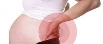
In the photo, the same painful area in women is highlighted in yellow.
Reasons for development
The main etiological factor that can provoke genital neuropathy is pinching of the pudendal nerve, which occurs in the Alcock canal. It is in this regard that experts refer to the disease as “Alcock canal syndrome.”
The disease can progress due to the formation of a hernia or injury to the groin area. Damage to the inguinoiliac nerve is the result of muscle scars beginning to form. As a rule, this occurs after injury or surgery.
Neuropathy can also develop as a result of the following factors:
- prolonged labor;
- presence of pelvic bone fractures;
- hypertonicity of the piriformis gland;
- herpes;
- tension in the obturator internus muscle;
- the presence of malignant neoplasms in the pelvic area;
- spasm of the muscles located in the anus;
- Nerve damage due to cycling or horseback riding.
An examination will help determine exactly whether there is a pathology. To do this, you need to contact a neurologist, he will prescribe diagnostic measures in order to establish a diagnosis.
Definition of disease
Pelvic neuralgia is a pinched nerve in the hip joint. In this condition, the nerve that is located between the bones, ligaments and tendons is pinched, which causes severe pain.
Sometimes the pain lasts only a few minutes, and sometimes it drags on for 2-3 days. Pathology develops in both men and women. The danger is that this disease can disguise other ailments. Therefore, the treatment is carried out incorrectly.
Disease code according to ICD-10: M79.2. Pelvic neuralgia also includes anorectal neuralgia. This is an inflammation of the nerves in the coccyx and anus, as well as in the perineum. The inflammatory process is accompanied by acute pain. Also a type of pelvic neuralgia is pudendal neuralgia.
Main symptoms
Symptoms of neuropathy vary. The main symptom is a change in the function of organs that are located in the pelvis. A person may have a sensitivity disorder or problems with autonomics.
Moreover, a change in sensitivity can be accompanied by a pronounced pain syndrome, and as for autonomics, there will be dysfunction of the glands and other structures that consist of smooth muscle fibers.
Trophic disturbances in the epidermis of the scrotum, perineum, and area near the anus are also a symptom that infringement may have occurred.
Consider the symptoms of the pathology:
- dysfunction of the genital organs;
- feeling of foreign bodies in the groin area;
- unpleasant sensations in the groin area - burning sensation, severe itching;
- pain that is aching in nature - in the pelvic area;
- excessive sensitivity of the epidermis in the groin area;
- after urination, the patient experiences discomfort accompanied by pain;
- There may be a false urge to empty the bladder.
Less commonly, the following symptoms can be noted in pathology:
- constipation;
- pain during coitus;
- feeling as if the muscles of the perineum were numb.
Experts have also found that the disease increases anxiety, and in some cases even causes depression. As a concomitant disease, pathology can also accompany Alzheimer's disease.
Treatment for a pinched nerve in the lower back: what you need to know
TO
As we know, there is no exact definition of the causes and symptoms of the disease, since medical experts have not yet come to a common opinion.
Source: //medspina.ru/nevralgiya/nejropatiya-polovogo-nerva.html
Complications and prognosis
In most cases, doctors are able to relieve compression of the ilioinguinal nerve. Treatment leads to the disappearance of pain and discomfort. However, a favorable prognosis is possible only if the patient consults a doctor in a timely manner. A complication of an advanced form of neuropathy is the chronicity of the process. In this case, persistent disorders of sexual function and frequent urinary incontinence occur. Patients suffer from chronic constipation. In such cases, surgical treatment of neuropathy is indicated, which requires long-term recovery after surgery.
How to recognize a pinched sciatic nerve and what to do about it
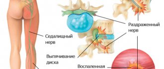
The sciatic nerve is the longest nerve in the human body. It begins in the lower back, in the lumbar region, and, splitting into two parts, stretches through the buttocks, thighs, calves to the feet. It is this nerve that ensures the mobility of the legs, connecting the muscles located in them with the central nervous system.
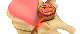
But everything is fine only until one day the sciatic nerve becomes irritated or pinched. Most often, the cause of this is an intervertebral hernia.
Designua/Shutterstock
The bulging part of the intervertebral disc presses on the sciatic nerve. This leads to pain in the lower back or along the entire length of the leg. This situation is called pinched sciatic nerve, or sciatica.
However, sciatica can be caused in other ways. Trauma, a bone spur (excessive bone growth) on the vertebrae, diabetes (this disease damages the nerves) or, say, a tumor - all this can also cause pinching.
How to tell if you have a pinched sciatic nerve
Sciatica can be suspected based on the following symptoms:
- Painful sensations affect only one leg (side of the body).
- The pain seems to spread from the lower back into the buttock and down the leg. If we talk about the legs, discomfort most often appears in the back of the thigh and lower leg.
- The pain varies in nature - from a weak aching to a burning sensation or sudden shooting. Sometimes it feels like an electric shock.
- In addition to pain, and in some cases instead of it, there may be numbness, tingling or muscle weakness in the affected leg.
- Your condition depends on your posture or movements. Thus, the pain worsens when you cough or sneeze. And after some time spent sitting, things in the lower back or legs clearly become worse.
When to see a doctor
As a rule, sciatica attacks do not last long, and their pain gradually decreases and disappears completely within a few weeks. This process is accelerated if you perform stretching exercises for the lower back and hips (more on them later). But this doesn't always happen.
Be sure to consult a therapist if:
- painful sensations last longer than a week without decreasing or responding to home care;
- the pain is so severe that it significantly reduces your quality of life and you are forced to take painkillers daily;
- every day attacks of sciatica become more frequent, and the pain becomes stronger.
Pinching is not treated by a therapist, but after listening to complaints and conducting an examination, he will refer you to the right specialist: a neurologist, surgeon, orthopedist, or, say, an endocrinologist. And these doctors will make the main diagnosis and help cope with the disease and physical discomfort.
Call an ambulance or go to the emergency room if:
- you have sudden and very severe pain in the lower back or leg, while you feel distinct numbness and muscle weakness;
- the pain occurred immediately after a serious injury, such as an accident or a fall from a height;
- in addition to pain, there were problems with bladder or bowel control.
How to treat a pinched sciatic nerve
Treatment is only required if you have the dangerous symptoms listed above. Depending on your diagnosis, your doctor will prescribe you medications (this could include anti-inflammatory drugs, anticonvulsants, muscle relaxants, corticosteroid injections), send you to physical therapy, or recommend that you consider surgery.
But most often, it is quite possible to get rid of the discomfort of sciatica using home methods. This is what experts from the American research organization Mayo Clinic recommend.
- Apply cold compresses. They can be used in the first day or two after the sciatic nerve is pinched. Apply a heating pad with cold water or an ice pack (frozen vegetables) wrapped in a thin cloth to the painful area for 20 minutes several times a day.
- Apply warm compresses. They are allowed to be used 2-3 days after the first attack. Apply a heating pad with hot (but not scalding) water to the affected area for 10–15 minutes. Repeat several times throughout the day as needed. To reduce pain, you can try alternating cold and warm compresses.
- Take an over-the-counter pain reliever. For example, based on ibuprofen.
- Move more. The first desire that arises with sciatica is to sit down or lie down, but this is a bad option. Immobility increases nerve irritation. But movement, on the contrary, helps get rid of pinching.
- Do regular stretching exercises for your lower back and hips. This is the easiest way. There are dozens of exercises, choose those that make it easier for you. Try arching your lower back, pushing your chest forward and pulling your shoulders back.
- Do a butterfly stretch: sit on the floor with your knees bent so that your feet are pressed tightly together; Press your hips as close as possible to the floor and slowly lean forward, trying to lie on your legs with your chest.
- Perform spinal twists: feet shoulder-width apart, pelvis motionless, body turning left and right as far as possible. You should feel your back stretching in the lumbar region - these are the movements that will help relieve pain.
- Hang on the horizontal bar with the muscles of your legs and torso as relaxed as possible.
Source: https://Lifehacker.ru/zashhemlenie-sedalishhnogo-nerva/
Diagnostics
To detect neuropathy of the ilioinguinal nerve, doctors prescribe an ultrasound examination. In the presence of pathology, a deterioration in blood flow in the pudendal artery is determined. This vessel is usually compressed along with the inguinal branch.
A reliable diagnostic method is nerve block. The patient is injected into the affected area with a solution of analgesics and corticosteroids. If after this the discomfort goes away, it means that the cause of the pain was compression of the inguinal nerve. In this case, the patient is prescribed a course of treatment.
Diagnostic techniques
A neurologist is involved in the diagnosis and treatment of PPN neuritis. The diagnostic process begins with an external examination and questioning of the patient in order to clarify the clinical picture of the pathology. The doctor checks the dynamic qualities of the lower limb, while simultaneously interviewing the patient - this helps to clarify the diagnosis.
With tension in the abdominal muscles, extension and abduction of the hip joint, pain in the groin area increases, radiating to the upper inner surface of the thigh, and with flexion and adduction it decreases. Palpation reveals an area of the most severe pain, located one transverse finger inward from the anterior superior iliac spine. It is here that the RPN branch passes through the extrinsic abdominal muscle.
Subsequently, the patient is sent for examination using special diagnostic equipment - magnetic resonance imaging (MRI), computed tomography (CT), ultrasound (ultrasound). The data from these examinations help to detect the locations and causes of nerve compression, based on the presence of inflammatory processes in nearby tissues, impaired blood flow in them, and make an accurate diagnosis. After this, the doctor selects tactics and strategies for therapeutic procedures.
Conservative treatment
When treating a pinched ilioinguinal nerve, the patient is prescribed the following medications:
- The anticonvulsant Gabapentin is used to relieve pain.
- To eliminate spasms, the muscle relaxant Mydocalm is prescribed.
- It is recommended to take the Neuromultivit multivitamin complex.
For severe pain, nerve blocks are performed using analgesics and steroid hormones. For discomfort in the genital or anal area, the use of vaginal and rectal suppositories based on diazepam is indicated.
Drug treatment is supplemented with physiotherapy. When a nerve is pinched, procedures such as phonophoresis and electrophoresis are useful.
Physical therapy is also recommended. It is useful to do exercises to open and close the muscles of the perineum. However, the exercise therapy complex should be performed only after pain relief.
Causes and treatment of a nerve in the groin when pinched
When the nerve of the inguinal canal is pinched, the supply of nerve cells to organs and tissues deteriorates. A painful syndrome occurs in the pelvic area, which can radiate to the back, buttocks or thighs. This phenomenon occurs due to injury or pathologies of internal organs. It is necessary to start timely treatment to avoid negative consequences.
Causes of pinching
The main cause of pinched nerve bundle on the right or left side is surgery to remove an inguinal hernia.
The carelessness of the surgeon can lead to damage to the branches of the nerves or their pinching by scar tissue. There are other provoking factors:
- increased tone of the piriformis muscle;
- oncological diseases;
- consequences of herpetic infection;
- spasm of the perineal muscles;
- inflammation of the pelvic organs.
If the nerve is pinched, the internal organs of the pelvis could be injured. Prolonged labor or prolonged cycling can lead to pinched nerve bundles. Another reason is diseases of the internal organs. These include: tuberculosis, cholelithiasis, rheumatic diseases, disorders of the cardiovascular system, varicose veins of the testicle (varicocele).
How to recognize symptoms
A pinched nerve in the groin can cause certain symptoms. They manifest themselves with varying intensities: from decreased sensitivity to acute pain in the groin area. It all depends on the progress of the process.
At the initial stage, patients complain of aching pain that intensifies at night. In women, the pain syndrome is felt acutely in a sitting position. The following symptoms are characteristic of this pathological condition:
- painful sensations intensify with pressure;
- there is itching and burning;
- there is a feeling of tingling, numbness in the groin area;
- there is discomfort in the anal area.
In some cases, sensitivity in the upper buttock, pubis (with pinched iliohypogastric nerve) or external genitalia (pinched ilioinguinal nerve) is reduced. Painful sensations always occur on the left or right.
If it lasts for a long time due to stress, urination becomes disordered and the process of bowel movement becomes difficult (constipation).
Therapeutic measures
To begin with, a diagnosis is made, taking into account the condition of the obturator nerve and the external cutaneous nerve. After the examination, the patient is prescribed complex treatment. Injection therapy is used to relieve pain. Drug blockades are injected into the affected area, which begin to act immediately. Other therapeutic measures are used:
- Make compresses with Dimexide. They help relieve spasms.
- Steroid hormones are prescribed. These include Dexamethasone.
- Anticonvulsants are used to relieve acute pain.
- B vitamins and antioxidants are prescribed. Vitamins are administered intramuscularly.
- Xefocam helps relieve inflammation in the initial stages.
- Rectal or vaginal suppositories with anesthetics are administered.
When acute manifestations are eliminated, physiotherapy is prescribed. This can be electrophoresis, ultrasound and manual therapy. Subsequently, therapeutic exercise will help improve muscle tone and prevent pinching. The muscles need to be compressed and relaxed in the hip area.
Due to vague symptoms, patients may not be aware of damage to the nerve bundle. The disease tends to develop into a chronic form and treatment may not be limited to medication. In more advanced stages, surgical intervention is used.
Surgical intervention
If there is no effect from conservative therapy, surgical treatment is indicated. The doctor performs decompression surgery under general anesthesia. This eliminates pinching of the nerve by nearby tissues.
This operation effectively eliminates all unpleasant symptoms. However, the rehabilitation period can last several months. During this period, it is necessary to follow a gentle regimen: avoid physical activity and prolonged sitting.
It is important to remember that even after surgery, relapses of the disease are possible. When exposed to unfavorable factors, re-pinching of the inguinal nerve is possible. Therefore, preventive measures and recommendations of the attending physician should be followed.
Prevention
How to prevent inguinal branch neuropathy? First of all, it is necessary to promptly cure such pathologies as hernia, pelvic fractures, herpes zoster, and gynecological diseases. If even minor aching pain persists for a long time after surgery in the pelvic area or difficult childbirth, you should consult a doctor.
A comfortable riding seat should be used. This will reduce stress on the inguinal nerve. When cycling for long periods, you need to take breaks periodically. This will reduce the risk of nerve damage.