Causes of the disease
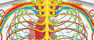
At the level of the thoracic part of the spine, nerves emerge from the spinal cord symmetrically on both sides, following under each rib. They innervate the skin and muscles of the corresponding area, and have connections with nerves going to the internal organs.
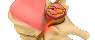
Osteochondrosis is the most common cause of a pinched nerve in the chest. With this disease, the height of the intervertebral discs decreases, and the shock-absorbing properties of the cartilage are lost. There are often bone deformities that increase pressure on the nerve. Often the disease develops against the background of scoliosis – a sideways curvature of the spine.
Diabetes mellitus, atherosclerosis, arterial hypertension, chronic renal and endocrine diseases, age-related changes lead to damage to blood vessels and nerves. This also worsens the working conditions of the muscles, ligaments, discs, and joints of the spine.
Prolonged stay in a forced position, heavy lifting, uneven load on the back (carrying a heavy bag in one hand) provoke the appearance of pain. A pinched nerve in the thoracic region can be on the left, right, or both sides.
Types of massage
For treatment and prevention purposes, the following massage techniques are used:
- point;
- general (therapeutic, classical massage);
- honey;
- canned.
Important! Vacuum cupping massage is very convenient to use at home. Thanks to the procedure, muscle spasms and severe pain disappear.
Vacuum cans help better than strong pills
Positive dynamics should appear after the first session; the patient should note changes in his own condition and feel lightness in the lumbar region. To achieve a sustainable result, about 10 sessions are required. The course is repeated once a year.
Great article on the topic:
How to relax your back muscles: TOP 12 best ways, poses and exercises, quickly at home, massage
Clinical manifestations
A pinched nerve in the thoracic spine is accompanied by the following symptoms:
- Pain in the back, chest, which spreads between the ribs. It can radiate to the lower back, under the shoulder blade, or into the arm. It happens in the form of attacks, very strong, burning, stabbing or barely noticeable, aching, but is constantly present. The pain intensifies with movement, breathing, coughing, sneezing. Sometimes it is difficult for a person to raise his arms or bend over.
- Loss of sensitivity along the nerve. A person may feel numbness, a crawling sensation, or, conversely, hyperesthesia, that is, increased sensitivity of the skin. Any touch then causes discomfort or pain.
- Considering that movements are accompanied by pain, breathing becomes shallow, which can further lead to problems with the respiratory system.
- Severe constant pain exhausts a person, leads to fatigue, irritability, and a general decrease in tone and immunity.
Diagnostics
A tightness in the chest on the left is often disguised as heart disease. Some people mistake the acute pain for an angina attack or even a heart attack, given the intensity of the pain. A doctor can distinguish these diseases by conducting an examination, including an ECG, echocardiography, and blood tests. It is characteristic that the pain that occurs when a nerve is pinched in the thoracic spine on the left is not relieved by cardiac medications (validol, nitroglycerin).
Often the pain radiates to the epigastric region, hypochondrium, and lower back. Then diseases of the stomach, intestines, liver, and kidneys should be excluded.
The osteopath conducts a detailed survey of the patient and palpates points along the spine. When a nerve is pinched in the thoracic spine, it is these zones (1.5 cm to the side of the spinal column) that are characterized by maximum pain. Because this is where the nerve exits under the skin.
X-ray methods help to identify abnormalities in the structure of the spine. The most accurate additional study for pathology of soft tissues is MRI, bones - CT. A consultation with a neurologist is required. He diagnoses at what level the nerve is pinched in the thoracic region and prescribes treatment.
Principles of treatment
Pain relief is aimed at reducing pain and eliminating muscle spasms in the affected area. If taking NSAIDs orally or by injection does not help, blockades are performed. This is the introduction of drugs into the most painful points, which are projections of the exit of nerves. Local anesthetics, hormones, NSAIDs, and muscle relaxants are used. Diuretics reduce swelling of the compressed nerve and eliminate pain. Drugs are prescribed that improve the conductivity of nerve tissue.
In addition to relieving acute pain, it is necessary to influence the cause of pinching of the intercostal nerve in the thoracic region. Medicines, massages, manual therapy, physiotherapeutic procedures, reflexology, and physical therapy help reduce the destruction of bones and joints. Medicines prescribed:
- vascular drugs - improve blood circulation;
- chondroprotectors – restore the properties of the cartilage layer between the vertebrae;
- calcium supplements, vitamin D fight osteoporosis;
- multivitamin complexes with minerals.
Acupressure, lymphatic drainage and classic types of massage are used. The impact on the back muscles helps eliminate spasms, improves blood flow and tissue nutrition. Thanks to this, it is possible to break the pathological circle, when pain causes muscle contraction, which compresses nerve fibers and blood vessels, disrupting blood supply and increasing pain.
In some cases, surgical treatments may even be necessary. For example, to eliminate the consequences of an injury, remove a significant hernia, tumor, or perform spinal stabilization.
Every person should be attentive to their health and not ignore the symptoms of a pinched nerve in the thoracic region if they appear. It is important to seek qualified help immediately. After all, the earlier the problem is identified, the greater the chances of achieving complete restoration of the function of the nerves and all structures of the spinal column, and of avoiding another pinching under the breast in the future.
content .. 180 181 182 183 184 185 186 187 188 189 ..Intercostal nerves (human anatomy)
Intercostal nerves
, nn. intercostales, are the abdominal branches of the I - XII thoracic spinal nerves. Upon departure from nn. spinales intercostal nerves enter the intercostal spaces and extend ventrally between the internal and external intercostal muscles to the sternum. On their way, the intercostal nerves give off cutaneous branches: lateral, rami cutanei laterales, on the side wall of the body and anterior, rami cutanei anteriores, at the sternum. According to the position of the nerves, the cutaneous branches are called thoracic or abdominal. Each lateral branch is divided into anterior and posterior branches, and the anterior branch is divided into medial and lateral branches. The upper 6 intercostal nerves are located throughout the intercostal spaces, and the lower 6 extend onto the anterior abdominal wall (Fig. 223).
Rice. 223. Diagram of the structure of intercostal nerves
The intercostal nerves innervate the intercostal muscles, the transverse thoracic muscle, the muscles of the anterior abdominal wall and some back muscles: mm-serrati posteriores superiores et inferiores, levatores costarum. In addition, they participate in the innervation of the pleura and peritoneum.
Cutaneous nerves innervate the skin of the body, maintaining a clear segmentation of the innervation territories, which look like horizontal stripes. The territory of innervation of the cutaneous branches of the V intercostal nerve corresponds to the level of the nipple, VII - the xiphoid process, X - the umbilicus, XII - the supra-inguinal zone. The division of the zones of innervation of the anterior and lateral branches occurs somewhat laterally from the lateral edge of the rectus abdominis muscles. Cutaneous nerves give branches to the mammary gland.
Lumbosacral plexus nevus (human anatomy)
The pelvic girdle and lower limb are innervated by the most powerful nerve plexus - the lumbosacral, plexus lumbosacralis, which consists of two plexuses - the lumbar, plexus lumbalis, and the sacral, plexus sacralis, connected through the lumbosacral trunk, truncus lumbosacralis.
Lumbar plexus of nerves (human anatomy)
Lumbar plexus
, plexus lumbalis, consists of the abdominal branches of the I, II, III and partially IV lumbar spinal nerves (Fig. 224).
Rice. 224. Lumbar plexus. 1 - quadratus lumborum muscle; 2 - XII intercostal nerve; 3 - iliohypogastric and ilioinguinal nerves; 4 - lateral cutaneous nerve of the thigh; 5 - femoral nerve; 6 - lateral cutaneous nerve of the thigh (in the thigh area); 7 - anterior cutaneous branch of the femoral nerve; 8 - femoral branch of the genital-femoral nerve; 9 - sacral plexus; 10, 11 — lumbosacral trunk; 12 - lumbar node of the sympathetic trunk; 13 - anterior branch of the II lumbar nerve
The abdominal branches that form the plexus are connected to each other by loop-like connections and run anterior to the transverse processes of the lumbar vertebrae between the two initial parts of m. psoas major, where the plexus gives off its branches.
1. Iliohypogastric nerve
, n. iliohypogastrics (from LI), comes out of m. psoas major on the anterior surface of the quadratus lumborum muscle, pierces the tendon stretch of the m. transversus abdominis and runs anteriorly above the iliac crest between the transverse and internal oblique abdominal muscles, giving them muscle branches. With its lateral cutaneous branch, ramus cutaneus lateralis, it innervates the skin above the greater trochanter of the femur, and the final one, the anterior cutaneous branch, ramus cutaneus anterior, innervates the skin in the area of the superficial ring of the inguinal canal.
2. Ilioinguinal nerve
, n. ilioinguinalis, starts from the abdominal branch of the first lumbar spinal cord or from the iliohypogastric nerve and goes below the latter into the inguinal canal, appearing in the subcutaneous tissue at its superficial ring. Along its length, the nerve sends branches to the muscles of the abdominal wall.
The terminal section of the nerve is divided into the lateral cutaneous branch, ramus cutaneus lateralis, which innervates the skin of the medial inguinal region, and the anterior scrotal branch, nn. scrotales anter lores, - in men and anterior labial nerves, nn. labiates anteriores, - in women, innervating the skin of the scrotum or labia majora.
3. Genitofemoral nerve
, n. genitofemoralis (from LI-LII) pierces the psoas major muscle and runs down its anterior surface, dividing here into two terminal branches: 1) the genital, ramus genitalis, which joins the spermatic cord in the inguinal canal, the formation of which and the shell of the testicle it innervates; 2) femoral, ramus femoralis, innervating the skin of the thigh below the inguinal fold.
4. Lateral cutaneous nerve of the thigh
, n. cutaneus femoris lateralis (from LII-LIII), emerges from m. psoas major at its lateral edge on the anterior surface of the iliacus muscle. Then it descends downwards and outwards, passes through the anterior abdominal wall medial to the anterior superior iliac spine and on the thigh, penetrating through the fascia lata, branches into the skin of the lateral surface of the thigh.
5. Femoral nerve
, n. femoralis (from LI-LIV), formed on the anterior internal surface of m. psoas major, then crosses it and goes between it and m. iliacus in the lacuna musculorum on the anterior surface of the thigh, separating from the femoral artery fascia iliopectinea. On the thigh it branches, giving: 1) muscular branches, rami musculares, to mm. quadriceps, restineus and sartorius; 2) anterior cutaneous branches, rami cutanei anteriores, innervating the skin of the anteromedial surface of the thigh. One of the cutaneous branches is the hidden nerve, n. saphenus, goes along with the femoral artery to the canalis adductorius, exits through its anterior opening with a. genus descendens, descending under the sartorius muscle to the medial surface of the tibia, where it spreads down near the v. saphena magna, innervating the skin of the medial surface of the leg (rami cutanei mediales). It gives off the infrapatellar branch, ramus infrapatellaris, which innervates the skin of the knee joint area below the patella.
6. Obturator nerve
, n. obturatorius (from LI-LIV), located initially behind and inwardly m. psoas major, medial to the femoral nerve, then goes to the pelvis, where it passes near the insertion of m. levator ani to the obturator canal, through which it enters the medial osteofascial space of the thigh, dividing into anterior and posterior branches. The posterior branch, ramus posterior, innervates mm. obturatorius externus, adductor magnus and gives branches to the hip-femoral joint. The anterior branch, ramus anterior, supplies nerves mm. pectineus, adductor brevis et longus, gracilis. In addition, it gives off a cutaneous branch, ramus cutaneus, to the skin of the medial surface of the thigh.
content .. 180 181 182 183 184 185 186 187 188 189 ..
