March 19, 2012
Scientists are still arguing about how the human brain works. However, they already know something well.
The brain is the most complex system of the human body, which controls all its activities. With the help of this system, not only conscious processes are controlled: speech, movement, emotions. The brain also regulates all processes that occur automatically in the body: movement, blood circulation, maintaining balance and many others. Scientists are still arguing about how the human brain works. However, they already know something well.
How to kill your brain?
Alcohol and nicotine are neurotoxic poisons. Find out how quickly they can kill a person's brain.
Medulla oblongata and midbrain: structure and functions
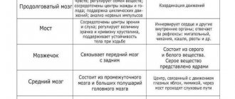
The medulla oblongata and midbrain are collectively called the brainstem . They contain several vital centers:
- protective reflexes (coughing, sneezing);
- regulation of breathing;
- regulation of vascular tone;
- regulation of the respiratory system;
- orientation reflexes.
So, the medulla oblongata is a vital organ. Accordingly, if an injury to the medulla oblongata occurs, the person dies very quickly due to damage to the respiratory center.
This is interesting: human chromosomes, their number in a healthy person?
Structure
The membranes of the brain and the spaces between the shells reliably protect the tissues of the central nervous system from damage to the hard bones of the skull. The first ones differ from each other in the structures that form them and in their functional affiliation:
- Hard – the densest. It is formed by thick collagen and elastic bundles of fibers tightly adjacent to each other, which form a kind of network. They are arranged randomly, which determines the strength and elasticity of the structure. In some areas, the dura mater extends into the fissures of the cerebral hemispheres in the form of processes. The processes of the dura mater of the brain, at the sites of penetration, split into 2 parts, forming sinuses. Their walls are so dense that they do not deform or fall off when cut. Through the sinuses, venous blood is removed from the circulatory system of the head.
- Arachnoid. It is formed by several layers: the outer layer of the endothelium, a layer of argyrophilic and collagen fibers that form the base and the inner cellular layer of the endothelium. The cells of the outer layer form cellular spots, mounds and arachnoid granulations.
- Soft. It is located closest to the cerebral hemispheres, enveloping them on all sides. It has an outer layer, covered with endothelial-like cells, and an inner one. Between the layers is the circulatory system of the central nervous system. Its blood vessels protrude through the upper layer into the subarachnoid space.
Between the protective membranes there are intershell spaces of the brain: subdural and subarachnoid. Liquor moves in them, which serves as a shock absorber. The subarachnoid space of the GM connects through the foramen magnum to the subarachnoid space of the spinal cord. According to modern concepts, there should not be a free subdural space.
Cerebellum
The cerebellum is a specialized department that coordinates movements.
It receives a large amount of information from the balance organs, orders from the cerebral cortex and implements movement. The cortex communicates that you need to walk, and then the cerebellum controls your gait. The cortex is usually not involved in this process - you go automatically. In addition, the cerebellum regulates balance.
For example, when you haven't slept for a very long time and fall asleep while sitting, your head begins to tilt to one side - this means that the cortex stops telling the cerebellum to maintain balance.
This is interesting: the structure and functions of cell organelles.
The cerebellum also regulates muscle tone. In order to sit or simply hold your head up, you need some constantly tense muscles. The cerebellum does this too. And muscle memory: probably many people are familiar with the fact that some movement that you have not done before is difficult to do the first time. But then it becomes easier and easier, and over time it begins to happen automatically due to the fact that the cerebellum begins to do this.
Involuntary movements, that is, for example, withdrawing a hand from something hot, are made quick by the cerebellum due to the fact that it takes control of them.
Thanks to the cerebellum, you can do voluntary movements not quickly, but precisely, for example, take something specific from the table.
This is interesting: the main property of the plasma membrane is... what?
So, the cerebellum provides:
- speed of involuntary movements and accuracy of voluntary ones;
- coordination of movements;
- regulation of balance;
- regulation of muscle tone;
- muscle memory.
Lobes of the cerebral cortex
Among them:
- Frontal lobe. This is the main part of the cortex in which the motor centers, mental functions and speech center are located. Analytical activities and the area responsible for speech motor skills are also located here.
- Temporal lobes. These areas are located on the sides of the cortex. They contain the main centers of feelings, the center for understanding speech, as well as emotional centers responsible for joy, fear, pleasure and other emotions.
- Occipital lobe. It processes visual data.
- Parietal lobe. Contains sensory activity centers as well as a center for musical understanding.
Diencephalon
These are several departments:
- First, the pituitary gland and pineal gland, which are endocrine glands.
- Secondly, the hypothalamus, which controls the endocrine glands themselves.
- And thirdly, the thalamus, which we will talk about in more detail.
Thalamus means hillock. The hypothalamus is under the hillock. It is always located under the thalamus. The diencephalon is already a fairly high level of control, and here are the centers of various emotions and instincts: the pain center, the pleasure center, the centers of thirst, hunger and satiety, the center of sleep and wakefulness, the center of thermoregulation.
The thalamus is a set of structures that do very important things. Try now to realize how much information you receive from your senses every split second. You feel the temperature at every point of your body. You feel the touch of all clothing at every point with which it comes into contact, the heat and cold emanating from objects. You hear an insane amount of sounds. You smell a lot of smells. You understand where your arms, legs and head are in space. You see many objects. You know the distance to each of them, their color, their shape.
And all this happens all the time. This is a huge amount of information. If you received information in the form of raw data, you would go crazy with the need to process it. Therefore, 90% of all this information does not reach your consciousness. And a small part of it arrives in the form of already processed data. The thalamus does just that. It’s like a funnel: it takes a huge amount of information and filters out everything that’s irrelevant.
The thalamus processes all types of information except smell. The sense of smell immediately enters the cerebral hemispheres. It doesn’t just filter the rest of the information, but processes and summarizes it. For example, you see a person’s face, but you perceive it not as a set of individual features, but as a whole. But it will be difficult for you to describe the face of another person: you will have to imagine it and only then describe it. That's why the police use identikit photos: they don't ask you to tell what shape your ears are. They ask you to choose the best of different options. It's easier - you compare images. The thalamus is the most important organ that allows us to work with information much more efficiently.
General information
The head and spinal regions of the central nervous system require additional protection. In addition to the bone frame, they are shrouded in meninges. There are only 3 of them:
- Solid (TMO). It differs from others in density and strength. Its outer side is rough, covered with a developed blood network, and loosely adheres to the bones of the skull, forming the epidural space in the cranial vault. Formed by fibrous tissue cells.
- Translucent arachnoid (arachnoid). Located under the hard layer. It looks like a thin web, which is why it got its name. Formed by cells of loose connective tissue. It consists of 2 layers, between which there are a large number of partitions.
- Soft. It is the lowest layer that fits tightly to the brain and spinal cord.
In some pathologies of the central nervous system, a subdural space appears between the hard and arachnoid membranes, which is filled with a small amount of cerebrospinal fluid. Normally it shouldn't be there.
Forebrain
Specifically, the cerebral cortex is a large part of the volume of our brain, and it is divided into lobes.
Each of the lobes is paired, because we have two hemispheres, and each has one of these lobes: the frontal lobes, the seminal lobes, the temporal lobes and the occipital lobes. Here are the highest centers, which:
- process sensations;
- give orders for movements.
Let's look at which parts of the cortex receive signals from where.
- The occipital lobe deals with visual images. It receives signals from the eyes after they are processed by the thalamus, and a picture is formed here.
- The parietal lobe receives information about the sense of touch—that is, the sense of touch and pain.
- The temporal lobe receives information about sounds, tastes, smells, and the sense of balance. The brain itself does not feel pain - there are no nerve endings in it.
- The frontal lobe is actually the place where consciousness lives and a holistic picture of the world is formed.
If certain parts of the brain are damaged, certain body functions will also suffer. Thus, destruction of the occipital lobe will lead to loss of vision. The eyes will see something, but you will not be able to perceive the picture.
If one feeling is missing, then the others will be more developed. The part of the brain that was involved in vision begins to deal with something else - hearing or tactile sensations, and in a person who is blind from birth, the remaining senses will be much more developed than in the ordinary person.
But if an adult already loses vision, not part of the brain, but, for example, the eyes due to injury, you can put a mechanical implant there, roughly speaking, a camera whose outputs are connected to the nerves and the signal is decoded so that the nervous system can understand it. A person will be able to see because there is a part of the brain that analyzes vision. Only the organs of vision are missing. Eye implants already exist, they don't have very good resolution, but they work.
The brain of humans and other vertebrates is symmetrically divided into right and left parts. In this case, the left side mainly controls the right side of the body and vice versa. There is a common misconception that the left hemisphere is “logical” and the right hemisphere is “emotional.” This is just a popular myth. In fact, their functions are somewhat different, but this is not so fundamental.
Functions
Each shell has an individual structure and functional purpose:
- The dura mater (DRM) forms processes through which the brain is divided into several important areas. They stabilize and fix the organ in the cavity of the cranium.
- The arachnoid supports the circulation of cerebrospinal fluid and normalizes metabolic reactions in the brain. Liquor washes it on both sides: in the subdural and subarachnoid space.
- The pia mater of the brain is rich in blood vessels through which arterial blood flows to the structures of the central nervous system.
In addition to protection, they provide nutrition to brain cells and take part in removing waste products from the body.
Cranial nerves
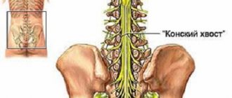
Like the spinal cord, nerves extend from the brain, and there are 12 pairs of them:
- olfactory nerve;
- optic nerve;
- oculomotor nerve;
- trochlear nerve;
- trigeminal nerve;
- abducens nerve;
- facial nerve;
- vestibulocochlear nerve;
- glossopharyngeal nerve;
- nervus vagus;
- accessory nerve;
- hypoglossal nerve.
Brain
Departments
What does the brain consist of?
The structures and basic functions of the brain are carried out by its different parts. From an anatomical point of view, the organ consists of five sections that were formed during the process of ontogenesis.
Various parts of the brain control and are responsible for the functioning of individual human systems and organs. The brain is the main organ of the human body; its specific departments are responsible for the functioning of the human body as a whole.
to contents ^

