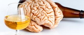
Today we will talk about the human brain, what parts it consists of, and how they work. To begin with, remember that the central nervous system consists of the spinal cord and the brain. At the same time, it can be divided into lower sections - this is the spinal cord itself.
Its main task is to conduct signals. He does very little management, and where he does, these are very simple management functions, such as the simplest reflexes. ... The middle sections nervous system are part of the brain. They are mainly involved in regulating the activities of organs and systems and communicating between them. The highest part of the central nervous system is the cerebral cortex and most of our brain. But this is typical only for a very small number of living creatures: humans, other great apes, many dolphins, whales, killer whales, dogs and wolves. In most other mammals, the cortex is thin and does not take up as much space as in humans.
This is interesting: a human skeleton with the name of the bones, description.
The cortex is a department that creates a kind of holistic picture of the world, where consciousness arises, and it controls the body as a whole. Connecting the central nervous system to the rest of the body is the peripheral nervous system , which is simply nerves that transmit various data. The peripheral nervous system connects the central nervous system to the organs and limbs.
The higher department - the cerebral cortex - regulates the connection and relationship of the body as a whole with the environment.
Structure
The bulk of the brain is made up of cells called neurons. They are capable of creating electrical impulses and transmitting data. In order for neurons to function, they require neuroglia, which collectively are support cells and make up half of all cells in the central nervous system. A neuron consists of two parts:
- axons - cells that transmit impulses;
- dendrites are cells that receive impulses.
Brain structure:
- Diamond-shaped.
- Oblong.
- Rear.
- Average.
- Front.
- Finite.
- Intermediate.
The main functions of the cerebral hemispheres are the interaction between higher and lower nervous activity.
Brain tissue
The structure of the human brain consists of the cerebral cortex, thalamus, cerebellum, brainstem and basal ganglia. The collection of nerve cells is called gray matter. Nerve fibers are white matter. The white color of the fibers comes from myelin. When the amount of white matter decreases, serious disorders such as multiple sclerosis occur.
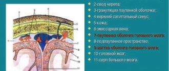
The brain includes shells:
- The dura attaches to the skull and cerebral cortex.
- The soft tissue consists of loose tissue, is located on all hemispheres, and is responsible for saturation with blood and oxygen.
- The arachnoid is located between the first two and contains cerebrospinal fluid.
Liquor is located in the ventricles of the brain. When it is in excess, a person experiences headaches, nausea, and hydrocephalus occurs.
Table “Structure and functions of the brain”
| Department | Structural features | Functions |
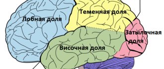
| End or front | It consists of two hemispheres, which are connected to each other by the corpus callosum. The surface of the hemispheres has many grooves and convolutions. | The right hemisphere is responsible for the left side of the body, and the left hemisphere is responsible for the right side. The centers for the regulation of voluntary movements, the centers of conditioned reflexes and mental functions are located here. The temporal lobe of the cerebral cortex regulates hearing, taste and smell, the occipital lobe regulates vision, the parietal lobe regulates touch; frontal – speech pronunciation. |
| Intermediate | Consists of the hypothalamus and thalamus. | The thalamus is a mediator in the transmission of stimuli to the hemispheres and helps to adequately adapt to changes in the environment. The hypothalamus regulates the functioning of metabolic processes and endocrine glands. Manages the work of the cardiovascular and digestive systems. Regulates sleep and wakefulness, manages food and drink needs. |
| Rear | It consists of the cerebellum and the pons, which is presented in the form of a white thick cushion located above the oblongata. The cerebellum is located behind the pons and has two hemispheres, the inferior and superior surfaces and the vermis. | This department provides the function of an intermediary in the transmission of impulses to higher parts of the brain. The cerebellum is responsible for coordination and precision of movements. |
| Average | Located from the anterior edge of the bridge to the optic tracts. | Responsible for hidden vision, as well as the implementation of orientation reflexes (turning the body in the direction of sound). |
| Oblong | It is a continuation of the spinal cord. | Controls balance, contains the centers of breathing, swallowing, sneezing and coughing, vomiting, cardiovascular and digestive. |
Brain cells
The main cells are called neurons. They process information, their number reaches 20 billion. There are 10 times more glial cells.
The body carefully protects the brain from external influences by placing it in the skull. Neurons are located in a semipermeable membrane and have processes: dendrites and one axon. The length of dendrites is small compared to the axon, which can reach several meters.
To transmit information, neurons send nerve impulses to an axon, which has many branches and is connected to other neurons. The impulse originates in the dendrites and is sent to the neuron. The nervous system is a complex web of neuron processes that are interconnected.
The structure of the brain and the chemical interaction of neurons have been studied superficially. At rest, a neuron has an electrical potential of 70 millivolts. Excitation of a neuron occurs through the flow of sodium and potassium across the membrane. Inhibition occurs as a result of the action of potassium and chlorides.
The job of a neuron is to interact between dendrites. If the excitatory effect prevails over the inhibitory effect, then a certain part of the neuron membrane is activated. Thanks to this, a nerve impulse arises that moves along the axon at a speed of 0.1 m/s to 100 m/s.
Thus, any planned movement is formed in the cortex of the frontal lobes of the cerebral hemispheres. Motor neurons give commands to parts of the body. Simple movement activates the functions of parts of the human brain. When talking or thinking, large parts of the gray matter are involved.
Finite brain
The parts of the human brain directly depend on the functioning of the hemispheres, or telencephalon. The two hemispheres, which make up up to 80% of the mass of the entire brain, are connected through the corpus callosum and other commissures. The cortex covering the elements of the department consists of several layers of gray matter. It is thanks to it that the realization of higher mental activity is possible.
The structure of the brain cannot be briefly described, since without studying its structure it is impossible to understand its functions.
The telencephalon extends from the occipital to the frontal bone.
It distinguishes 2 large hemispheres: left and right.
It differs from other parts of the brain by the presence of a large number of convolutions and grooves.
The structure and development of the brain are closely interrelated.
ancient, which includes the olfactory tubercle;
perforated anterior substance;
semilunar, subcallosal and lateral subcallosal gyri;
old, which includes the hippocambus and dentate gyrus (fascia);
new, represented by the rest of the cortex.
they are separated by a longitudinal groove, in the depths of which the fornix and corpus callosum are located.
They connect the hemispheres of the brain.
The corpus callosum is a new cortex made up of nerve fibers.
There is a vault underneath it.
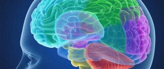
The structure of the cerebral hemispheres is presented as a multi-level system. So they distinguish between lobes (parietal, frontal, occipital, temporal), cortex and subcortex.
The cerebral hemispheres perform many functions. The right hemisphere controls the left half of the body, and the left hemisphere controls the right. They complement each other.
Functions of departments
The largest part of the brain is the cerebral hemispheres. They must be symmetrical and connected to each other by axons. Their main function is to coordinate all parts of the brain. Each hemisphere can be divided into frontal, temporal, parietal and occipital lobes. People don’t think about which part of the brain is responsible for speech. The temporal lobe contains the primary auditory cortex and the center, which, when disrupted, causes loss of hearing or problems with speech.
Based on the results of scientific observations, scientists have determined which part of the brain is responsible for vision. This is done by the occipital lobe, located under the cerebellum.
The associative cortex is not responsible for movements, but ensures the performance of functions such as memory, thinking and speech.
The trunk is responsible for connecting the dorsal and forebrain, and consists of the medulla oblongata, midbrain and diencephalon. The oblong part contains centers that regulate the functioning of the heart and breathing.
Front
Main functions:
- innate instincts;
- developed sense of smell;
- emotions, memory;
- reactions to stimuli.
The forebrain is one of the most extensive sections, consisting of the diencephalon and hemispheres (right and left), which are separated in the form of a gap, in the depth of which there are jumpers (corpus callosum).
The cerebral cortex is covered with nerve fibers - a white substance that forms the connection between neurons and parts of the brain. The hemispheres are covered with a cortex that contains gray matter. The cell bodies of neurons are components of the gray matter, arranged in columns in several layers. From the gray matter inside the hemispheres, connections are formed from nuclei located in the middle of the white matter, thereby forming the subcortical centers.
In the cerebral hemispheres, neurons are involved in processing nerve signals emanating from the sensory organs. This process occurs in the middle and posterior regions of the brain. Each lobe of the hemisphere is responsible for certain zones:
- the occipital lobe is responsible for visual functions;
- in the lobes of the temples there are neurons of the auditory zone;
- The parietal lobe controls muscle and skin sensation.
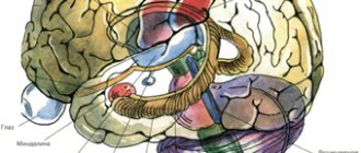
Subcortical structures
Beneath the main cortex is a collection of neurons: the thalamus, basal ganglia, and hypothalamus.
The thalamus is necessary for connecting the senses with parts of the sensory cortex. Thanks to it, the processes of wakefulness and attention are supported.
The basal ganglia are responsible for the initiation and inhibition of coordination movements.
The hypothalamus regulates the functioning of hormones, the body's water metabolism, the distribution of fat reserves, sex hormones, and is responsible for the normalization of sleep and wakefulness.
Forebrain
The functions of the forebrain are the most complex. It is responsible for mental activity, learning ability, emotional reactions and socialization. Thanks to this, it is possible to predetermine the characteristics of a person’s character and temperament. The anterior part is formed at 3-4 weeks of pregnancy.
To the question of which parts of the brain are responsible for memory, scientists have found the answer - the forebrain. His cortex is formed during the first two to three years of life, for this reason a person does not remember anything before this time. After three years, this part of the brain is able to retain any information.
A person's emotional state has a great influence on the front part of the brain. Negative emotions have been found to destroy it. Based on experiments, scientists answered the question of which part of the brain is responsible for emotions. They turned out to be the forebrain and cerebellum.
The front part is also responsible for the development of abstract thinking, computational abilities and speech. Regular mental training can reduce the risk of developing Alzheimer's disease.
Diencephalon
It responds to external stimuli, is located at the end of the brain stem and is covered by the cerebral hemispheres. Thanks to it, a person can navigate in space, receive visual and auditory signals. Participates in the formation of all types of feelings.
All functions of the human brain are interconnected. Without an intermediate, the functioning of the entire organism will be disrupted. Damage to part of the midbrain leads to disorientation and dementia. If the connections between the lobes of the hemispheres are disrupted, speech, vision or hearing will be disrupted.
The diencephalon is also responsible for pain sensations. A malfunction increases or decreases sensitivity. This part forces a person to show emotions and is responsible for the instinct of self-preservation.
The diencephalon controls the production of hormones, regulates water metabolism, sleep, body temperature, and sexual desire.
The pituitary gland is part of the diencephalon and is responsible for height and weight. It regulates procreation, sperm and follicle production. Provokes skin pigmentation and increased blood pressure.
Intermediate department
It consists of a ventral (hypothalamus) and dorsal (metathalamus, thalamus, epithalamus) part. The thalamus is a mediator in which all received stimuli are sent to the cerebral hemispheres. It is often called the optic thalamus. Thanks to it, the body quickly adequately adapts to a changing external environment. The thalamus is connected to the cerebellum by the limbic system.
The hypothalamus is a subcortical center in which the regulation of autonomic functions occurs. Its influence occurs through the endocrine glands and the nervous system. It is involved in the regulation of the functioning of some endocrine glands and metabolism. Below it is the pituitary gland. Thanks to it, body temperature, digestive and cardiovascular systems are regulated. The hypothalamus regulates wakefulness and sleep, shapes drinking and eating behavior.
This section is located between the midbrain and the corpus callosum. There are visual hillocks here that have a number of important functions, in particular the processing of centripetal impulses coming from the outside world and their transmission to the brain. In addition, they are responsible for such parameters of emotional behavior as pulse, breathing, blood pressure, facial expressions, etc.
Midbrain
The midbrain is located in the stem part. It is a conductor of signals from the front to various departments. Its main function is to regulate muscle tone. It is also responsible for the transmission of tactile sensations, coordination and reflexes. The functions of parts of the human brain depend on their location. For this reason, the midbrain is responsible for the vestibular apparatus. Thanks to the midbrain, a person can simultaneously perform several functions.
In the absence of intellectual activity, brain function is disrupted. People over 70 years of age are susceptible to this. When the middle part malfunctions, coordination failures occur and visual and auditory perception shifts.
Bark
The cerebral cortex is a 3 mm thick superficial layer covering the hemispheres. It consists of vertically oriented nerve cells with processes. It also contains afferent and efferent nerve fibers, neuroglia. What is the cerebral cortex? This is a complex structure with horizontal layering.
The structure of the cerebral cortex: it has 6 layers (outer granular, molecular, outer pyramidal, inner granular, inner pyramidal, spindle cells), which have different density, width, size and shape of neurons. Due to the vertical bundles of nerve fibers, neurons and their processes present in the cortex, it has vertical striations. The human cerebral cortex, which contains more than 10 billion neurons, has an area of about 2200 sq.cm.
temporal lobe – hearing and smell;
occipital – vision;
parietal – touch and taste;
frontal – speech, movement, complex thinking.
association (connecting different cortical areas in one hemisphere);
commissural (connecting the hemispheres);
located in the convolutions between the furrows;
present in the outer parts of the hemispheres;
part of the internal capsule;
located in the corpus callosum.
The white matter of the brain is formed by nerve fibers that connect the gyral cortex of both hemispheres and the underlying formations. The subcortex of the brain consists of subcortical nuclei. The telencephalon controls all processes important for human life and our intellectual abilities.
Medulla
It is located on the border of the spinal cord and the bridge and is responsible for vital functions. The oblong part consists of elevations called pyramids. Its presence is characteristic only of erectus. Thanks to them, thinking appeared, the ability to understand commands, and small movements were formed.
The pyramids are no more than 3 cm long; they are flanked by olive trees and posterior pillars. They have a large number of pathways throughout the body. In the neck region, motor neurons on the right side of the brain go to the left side and vice versa. Therefore, the loss of coordination occurs on the opposite side of the problem area of the brain.
The cough, respiratory and swallowing centers are concentrated in the medulla oblongata, and it becomes clear which part of the brain is responsible for breathing. When the ambient temperature drops, skin thermoreceptors send information to the medulla oblongata, which reduces the breathing rate and increases blood pressure. The medulla oblongata forms appetite and thirst.
Suppression of the function of the medulla oblongata may be incompatible with life. There is a violation of swallowing, breathing, and heart activity.
hindbrain
This section consists of the pons located in front and the cerebellum located behind it. The structure of the cerebral pons: its dorsal surface is covered by the cerebellum, and its ventral surface has a fibrous structure. These fibers are directed transversely. On each side of the bridge they pass into the cerebellar middle peduncle.
The bridge itself looks like a white thick roller. It is located above the medulla oblongata. The nerve roots emerge from the bulbar-pontine groove. Hindbrain: structure and functions - on the frontal section of the bridge, it is noticeable that it consists of a large ventral (anterior) and a small dorsal (posterior) part. The border between them is the trapezoidal body. Its thick transverse fibers belong to the auditory tract. The hindbrain provides the conductive function.
The cerebellum, often called the cerebrum, is located posterior to the pons. It covers the rhomboid fossa and occupies almost the entire posterior fossa of the skull. Its mass is 120-150 g. The cerebral hemispheres hang above the cerebellum, separated from it by a transverse fissure of the brain. The inferior surface of the cerebellum is adjacent to the medulla oblongata.
It distinguishes 2 hemispheres, as well as the upper and lower surfaces and the worm. The boundary between them is called a deep horizontal gap. The surface of the cerebellum is cut by many slits, between which there are thin ridges (gyri) of the medulla. The groups of gyri located between the deep grooves are lobules, which, in turn, make up the lobes of the cerebellum (anterior, flocnonodular, posterior).
There are 2 types of substance in the cerebellum. Gray is on the periphery. It forms the cortex, which contains the molecular, pyriform neurons and granular layer. The white matter of the brain is always located under the cortex. Likewise, in the cerebellum it forms the brain body. It penetrates into all convolutions in the form of white stripes covered with gray matter.
Posterior
The structure of the hindbrain includes:
- cerebellum;
- bridge.
The hindbrain controls most of the autonomic and somatic reflexes. If it is disrupted, the chewing and swallowing reflex will cease to function. The cerebellum is responsible for muscle tone, coordination, and transmission of information throughout the cerebral hemispheres. If the functioning of the cerebellum is impaired, then movement disturbances appear, paralysis, nervous walking, and swaying occur. Thus, it becomes clear which part of the brain provides coordination of movement.
The posterior pons controls muscle contractions during movement. Allows the transmission of impulses between the cerebral cortex and the cerebellum, where the centers that control facial expressions, chewing centers, hearing and vision are located. Reflexes that are controlled by the bridge: coughing, sneezing, vomiting.
The front and rear axles function with each other so that the whole body works smoothly.
Intermediate department
The structure of the hindbrain includes two generally recognized elements - the cerebellum and the pons.
The pontine component is the dorsal and ventral surfaces; this entire system is located below the cerebellum. The muscle component of the pons fibers is located transversely, which simplifies the transition from the pons to the middle part of the cerebellar peduncle. The main functions of the hindbrain are conductive. The cerebellum occupies the back of the cranial fossa almost completely. Its mass reaches 150 g. It is separated by a transverse slit from the hemispheres hanging above it. Being part of the structure of the hindbrain, the cerebellum also consists of the white body. It also secretes gray matter, which forms the basis of the cortex and, in turn, consists of:
- molecular layer;
- piriform neurons;
- granular layer.
How well the cerebellar function is performed, the functions of the human motor system will be so harmonious.
If we consider the structural features of the brain without characteristics of the diencephalon, structure and its functions, the picture would be incomplete. The intermediate department consists of:
- thalamic (visual);
- third ventricle;
- hypothalamus.
The entire structure is located under the corpus callosum.
The functions of the diencephalon include the regulation and distribution of signals received by it to other parts. The thalamus plays the main role in this process, acting as an intermediary between the stimulus and the cerebral hemispheres. Thanks to the visual thalamus, the body easily adapts to changes in the environment.
The main functions of the system include:
- extrapyramidal sensitivity wire;
- control over the motor system;
- regulation of the autonomic system.
The intermediate department has another important function. This is giving sensations an emotional coloring of any character.
With a detailed examination of the parts of the brain and their functions, we can safely say that this organ is a block of programming, control and regulation of all human activity.
Our well-being will depend on its condition. It is the main regulator of all processes of a living organism, as well as one of the significant elements of the central nervous system.
There are six main divisions.
- The medulla oblongata is responsible for connecting the brain with the spinal cord.
- The pons controls the contractions of all muscles during complex movements.
- The midbrain is responsible for hearing, vision and muscle tone.
- The diencephalon is responsible for interaction with the outside world.
- The cerebellum is responsible for coordination of movements, as well as orientation in space.
- The cerebral hemispheres are responsible for thought processes.
In addition to the fact that each part of the brain has its own tasks, the holistic structure determines consciousness, character, temperament and other psychological characteristics of behavior. The formation of certain types is determined by varying degrees of influence and activity of one or another segment of the brain.
The first psychotype or choleric. The formation of this type of temperament occurs under the dominant influence of the frontal lobes of the cortex and one of the subsections of the diencephalon - the hypothalamus. The first generates determination and desire, the second section reinforces these emotions with the necessary hormones.
The characteristic interaction of the departments that determines the second type of temperament - sanguine - is the joint work of the hypothalamus and hippocampus (the lower part of the temporal lobes). The main function of the hippocampus is to maintain short-term memory and convert acquired knowledge into long-term memory. The result of such interaction is an open, inquisitive and interested type of human behavior.
Melancholic people are the third type of temperamental behavior. This variant is formed due to increased interaction between the hippocampus and another formation of the cerebral hemispheres - the amygdala. At the same time, the activity of the cortex and hypothalamus is reduced. The amygdala takes on the entire “blow” of exciting signals. But since the perception of the main areas of the brain is inhibited, the reaction to excitement is low, which in turn affects behavior.
In turn, by forming strong connections, the frontal lobe is able to set an active pattern of behavior. When the cortex of this area interacts with the tonsils, the central nervous system generates only highly significant impulses, while ignoring unimportant events. All this leads to the formation of a Phlegmatic model of behavior - a strong, purposeful person with an awareness of priority goals.
Functions and structure of the diencephalon
Even knowing which parts of the brain are responsible for what, it is impossible to understand the work of the body without determining the function of the diencephalon. This part of the brain includes:
- thalamus;
- hypothalamus;
- pituitary;
- epithalamus.
The diencephalon is responsible for regulating metabolism and maintaining normal conditions for the functioning of the body.
The thalamus processes tactile and visual sensations. Detects vibration and responds to sound. Responsible for the change between sleep and wakefulness.
The hypothalamus controls heart rate, body thermoregulation, blood pressure, the endocrine system and emotional mood, produces hormones that help the body in stressful situations, and is responsible for feelings of hunger, thirst and sexual satisfaction.
The pituitary gland is responsible for sex hormones, maturation and development.
The epithalamus controls biological rhythms, secretes hormones for sleep and wakefulness, reacts to light when eyes are closed and secretes hormones for awakening, and is responsible for metabolism.
General information about the structure of the brain
They have been trying to study it for a long time, but for all this time scientists have not been able to accurately and unambiguously answer the question 100% of what it is and how this organ works. Many functions have been studied, for some there are only guesses.
Visually, it can be divided into three main parts: the brain stem, the cerebellum and the cerebral hemispheres. However, this division does not reflect the full versatility of the functioning of this organ. In more detail, these parts are divided into departments responsible for certain functions of the body.
Oblong section
The human central nervous system is an inextricable mechanism. A smooth transitional element from the spinal segment of the central nervous system is the medulla oblongata. Visually, it can be imagined as a truncated cone with the base at the top or a small onion head with thickenings diverging from it - nerve tissues connecting to the intermediate section.
There are three different functions of the department - sensory, reflex and conductive. Its tasks include controlling the basic protective (gag reflex, sneezing, coughing) and unconscious reflexes (heartbeat, breathing, blinking, salivation, secretion of gastric juice, swallowing, metabolism). In addition, the medulla oblongata is responsible for such senses as balance and coordination of movements.
Midbrain
The human midbrain is responsible for such an important ability of the body as sleep.
The middle section has a complex structure. There are 4 clusters of nerve cells - tubercles, two of which are responsible for visual perception, the other two for hearing. Nerve clusters are connected to each other and to other parts of the brain and spinal cord by the same nerve-conducting tissue, visually similar to legs. The total size of the segment does not exceed 2 cm in an adult.
Diencephalon
The department is even more complex in structure and functions. Anatomically, the diencephalon is divided into several parts: Pituitary gland. This is a small appendage of the brain that is responsible for the secretion of necessary hormones and regulation of the body's endocrine system.
The pituitary gland is conventionally divided into several parts, each of which performs its own function:
- The adenohypophysis is a regulator of peripheral endocrine glands.
- The neurohypophysis is connected to the hypothalamus and accumulates the hormones it produces.