Carpal tunnel syndrome is a set of clinical signs caused by pinching and inflammation of the nerve fibers passing inside the carpal tunnel. The pathology is manifested by a sensitivity disorder in the skin of the hand and functional disorders of the upper limb. The disease develops in people who constantly strain their wrists.
The carpal or carpal tunnel is designed to carry nerves, blood vessels and tendons to the fingers and palms. The canal is bounded by the carpal bones and the carpal transverse ligament. This tunnel is very narrow, especially where the ligament is located. This anatomical feature is fertile ground for the development of pathology. Any narrowing of the carpal tunnel leads to disruption of the blood supply to the nerve fibers and their compression. As a result of such changes, compression-ischemic neuropathy of the median nerve develops with characteristic clinical signs. The pathological process develops gradually, starting with sensory disturbances and ending with motor-trophic disorders.
Compression, overstretching or traumatization of the carpal nerve disrupts its nutrition, which is clinically manifested by sensitive, motor and autonomic symptoms: trembling, itching, pain, uncontrolled mobility of the fingers and their numbness.
The syndrome predominantly affects mature and elderly women aged 45-55 years. The pathology occurs mainly in people who perform constant work with their hands. It has a code according to ICD 10 - G56.0.
This disease requires mandatory conservative or surgical treatment. In its absence, the functions of the hand are irreversibly lost, which significantly reduces the quality of life of patients. This determines the need for early detection of the syndrome. Degenerative-dystrophic processes and persistent numbness of the hand are serious consequences of the pathology.
Introduction
Carpal tunnel syndrome is the most common compressive neuropathy of the upper extremity. Patients who do not follow conservative treatment often require surgical treatment to eliminate or reduce the symptoms that characterize the disease. The first publication on carpal tunnel was made in 1924 by Herbert Galloway (1). Since then, various incisions have been described to access the carpal ligament and avoid injury to the underlying median nerve. Open carpal tunnel decompression surgery is still the gold standard for releasing the median nerve, but complaints of pain with the classic volar incision have led to the development of endoscopic techniques. There are two main endoscopic approaches for carpal tunnel decompression: single-portal or double-portal methods. This chapter discusses endoscopic carpal tunnel release using the distal single-portal approach. This technique was developed following reports of injuries to anatomical structures in the distal transverse carpal ligament. It allows direct visualization of the superficial palmar arch, median nerve, and flexor tendons (2).
Symptoms
Carpal tunnel syndrome is manifested by specific clinical signs:
- Changes in hand sensitivity are the very first symptom of pathology. Paresthesia in the form of numbness of the fingers occurs in the morning. Immediately after waking up, hypalgesia, hyperpathia, or a combination of hypo- and hyperalgesia appears. At the same time, sensitivity may increase in some areas of the hand, while in others it may sharply decrease. In some patients, numbness of the fingers is often accompanied by a complete loss of pain perception. Patients with the syndrome complain of tingling, burning and coldness of the hand. Closer to noon, these signs disappear, and sensitivity is restored. The appearance of paresthesia can be triggered by performing simple actions. As the syndrome progresses, paresthesia intensifies, numbness spreads to almost the entire hand, pain appears, and a feeling of cold or heat appears at the fingertips. The sensitivity of the skin is completely disrupted.
Initially, it is localized in the zone of innervation of the affected nerve, and then spreads throughout the entire limb to the shoulder. Depending on the etiology, symptoms can be unilateral or bilateral. Systemic diseases accompanied by tissue swelling lead to a narrowing of the canal on both arms. Local processes - injuries, arthritis, tendonitis affect the nerve on one side. Unilateral lesions are more common on the dominant hand: in left-handed people on the left, in right-handed people on the right.
The second symptom of the pathology is a burning pain shooting into the fingers.- Due to the development of muscle weakness, the motor activity of the hand is impaired. Hand movements become limited and uncoordinated: all things literally fall out of the hands, which quickly get tired. Patients cannot grasp and hold an object in their hand, perform small purposeful actions - fasten buttons, tie shoelaces, use cutlery. First of all, the function of grasping and holding is disrupted, grip strength decreases, and inaccuracy in hand movements appears. These pathological phenomena worsen the patient's quality of life. Muscle weakness and numbness interfere with work activity, reducing performance. Atrophic processes in the muscles gradually develop, and the hand becomes deformed.
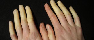
- Signs of damage to the autonomic nervous system are cold hands, acrocyanosis, pale skin and thickening of the palm, dyshidrosis, brittleness and splitting of nails, poor coordination of movements. Due to the unpleasant sensations in the hand, the discomfort increases, especially at night, and insomnia occurs.
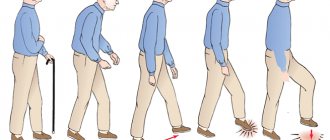
Light gymnastics with fingers and hands, restoring impaired blood supply, helps relieve symptoms and provide short-term relief to patients. Patients rub and knead the hand, change the position of the hand, shake and wave it. The discomfort disappears briefly if you lower your hand down and move your fingers slightly.
A distinctive sign of pathology, which makes it easy to carry out differential diagnosis with similar processes, is the preservation of the functions of the little finger. In carpal tunnel syndrome, the fifth finger of the hand is not affected.
Carpal tunnel syndrome is a sluggish pathological process that does not threaten the patient’s life and has a favorable prognosis for recovery, but in some cases leads to limitation of motor activity of the affected limb. Only correct and timely treatment will return the patient to a full life. In the absence of medical care, the median nerve will gradually atrophy, irreversible processes will begin in the hand, which will ultimately end in its complete immobility.
Operation technique
Positioning the patient
The patient lies supine with the wrist in a neutral position. The surgical field is marked. Two initial lines of incisions: one line is drawn along the longitudinal palmar, and the other transverse along the axis of the abducted first finger. A 1.5 cm incision is made proximally at the intersection of these two lines. An additional landmark for the access point to the carpal ligament is where the tip of the 4th finger touches when it is flexed at the metacarpophalangeal and proximal interphalangeal joints. In the distal forearm, two additional longitudinal lines are drawn: the first along the outer edge of the flexor carpi ulnaris tendon, and the second along the tendon of the palmaris longus muscle. The midpoint between these lines is indicated by the letter "x" for placing the endoscope cannula between the median nerve and the ulnar neurovascular bundle (Fig. 1).
Operation technique
The skin is incised in the marked area. Ragnell hooks spread the edges of the wound, retracting the palmar fascia from the underlying neurovascular bundle. The palmar fascia is incised longitudinally to expose the palmar medial space. The median nerve, superficial palmar arch, and carpal ligament are then visualized (Figure 2).
The hooks are moved deeper to retract the palmar fascia. A mosquito or narrow clamp is then inserted under the carpal ligament in the direction of the “x” point on the distal forearm (previously marked with a marker), creating a cannula space. A roller is then placed under the wrist to straighten the hand for insertion of the obturator. It is inserted between two lines marked on the forearm (Figure 3).
During insertion, the tip of the seal must always rest against the lower surface of the carpal ligament so as not to damage the structures of the carpal tunnel (nerve and tendon). Next, the cannula is inserted, as soon as the tip of the cannula is felt under the skin at point “x”, the obturator is removed, and the cannula remains in place with a slot facing slightly to the ulnar side. A standard endoscope with a diameter of 4 mm 30 degrees is inserted through the cannula (Fig. 4 A, B).
If everything is done correctly, we will see the carpal ligament (Fig. 5). When the cannula is rotated laterally, the median nerve is visualized (Fig. 6).
Fig 5. Fig 6.
After we have verified the correct placement of the cannula, it is rotated again to the ulnar side (medial). Once a clear image of the carpal ligament is obtained, the endoscope is removed and a blade is attached to its tip using a locking device (Fig. 8). The carpal ligament is incised by advancing the blade under the straight line with endoscopic visualization through the cannula from distal to proximal (Fig. 9 A, B).
Fig 8. Fig 9.
Incision of the carpal ligament is complete when the blade is palpated through the skin of the distal forearm. The blade is then removed and an endoscope is inserted to visualize the cut edges of the carpal ligament. (Fig. 10). The median nerve and flexor tendons should also be visualized as the cannula is rotated. Next, all instruments are removed from the canal, hemostasis is performed, the wound is sutured and an aseptic dressing is applied.
Gymnastics in disease prevention
Physical exercises are more suitable for the prevention of carpal tunnel syndrome, which can be performed at home
. It is recommended to do exercises before starting work, which will help prevent injury. You can do the following hand exercises at home:
Performing exercise 1 – Arms in a straight position in front, palms turned towards you vertically. Maintain the pose for five seconds.
Performing exercise 2 - Hands in a straight position in front of you, fingers relaxed. Hold for ten seconds.
Performing exercise 3 – Hands in front of you, palms bent into fists. Bend your wrists down without unclenching your fists. Hold for five seconds.
Performing exercise 4 – Hands are placed along the line of the body, while shaking the hands.
The doctor can select exercises individually.
results
Since the development of endoscopic techniques for carpal ligament dissection, numerous articles have appeared in the hand surgery literature reporting the results, outcomes, and complications of these techniques. Enthusiasts highlight lower postoperative relapse rates, improved short-term functional outcomes, and shorter disability periods. Opponents focused on the higher incidence of complications that arise with the endoscopic technique, especially when mastering the surgical technique. Dr. Mirza published an article on 280 cases of endoscopic surgery using the above technique. Mean grip strength was essentially equal to preoperative values 1 month after surgery, and patients returned to work after an average of 14 days. No patients reported pain in the scar area [2]. Since then, several prospective randomized studies have been conducted comparing open and endoscopic carpal ligament dissection. Trumble et al performed a multicenter randomized comparative study of the single-portal Agee technique and traditional open access [3]. They found that patients who had endoscopic surgery had better functional outcomes in the first three months after surgery and returned to work more quickly. Other randomized studies have shown similar results and return to work after 2 weeks in both groups of patients [4,5]. In addition, some articles reported more frequent relapses in patients after endoscopic surgery, negating the beneficial effect of a faster return to daily life. A meta-analysis of 13 randomized controlled trials was published in 2004 [6]. The study confirmed the conclusion that patients operated on with endoscopic methods had better scar sensitivity and grip strength. They also found a higher incidence of reversible nerve damage among these patients. Results in terms of pain and return to work were inconclusive [6].
Causes
Any factors that cause the carpal tunnel to become narrowed or swollen or cause fluid retention can cause carpal tunnel syndrome. Among the many possible reasons are the following:
- hormonal changes;
- obesity;
- diabetes;
- acromegaly - enlargement of bones due to dysfunction of the pituitary gland;
- decreased activity of the thyroid gland;
- renal failure;
- alcoholism;
- amyloidosis - deposition of abnormal proteins in tissues and organs;
- rheumatoid arthritis and gout;
— Paget's disease is a chronic bone disease in which the bones thicken and become deformed;
- tumors - lipomas (wen), ganglia (fluid-filled cysts formed in tendon sheaths), deformation of the wrists after fractures;
— use of hand-held vibrating tools.
Complications
The incidence of reported complications when using endoscopic techniques ranges from 0.2% to 5% [7]. However, many of the most serious complications arose in the early stages of development of endoscopic technology and were eliminated by changes in the design of the equipment. Dr. Agee's original technique has resulted in several cases of nerve transection [8]. Since then, the blade shape has been redesigned and a large multicenter trial has been conducted using the new device. The complication rate was found to be 1.8% [9]. Due to incomplete visualization of the carpal ligament, Dr. Chow changed his original transbursal technique to an extrabursal approach. Dr. Nagle compared the two methods and found that the complication rate of 11% with the original technique was reduced to 2.2% once a new access point was selected [10]. Damage to anatomical structures in the distal carpal ligament led Mirza to develop a uniportal technique that allows direct visualization of the superficial palmar arch, median nerve, and flexor tendons. During his early experience, Mirza reported two cases of transient neuropraxia of the ulnar nerve. After instrument redesign, a later report of 475 patients identified one case of reflex sympathetic dystrophy and one transient neuropraxia [11]. There are various reports in the literature on complementing endoscopic carpal ligament dissection with an open method. Today, the most common complications during endoscopic dissection of the carpal ligament are:
1) Median nerve injury: Dheansa and Belcher reported two cases of median nerve injury using the original Agee technique in anesthetized patients [12].
2) Ulnar nerve injury: Cases of ulnar nerve transection have been reported using the Chow dual portal approach
3) Damage to digital nerves: These range from transient digital neural neuropraxia to complete loss of sensation [15].
4) Trauma to the superficial palmar arch [16].
5) Damage to the flexor tendons.
6) Incomplete transection of the carpal ligament, leading to relapse and reoperation [7].
Underwater rocks
To date, our practice has made more than 40 attempts at uniportal endoscopic dissection of the carpal ligament using Dr. Mirza’s technique. To avoid complications, insertion of a blunt forceps, elevator and dissecting cannula under the ligament is always performed gently and without force. If at some point in the operation we encounter significant resistance, then a decision is made to switch to an open dissection technique. We also actively look for and isolate the recurrent motor branch to ensure that it is not damaged. In addition, when inserting a dissecting cannula into the distal forearm, care must be taken to avoid compression of the dissecting cannula (recurrent motor branch). Currently, the percentage of open operations in case of failure of instrument insertion during endoscopy is approximately 20%. By strictly following these recommendations, we have not had any complications so far.