Find out more about other diseases starting with the letter “C”: Compression of the brain; Senile chorea; Sensitive ataxia; Serous meningitis; "Rigid person" syndrome; Alien hand syndrome; Restless legs syndrome; Bogorad syndrome; West syndrome; Gaye-Wernicke syndrome; Guillain-Barre syndrome; Piriformis syndrome; Carpal tunnel syndrome; Carotid sinus syndrome; Kleine-Levin syndrome; Klippel-Feil syndrome; Cauda equina syndrome; Crumpy syndrome; Lambert-Eaton syndrome; Landau-Kleffner syndrome.
Description of West syndrome
West syndrome belongs to a group of neurological pathologies characterized by uncontrolled motor activity in certain muscle groups. Against the background of underdevelopment of the neuropsychic sphere, spasms of individual or large areas of the muscles occur. The first signs of the disease appear in early childhood (up to four years). The largest percentage of cases are recorded in the first year of a baby’s life in the form of individual symptoms, less often in a generalized form.
The syndrome got its name from the name of the doctor whose child suffered from this form of epileptic lesion. Until the introduction of computer and magnetic resonance scanning into clinical practice, it was generally accepted that the syndrome belonged to generalized epilepsy. After receiving the first visual evidence of damage to individual foci of the brain matter, experts decided to classify the pathology into a differentiated category (since 1964).
West epilepsy accounts for about two percent of all forms of childhood epilepsy. The anomaly occurs in 2-5 cases for every ten thousand children. Male infants are 10% more likely to get the disease than female infants. The vast majority of cases appear already 4-6 months after birth. Usually, by the age of three, uncontrolled spasms go away on their own. There are cases of transformation of West's anomaly into other forms of epileptic lesions.
A little history and statistics
The West syndrome we are considering was first noticed and described by a doctor with the same name in 1841. He observed the manifestations in his child and identified them as a separate symptomatic complex, which was subsequently made into a separate diagnosis. Since this pathology occurs in infancy, its characteristic spasmodic and paroxysmal manifestations are called infantile spasms. Initially, this syndrome was not classified as a separate category of diseases, but was considered a variant of the manifestation of a generalized form of epilepsy.
In the mid-20th century, based on data obtained from the study and analysis of EEG data from small patients, the specificity of hypsarrhythmic brain activity was identified, characterized by a chaotic alternation of slow-wave periods with high-amplitude spikes. Based on these specific EEG patterns, the disease was identified as a separate diagnostic criterion, characteristic specifically for young patients.
Causes of the disease
The root causes are both obvious pathogenic effects on the intrauterine development of the fetus, and cryptogenic (unexplained) ones that are not immediately obvious. Symptomatic factors include:
- Intrauterine infection (cytomegaly, herpes, encephalitis, etc.).
- Insufficient oxygen supply to brain structures as a result of hypoxia, intracranial injury, suffocation of a newborn during labor (umbilical cord entanglement), ischemic damage.
- Disturbances in the anatomical development of the head skeleton.
- Hereditary transmission of the disease from older relatives.
- Routine vaccination with DTP agents. This factor has not been clinically proven. It is possible that the age of onset of symptoms usually coincides with the usual timing of medical immunization.
As a result of exposure to various factors, the functionality of neurons that produce serotonin decreases. A number of experts prove that the connection between the brain and the glandular structures of the adrenal glands is disrupted, and myelin production is reduced. Hormonal imbalance leads to systematic spasms of muscle sections.
A decrease in symptoms and its complete disappearance by 3-4 years of age is associated with a period of relative maturation of the brain, a decrease in the degree of its excitability, and an increase in the concentration of myelin in the blood. If there is insufficient compensation for these processes, transformation of infant West syndrome into other forms of epilepsy may occur.
5.4. Epilepsy with prolonged spike-wave complexes during slow-wave sleep
general characteristics
Epilepsy with prolonged spike-wave complexes during slow-wave sleep debuts between the ages of 8 months and 14 years and is manifested by partial and generalized epileptic seizures, severe cognitive impairment, and a pattern of continued epileptiform activity during slow-wave sleep.
Seizures are represented by motor paroxysms (myoclonic absences, generalized clonic seizures, orofacial paroxysms) or generalized tonic-clonic seizures in combination with atypical absences and atonic paroxysms. A number of patients initially experience unilateral partial motor seizures or generalized tonic-clonic seizures, and later atypical absence seizures occur.
In some patients, epileptic seizures may be absent [Continuous..., 1995; Epilepsy..., 2006].
Electroencephalographic patterns
On an interictal EEG in a state of quiet wakefulness, epileptiform activity may not be observed, or generalized “peak-wave” and “polypeak-wave” complexes with a frequency of 2–3 and/or 3–4 beats/s and/or are recorded against a moderately altered background. focal peaks, sharp waves, “Rolandic complexes” with predominant localization in the frontotemporal, central temporal or central parietotemporal regions [Schwarzmayr P., Mayer-Ewert K., 1999; Atlas..., 2006; Epilepsy..., 2006]. Similar changes can also be detected during naps (
).
In general, during slow-wave sleep, the EEG shows (usually with the exception of children under 4–5 years of age) prolonged (more than 85% of the entire recording), bilateral or diffuse peak-wave complexes [Electrical..., 2002, 2005] with complete or almost complete absence of physiological sleep patterns (
). During paradoxical sleep, epileptiform activity is fragmented, accounting for less than 25% of the curve, and may be absent or focused in the frontal regions [Atlas..., 2006].
During an attack, a pattern corresponding to the type of epileptic seizure is recorded on the EEG [Zenkov L.R., 2001].
Signs of pathology
The main signs of the development of neurological failure are systematic muscle spasms and impaired psychomotor skills in the child. In most cases, muscle spasms appear secondarily after the initial diagnosis of psychomotor development delay. The baby does not always show a grasping reflex and cannot fix his gaze on a moving object.
Next, short-term, symmetrical, serial (with an interval of no more than a minute) spasms of individual muscle groups appear. The number of attacks can range from several units to several hundred. The maximum predisposition to an attack occurs both during the period of relaxation (for example, before falling asleep) and during the beginning of daytime activity (immediately after waking up). Paroxysms are provoked by sensory or sound stimuli.
Rarely, a generalized contraction of all muscle groups of the body may occur. More often, the patient suffers from involuntary contractions in the neck (frequent head nods), shoulder area, abdominal area and upper limbs. Children who have already learned to walk may suffer from involuntary falls, while consciousness is fully preserved, the child can hear, see and speak.
Another form of external manifestations is a non-convulsive variant of the pathology, when instead of spasms there is a complete stop of the physical manifestations of the body. The movements stop. The disorder can affect absolutely all muscle groups: eyeballs, respiratory muscles (the breathing rhythm is disrupted), asymmetric combinations (throwing the head back to one side, looking away to the other).
Diagnostic methods
When diagnosing an anomaly, neurologists pay primary attention to three main areas of symptoms: short-term spasms, mental retardation, age of primary manifestations. If symptoms appear later than the first year of life, doctors need to rule out other diseases with similar manifestations. A pediatrician, an epileptologist and a geneticist join the work of the neurologist.
Instrumental methods for fixing the disease include:
- EEG. Measurements are taken both during wakefulness and during sleep. The presence of West syndrome is indicated by the disorder of the signature, the undulation of the graph both during physical activity and in a state of complete rest.
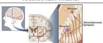
- Computer scanning. It is rarely carried out, since it cannot always detect the boundaries of damage to cerebral structures.
- MRI of the brain. It is the most informative way of diagnosing. It can be carried out several times in the process of monitoring the excitability of the GM cortex. The technique is the safest for young patients. It does not require lengthy preparation and is painless.
If the child has the above-described signs of the disease, the attending specialist will refer you to undergo magnetic resonance imaging. To quickly find a diagnostic center, a detailed list of tomography clinics has been created and posted on the Mrt-v-msk portal. On its pages you can find a medical organization in any area of the city, compare ratings, prices for procedures, read reviews, and leave your own assessment. Registration for diagnostics in any clinic is carried out by calling the hotline located at the top of the mrt-v-msk page. Consultants will guide you to the nearest medical centers, announce prices, and book free time for the session.
Etiological features
Most cases are symptomatic.
Unfortunately, there is no exact data regarding the etiology of the disease. The causes may be intrauterine infections in acute form (cytomegalovirus, herpetic), hypoxic lesions of the fetus, postnatal encephalitis, premature birth, asphyxia of the newborn, intracranial birth injury, postnatal ischemia, etc.
In addition, it is likely that West syndrome in children may be a consequence of anatomical abnormalities of the brain, such as, for example, agenesis of the corpus callosum, hemimegalencephaly.
Also, infantile spasms can have secondary symptoms, that is, they can be a consequence and symptom of other diseases, such as phakomatosis, neurofibromatosis, Down syndrome, and some gene mutations. There is also evidence of a connection between infantile spasms and phenylketonuria.
How to deal with West syndrome?
Until the mid-20th century, it was generally accepted that the disease was not amenable to therapeutic or drug treatment. Later it was discovered that the symptoms partially or completely disappear when taking steroid drugs. Experts still disagree on what dosage is optimal. Most tend to prescribe large dosages of drugs for a period of two to six weeks of use.
To reduce the intensity of spasms, nitrazepam, valperic acid and large doses of vitamin B6 are prescribed. If therapeutic methods do not show effectiveness, consultation with a neurosurgeon about excision of pathological foci of the cerebral cortex is recommended.
Treatment
A real breakthrough in the treatment of West syndrome in children has been the use of ACTH (adrenocorticotropic hormone) drugs to relieve seizures. The use of ACTH in combination with prednisolone leads to a reduction and even complete disappearance of muscle spasms. In turn, the EEG pattern confirms the absence of hypsarrhythmic characteristics. The only stumbling block in the treatment of West syndrome is that the selection of doses and duration of course treatment with these drugs is a purely individual factor and is done empirically. In 90% of cases, a good effect is achieved by using significant dosages of drugs.
In the early 90s of the 20th century, a positive effect of treatment with vigabatrin was discovered, but the advantage of this drug was proven only for patients with tuberous sclerosis. For the remaining category of patients, steroid therapy remained in first place. However, the disadvantage of steroid therapy is poorer tolerability of the drugs and the tendency of the disease to relapse.
Prognosis of the course of the disease
The disappearance of the syndrome occurs by the age of three, while the damage caused to the psychomotor area persists in 70-80% of cases. Movement disorders persist in half of the cases. Only 5-10% of patients undergo minimal changes. More than half of those suffering from the syndrome experience transformation of the pathology into other forms of episyndrome. With genetic brain abnormalities, mortality in the early stages accounts for a quarter of cases.
The best prognosis is for patients who did not exhibit delayed psychomotor development before the onset of spastic symptoms. About 45% of children who have suffered an anomaly retain full intellectual abilities. Timely diagnosis and correct treatment are important.
Diagnostics
The diagnosis of West syndrome is based on three factors:
- delayed mental and mental development;
- hypsarrhythmic EEG pattern;
- muscle spasms.
Of great importance is the age at which the disease begins to manifest itself, as well as the connection between spasms and sleep. In cases where the disease manifests itself at an atypically late age, diagnostic difficulties may arise.
If West syndrome is suspected, the child undergoes a consultation with a neurologist, with further examination by a geneticist and an epileptologist. It is important to timely differentiate diseases such as benign infant myoclonus, infant myoclonic epilepsy, Sandifer syndrome, in which the head tilt is pronounced like torticollis, as well as episodic manifestations of opisthotonus, which resemble spasmodic contractions, but, in fact, are not them.