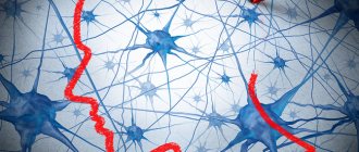
What is neuroglia
The collection of glial cells is called neuroglia. These are special cell populations that are found in the central nervous system and in the periphery. They maintain the shape of the brain and spinal cord and also supply it with nutrients. It is known that there are no immune reactions in the central nervous system due to the presence of the blood-brain barrier.
Neuroglia are brain structures designed to support the functioning of neurons. The term comes from the phrase “nerve glue.” The principle of its operation was first explained by the Italian biologist Emilio Lugaro in 1907. He proposed that glial cells exchange substances with the extracellular fluid and thus exercise control over the neural environment.
Given this, neuroglia outnumber neurons. It is present in invertebrates and vertebrates, and may differ from neurons in the absence of axons and the presence of only one type of process. Its cells do not form synapses and do not lose the ability to divide throughout their lives. While neurons and neuroglia are in close proximity to each other, there are no direct connections between these components.
Neuroglia are responsible for controlling human behavior. Some of its cells are located in the hypothalamus and control appetite, heart rhythms, and sleep cycles.
Pathologies
The central nervous system, like any other tissue in the body, can be damaged. Neuroglia experience pathological effects first. Protective functions allow you to take the blow yourself.
All viruses that can affect the nervous system begin their activity by changing glia. As a result, the cells give rise to benign neoplasms and form cysts in the spinal cord and brain.
With a strong impact on microglia, the myelin sheath of neurons begins to collapse, which contributes to the occurrence of such serious diseases as:
- amyotrophic sclerosis;
- parkinsonism;
- neuropathy;
- Alzheimer's disease.
The destruction of the protective barrier of glia leads to severe diseases of the nervous system and disorders of the brain. The latest research in this area allows us to hope for a breakthrough in the treatment of many pathologies associated with organic changes in neuroglial tissue.
Nervous system
This is a collection of organized cells that specialize in conducting electrochemical signals from sensory receptors through a network to the site of reaction.
They come in two types: diffuse and centralized. In the diffuse type, found in lower invertebrates, the brain is absent and the neurons are distributed throughout the body in a reticular pattern. In the centralized systems of higher invertebrates and vertebrates, some of them play a dominant role in processing information and regulating responses.
Breaking out of pathneuron networks
Is the participation of glia in the regulation of neuronal functions limited to the formation of the myelin sheath around axons? Apparently not. Richard Robitaille of the University of Montreal discovered that the magnitude of the electrical potential generated in frog muscle by stimulating a synapse increased or decreased depending on what chemicals he introduced into the Schwann cells surrounding the synapse. When Eric A. Newman of the University of Minnesota touched the retina of a rat, the “calcium signals” sent by the glia changed the firing rate of the visual neurons. And Maiken Nedergaard of New York Medical College, who studied slices of the rat hippocampus (an area of the brain involved in memory processes), observed increased electrical activity at synapses while surrounding astrocytes increased calcium uptake. Scientists view such changes in the effectiveness of synapses as a major factor in the plasticity of the nervous system, i.e., its ability to change responses based on past experience, and glia may thus play an important role in the cellular processes of learning and memory.
Immune defense of the brain
The brain, where many biochemical reactions take place, which means a lot of immunogenic substances are formed, must be protected from humoral immunity. It is important to understand that the neuronal tissue of the brain is very sensitive to damage, after which neurons are only partially restored. This means that the appearance of a place in the central nervous system where a local immune reaction will take place will also lead to the death of some surrounding cells or demyelination of neuron processes.
At the periphery of the body, this damaged somatic cells will soon be filled with newly formed ones. And in the brain it is impossible to restore the function of a lost neuron. And it is neuroglia that limits the brain from contact with the immune system, for which the central nervous system is a huge amount of foreign antigens.
Ependymal glial cell
Ependymal glial cells are found in specific areas of the central nervous system. They form the endothelial lining of the cerebral ventricles and the central spinal canal. They originate in embryogenesis from the ectoderm, and therefore represent a special type of neuroepithelium. It is multi-layered and performs a number of functions:
- supporting: constitutes the mechanical frame of the ventricles, which is also supported by the hydrostatic pressure of the cerebrospinal fluid;
- secretory: releases some chemicals into the cerebrospinal fluid;
- demarcating: separates the medulla from the cerebrospinal fluid.
Like all other neuroglial cells, they are created by the neuroectoderm. These units of the nervous system form the epithelial lining of the ventricular cavities in the brain and the central canal in the spinal cord. They also form an epithelial layer that protects the network of blood vessels located in the wall of the lateral ventricles of the cerebral hemispheres.
Pathological processes
Due to exposure to pathologies, neuroglial cells are exposed to various negative consequences.
The following changes may occur:
- swelling and swelling;
- hypertrophy and atrophy;
- hyperplasia;
- amoeboid degeneration;
- homogenizing metamorphosis.
This disease, due to which the cellular structure changes, also occurs in histological examination when it is necessary to identify other human diseases. For a long period of time to examine the nervous system, neuroglial substances were considered secondary. Now they are considered the main components of nervous tissue. Pathologies can cause complex diseases.
Astrocytes
Astrocytes are glial cells of the brain that act as a “recycler” of dysfunctional mitochondria that are produced by neurons. Astrocytes do not simply eliminate them, but replace them with their own formations.
There is a division into protoplasmic and fibrous. Fibrous glial cells are distributed in the white matter of the central nervous system among myelinated nerve fibers. They are characterized by the presence of numerous fibrils in the cytoplasm. The main processes diverge in a radial direction (hence the name astrocyte, which means “star-shaped cell”).
Unlike fibrous ones, protoplasmic ones predominate in the gray matter of the central nervous system. They contain fewer fibrils and organelles within their cytoplasm. Their processes come into contact with the capillaries, like the processes of fibrous ones.
The astrocyte system is thought to involve the action of neurotransmitters such as glutamate and gamma-aminobutyric acid (GABA). They act as a repository for the latter.
Astrocytes are glial cells in the brain that make up the medulla. They are star-shaped and small in size, although they are larger than microglial cells. However, there are only two types of astrocytes: fibrous and protoplasmic. The first type of cells is located in the white and gray matter of the brain, although there are much more of them in the white.
This means that they are most common in areas where there are a significant number of neuronal myelinated processes. Protoplasmic astrocytes are also glial cells: they are found in the white and gray matter of the brain, but they are much more numerous in the gray matter. This means that their function is to create support for the bodies of neurons and the structural organization of the blood-brain barrier.
GLIA - MORPHOLOGY AND FUNCTION
The human brain is made up of hundreds of billions of cells, with nerve cells (neurons) not making up the majority. Most of the volume of nervous tissue (up to 9/10 in some areas of the brain) is occupied by glial
(from Greek: glue together). The fact is that the neuron performs a gigantic, very delicate and difficult job in our body, for which it is necessary to free such a cell from
Rice. 2.2.
Types of glial cells in the brain.
1 - neuron; 2 - perineural oligodendrocyte; 3 - oligodendrocyte in the white matter; 4 - fibrous astrocyte; 5 - blood vessel; 6 - protoplasmic astrocyte.
everyday activities related to nutrition, removal of waste, protection from mechanical damage, etc. - this is provided by other servicing cells, i.e. glial cells (Fig. 2.2). There are three types of glial cells in the brain: microglia, oligodendroglia and astroglia, each of which provides only its intended function. Microglial cells participate in the formation of the meninges, oligodendroglia - in the formation of sheaths (myelin sheaths) around individual processes of nerve cells. Myelin sheaths around peripheral nerve fibers are formed by special glial cells called Schwann cells. Astrocytes are located around neurons, providing their mechanical protection, and in addition, they deliver nutrients to the neuron and remove waste products. Glia cells also provide electrical insulation of individual neurons from the influence of other neurons. An important feature of glial cells is that, unlike neurons, they retain the ability to divide throughout their lives. This division in some cases leads to tumor diseases of the human brain. The nerve cell is so specialized that it has lost the ability to divide. Thus, the neurons of our brain, once formed from precursor cells (neuroblasts), live with us throughout our lives. On this long journey, we only lose neurons in our brain.
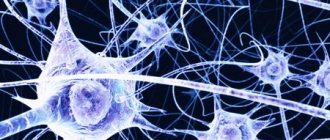
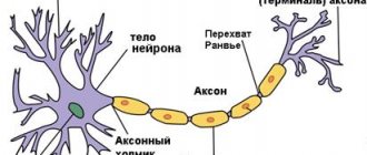
NEURON
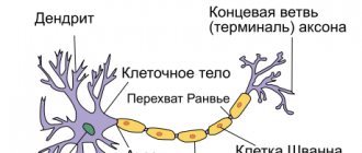
A neuron is the main cell of the central nervous system. The shapes of neurons are extremely diverse, but the main parts are the same in all types of neurons. A neuron consists of the following parts: soma
(bodies) and numerous branched processes.
Each neuron has two types of processes: an axon,
along which excitation is transmitted from a neuron to another neuron, and numerous
dendrites
(from the Greek tree), on which axons from other neurons end in
synapses
(from the Greek contact). The neuron conducts excitation only from the dendrite to the axon.
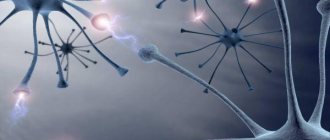
The main property of a neuron is the ability to excite (generate an electrical impulse) and transmit (conduct) this excitation to other neurons, muscle, glandular and other cells.
In Fig. Figure 2.3 shows a diagram of a neuron, in which its main parts are easily visible.
Neurons in different parts of the brain perform very diverse work, and in accordance with this, the shape of neurons from different parts of the brain is also diverse (Fig. 2.4). Neurons located at the output of a neural network of some structure have a long axon along which excitation leaves this brain structure. For example, neurons of the motor cortex of the brain, the so-called pyramids of Betz (named after the Kiev anatomist B. Betz, who first described them in the mid-19th century), have an axon of about 1 m in humans; it connects the motor cortex of the cerebral hemispheres with segments of the spinal cord. This axon carries “motor commands,” such as “move your toes.”
How is a neuron excited? The main role in this process belongs to the membrane, which separates the cell cytoplasm from the environment. The membrane of a neuron, like any other cell,
Rice. 2.3.
Neuron diagram.
1 - dendrite; 2 - spines; 3 - neuron body; 4 - dendrite; 5 - axon hillock; 6 - axon; 7 - synaptic cleft; 8 — axon end; 9 - postsynaptic neuron.
Rice. 2.4.
Neurons of various brain structures.
1 - grain cell; 2 - double pyramid of the hippocampus; 3 - pyramidal cell; 4 - Purkinje cell; 5 - large cell of the reticular formation: 6 - thalamic neuron.
Rice. 2.5.
Scheme of the structure of a biological membrane.
1 - lipids; 2 - integral proteins; 3 - peripheral proteins; 4 - glycoproteins. The number of polar “heads” of phospholipids is approximately 10 times greater than the number of molecules of integral proteins.
is very complicated. Basically, all known biological membranes have the same structure (Fig. 2.5): a layer of protein molecules, then a layer of lipid molecules and another layer of protein molecules. This whole structure resembles two sandwiches stacked with butter facing each other. The thickness of such a membrane is 7–11 nm. To imagine these dimensions, imagine that the thickness of your hair has decreased by 10 thousand times. A variety of particles are embedded in such a membrane. Some of them are protein particles and penetrate the membrane through (integral proteins); they form passage points for a number of ions; sodium, potassium, calcium, chlorine. These are so-called ion channels.
Other particles are attached to the outer surface of the membrane and consist not only of protein molecules, but also of polysaccharides.
These are receptors
for molecules of biologically active substances, such as mediators, hormones, etc. Often the receptor, in addition to a place for binding a specific molecule, also includes an ion channel.
The main role in neuron excitation is played by membrane ion channels. These channels are of two types: some work constantly and pump sodium ions out of the neuron and pump potassium ions into the cytoplasm. Thanks to the work of these channels (they are also called pumping channels
or
ion pump),
constantly consuming energy, a difference in ion concentrations is created in the cell: inside the cell the concentration of potassium ions is approximately 30 times higher than their concentration outside the cell, while the concentration of sodium ions in the cell is very small - approximately 50 times less than outside the cell .
The property of the membrane to constantly maintain the difference in ionic concentrations between the cytoplasm and the environment is characteristic not only of the nervous cell, but also of any cell in the body. As a result, a potential arises between the cytoplasm and the external environment on the cell membrane: the cell cytoplasm is charged negatively by an amount of about -70 mV relative to the external environment of the cell.
This potential can be measured in the laboratory with a glass electrode if a very thin (less than 1 micron) glass tube filled with a salt solution is inserted into the cell.
The glass in such an electrode plays the role of a good insulator, and the salt solution acts as a conductor. The electrode is connected to an electrical signal amplifier and this potential is recorded on the oscilloscope screen. It turns out that a potential of about -70 mV is maintained in the absence of sodium ions, but depends on the concentration of potassium ions. In other words, only potassium ions participate in the creation of this potential, and therefore this potential is called the “resting potassium potential”, or simply “resting potential”.
Thus, this is the potential of any resting cell in our body, including a neuron.
Oligodendrocytes
Oligodendrocytes are specialized neuroglial cells located in the central nervous system of invertebrates and vertebrates that function to produce myelin, the insulating sheath on the axons of nerve fibers.
They are divided into interfascicular and perineural, have few cytoplasmic fibrils and a fairly developed Golgi apparatus. They can be distinguished from astrocytes due to the greater density of both the cytoplasm and the nucleus, the absence of fibrils and glycogen in the cytoplasm and the large number of microtubules in the processes.
Interfascicular oligodendrocytes are built in rows between the nerve fibers of the white matter of the central nervous system. The perineurals are located in the gray matter, very close to the soma of neurons. Glial cells, equivalent to oligodendrocytes but located in the peripheral region of the nervous system, are called Schwann cells.
The type of axon determines whether loose or tight myelination will occur. In dense, the oligodendrocyte wraps itself, like a folded sheet, around the axon until the fiber is covered in several layers. Between the segments of the myelin sheath there are areas called nodes of Ranvier, which are important in the transmission of nerve impulses.
Oligodendrocytes are types of glial cells that surround the neuron and its processes. They are found both in the central nervous system and near the peripheral mixed and autonomic nerves. Oligodendrocytes themselves are polygonal cells equipped with 1-5 processes. They interlock with each other, isolating the neuron from the internal environment of the body and providing conditions for nerve conduction and generation of impulses. There are three types of oligodendrocytes, which differ in morphology:
- central cell located near the body of the brain neuron;
- satellite cell surrounding the neuron body in the peripheral ganglion;
- Schwann cell, enveloping the neuronal process and forming its myelin sheath.
Oligodendrocyte glial cells are found both in the brain and spinal cord and in peripheral nerves. Moreover, it is not yet known how the satellite cell differs from the central one. Considering that the genetic material of all cells of the body, except for the sex cells, is the same, it is likely that these oligodendrocytes can mutually replace each other. The functions of oligodendrocytes are as follows:
- supporting;
- insulating;
- separating;
- trophic.
Microglia - brain immunity
The collective name “glia” includes several types of cells with different functions. Oligodendrocytes and Schwann cells envelop nerve tissue and cover them with a myelin sheath, which insulates the electrical signal and accelerates its transmission, while astrocytes with numerous processes regulate water-salt metabolism, support the functioning of synapses and participate in the metabolism of neurotransmitters.
But the greatest interest in the last decade has been microglia.
Microglia were first described by Pio del Rio Ortega back in 1920, but then their study stalled for a long time - interest in them was revived only in the 1980s. Today, according to Amanda Sierra from the Basque Neuroscience Center of Achucarro, the study of microglia is rapidly gaining momentum.
Scientists already know that microglia play an important role in traumatic brain injuries, neurodegenerative diseases and inflammatory processes. In addition, microglial cells have recently been shown to act as macrophages of the immune system, neutralizing threats to the brain from microbes and cellular debris and removing unnecessary synapses.
Some of these functions are performed by several types of glia. Astrocytes and Schwann cells, for example, also remove unnecessary synaptic connections. But researchers are increasingly inclined to believe that, despite their common functions, there is no sufficient basis for combining different types of glial cells into one group. Moreover, in an article published in 2021, scientists advocated abandoning the term “glia” itself.
“Different glial cells have very little in common,” says Guy Brown, lecturer in biochemistry at the University of Cambridge. “I don’t think the label ‘glia’ has a future.”