Types of changes in vascular tone
Question No. 3134 (2010-04-29)
Hello! Help me please, I have the same problem. I live in the city of Essentuki and it is very difficult to get to a neurologist here. I have vegetative-vascular dystonia of mixed type. A year ago, attacks, headaches, dizziness, anxiety, panic and fears began. I contacted a neurologist and he prescribed cinnarizine.
I drank it, it seemed to feel better. And then it got even worse. I was admitted to the hospital. There I did an REG and here is the conclusion: Pulse blood flow to the vessels of both hemispheres is significantly reduced. The tone of large and medium arteries is increased, the tone of small arteries is within normal limits. Venous outflow is difficult. Elastic properties of the main arteries are within normal limits.
When performing functional tests with turning the head to the right, left, tilting the head back: an increase in blood supply in both hemispheres. When the head is tilted forward: blood filling without dynamics. Pulse blood flow in the vessels of the left IMS is within normal limits, and in the vessels of the right IMS it is significantly reduced.
blood filling without dynamics. And after this conclusion I was discharged from the hospital. Now I feel very bad too. I have a constant headache, especially in the back of the head, dizziness, I constantly yawn and want to sleep, lack of air, pain in the heart area. Arms and legs go numb, weakness, and sometimes blood pressure rises.
Frequent urination. Hands are constantly cold and feet too, heavy sweating. Anxiety, some kind of fear. Plus cervical osteochondrosis. Help me please, what should I do? I did an ultrasound on the kidneys, everything is fine, the cardiogram is also good, I checked the thyroid gland, the liver is fine. Help, I beg you. Thank you very much in advance!
There is always a reason for any symptoms. You list a lot of different complaints and not all of them fit into the diagnosis you indicated. As you understand, without an examination, which unfortunately is not possible, I cannot recommend taking any specific medications.
For this reason, I can only give more or less general recommendations.
Regarding problems with blood vessels and the results of the examination, you need a course of therapy with drugs that affect vascular tone and blood rheology.
Regarding panic attacks, the most effective would be to take one of the antidepressants for several months.
By the way, all this must be prescribed taking into account the patient’s age, and I don’t even have this information.
The tone of cerebral vessels can be decreased or increased. In the first case, there is hypotonicity, in the second - hypertonicity, the features of which are different.
Hypotonicity
Hypotonicity or hypotension is a decreased vascular tone, which is expressed in a decrease in blood flow volume and a drop in pressure in the arteries. When the tone of small vessels decreases, the flow of blood into the organ increases. The walls of the arteries are stretched during contraction of the ventricles of the heart (one of the states of the organ's muscle), thereby causing throbbing headaches.
If the tone of the small veins located in the brain decreases, there is difficulty in the outflow of blood from the organ. It is in the area of veins and venous sinuses that the largest volume of blood is localized. When a person is lying down, when the head is below the level of the neck (when bending forward or throwing it back), during physical exertion and emotional overload, blood flow is further disrupted, causing bursting headaches.
Hypertonicity
Hypertonicity of cerebral vessels is an increase in their tone, that is, the degree of wall tension, which causes an increase in resistance to blood flow. Hypertonicity is characterized by an increase in blood pressure and the occurrence of corresponding symptoms.
Hypertonicity, like hypotonicity, can occur against the background of the following pathological conditions of the body:
- constant lack of sleep and overwork;
- abuse of invigorating drinks (coffee, tea, etc.), alcohol, smoking;
- long stay in a stuffy room;
- frequent emotional experience, stress;
- development of hormonal disorders;
- VSD;
- development of atherosclerosis or osteochondrosis;
- heart and kidney diseases.
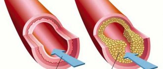
Spasm of the walls of blood vessels contributes to the narrowing of the lumen of the latter, causing a decrease in the volume of passing blood. Hypoxia develops, and with significant narrowing of the lumen, tissue ischemia.
What is cerebral vascular hypertonicity - treatment
20.03.2019
Vascular tone is the tension of the vascular walls maintained by the muscular wall in these vessels. This is a factor that determines the blood supply to brain tissue.
The tone of the muscle wall maintains a certain tension, allowing the lumen of the vessel to maintain the required diameter. Thanks to the muscular walls of the cerebral vessels, the brain tissue is protected from a lack or excess of blood, therefore, in a healthy body, the blood supply to the brain does not depend on pressure changes in other parts of the body.
However, with diseases of the internal organs or disorders of the nervous system, the tone of the arteries and veins may change: the muscle wall goes into a state of spasm - the lumen of the vessel decreases, and the supply of blood to the neurons decreases. This leads to tissue hypoxia, when the brain does not receive the proper amount of oxygen and nutrients. Vascular hypertonicity provokes functional and organic disorders in the brain.
What it is
Hypertonicity is a change in the tone of a vessel, in which the lumen of the latter decreases due to spasm of the smooth muscles of the arteries, and vice versa, if the tone decreases, this is called hypotonicity.
Increased tone can be of a physiological or pathological nature.
In the first variant, vascular tone naturally increases during temporary functional states, when a normal amount of adrenaline is released due to internal (pain, fear) and external (sharp loud sound) factors. Physiological hypertonicity will never lead to disorders in the long term and is considered a positive temporary condition.
Pathological hypertonicity of cerebral vessels is formed as a result of diseases of internal organs, glands and metabolic disorders. Severe arterial spasm can lead to ischemic stroke (acute cerebrovascular accident), which results in irreversible organic changes in tissue and subsequent loss of intellectual and motor capabilities.
Causes
The following reasons lead to spasm of cerebral arteries and veins, which are divided into psychological (neurological, psychiatric), somatic (bodily) and indirect.
Psychological reasons:
- Anxiety disorder, accompanied by excitement, fear due to constantly elevated levels of adrenaline.
- Vegetovascular dystonia. The disease is characterized by an imbalance between the functioning of the autonomic parts of the nervous system. In this disease, blood vessels spasm for no obvious reason.
- Stress, neuropsychic tension.
- Sleep disturbance: insomnia, taking a long time to fall asleep.
- Personality pathologies: psychopathy, accentuation. This is predominantly a hysterical, emotionally labile and cycloid personality type.
Somatic causes:
- Arterial hypertension - as a primary disease - a persistent increase in blood pressure over 140/80 for more than two weeks.
- Atherosclerosis is the presence of fatty deposits on the walls of the arteries.
- Inflammation of the walls of blood vessels.
- Diseases involving systems: rheumatoid arthritis, systemic lupus erythematosus or scleroderma.
- Diseases of the endocrine organs: pheochromocytoma is a tumor of the adrenal glands that sharply stimulates the glands and provokes the release of huge amounts of adrenaline.
- Dystrophic diseases of the musculoskeletal system: cervical osteochondrosis, spinal hernia.
- Hyperthyroidism. What it is? This is a disease of the thyroid gland, in which the release of T3-T4 hormones increases, which increases the sensitivity of blood vessels to adrenaline and norepinephrine.
- Inflammation of the tissue of the nerve ganglia of the sympathetic parts of the autonomic nervous system.
- In children, cerebral ischemia is observed due to pathologies during pregnancy and complicated childbirth. As a rule, a child’s pathology is diagnosed immediately in the maternity hospital.
Indirect reasons that increase the likelihood of hypertension:
- smoking;
- large doses of caffeine: more than three cups of coffee per day; caffeine temporarily stimulates the release of adrenaline into the blood;
- diabetes;
- heredity;
- age: from 50 years old, mostly men;
- prolonged stay in stuffy, unventilated rooms;
- weather sensitivity.
Symptoms
An increase in cerebral vascular tone refers to cerebral hypertonicity and the following symptoms develop:
- Headache in diffuse localization. The area of pain depends on the place where hypertonicity is most pronounced. Also, unpleasant sensations can spread throughout the entire head at once. The pain can spread to the neck, eyes, ears.
- Due to the deterioration of blood supply, intellectual and mnestic activity deteriorates: the pace of thinking slows down, the volume and concentration of attention decreases.
- Changes in physiological state: fatigue, exhaustion. Even simple work requires mental and physical effort.
- Emotional disorders: mood lability (sudden transitions from a bad mood to a good one and vice versa), irritability. Low threshold of excitability: quiet sounds and dim light can make a person angry.
- Nausea and vomiting.
- Hypertonicity, combined with difficulty in venous outflow, is manifested by a feeling of fullness in the head.
- Syncope is rarely observed in the clinical picture. However, especially sentimental people may faint from happiness.
Treatment
Complex therapy of the disease, how to restore brain function and how to increase the body’s performance:
- Treatment of the underlying illness that led to a disorder of brain tone.
- Psychohygiene: dosed mode of work and rest.
- Get a good night's sleep: remove bright light sources (phone, laptop) away from you an hour before bedtime.
- Aromatherapy. You can use scented sticks or scented oils.
- Balance your diet: reduce salt and alcohol intake.
- Drug treatment. Medicines that lower blood pressure: valerian, captopres or captopril.
- Folk remedies:
- rosehip tea;
- take a warm shower or bath.
Didn't find a suitable answer? Find a doctor and ask him a question!
Source: //sortmozg.com/zabolevaniya/gipertonus-sosudov-golovnogo-mozga
What are its features
Vascular tone refers to the tension of the walls, which is ensured under the influence of smooth muscles. The latter has different severity, based on where it is located (arteries, veins, capillaries):
- the greatest severity is seen in the area of the arteries: smooth muscles provide high resistance to blood pressure and constantly maintain the arterial lumen;
- the least pronounced is in the area of the veins: the layer cannot resist the pressure of the blood flow and maintain the venous lumen;
- there is no smooth muscle in the capillaries.
The tone of cerebral vessels (brain) is maintained by 2 mechanisms: neurogenic (nerve impulses) and myogenic (spontaneous contraction of the smooth muscle layer).
Trauma, surgery, stroke, pathologies of the ANS (for example, vegetative-vascular dystonia), intoxication syndrome, the development of certain diseases of infectious and endocrine etiology often become causes of disruption of neurogenic regulation of tone. In this case, the smooth muscle muscles contained in the vessels take over the implementation of normal blood circulation. This process is called basal tone. It is provided through the myogenic regulatory mechanism.
Hypertonicity
Hypertonicity of cerebral vessels is an increase in their tone, that is, the degree of wall tension, which causes an increase in resistance to blood flow. Hypertonicity is characterized by an increase in blood pressure and the occurrence of corresponding symptoms.
Hypertonicity, like hypotonicity, can occur against the background of the following pathological conditions of the body:
- constant lack of sleep and overwork;
- abuse of invigorating drinks (coffee, tea, etc.), alcohol, smoking;
- long stay in a stuffy room;
- frequent emotional experience, stress;
- development of hormonal disorders;
- VSD;
- development of atherosclerosis or osteochondrosis;
- heart and kidney diseases.
Spasm of the walls of blood vessels contributes to the narrowing of the lumen of the latter, causing a decrease in the volume of passing blood. Hypoxia develops, and with significant narrowing of the lumen, tissue ischemia.
searching results
No. 526 Chiropractor 14.01
Hello. Tell me, is it possible to do a massage if a child is crying? The child is 4 months old; at birth, depression syndrome was diagnosed as muscle hypotonicity. We suffered from streptoderma two months ago. But 1-2 small elements appear every two weeks in the form of a small abscess on the legs. They prescribed Cortexin injections and a course of massage. How to be. Thank you further
Olga Ustinova, St. Petersburg
We suggest you read: Elevated red blood cells in a child’s blood – what does this mean? Red blood cells - normal, primary and secondary erythrocytosis
Hello! I underwent rheoencephalography, and they wrote me the following conclusion: Moderate vascular hypertonicity in the VBB. Please tell me what this means. No one explained anything at the clinic, only after that they gave directions to undergo additional examinations (ultrasound of the heart) further
Hello, my name is Anna. I am 23 years old. I did an REG after an accident. Left FM lead: pulse blood flow to the vessels: not impaired. Tone of large and medium arteries: normal. Tone of arterioles and precapillaries: increased. Venous outflow: normal. Lead FM right: Pulse blood filling of vessels: moderately reduced.
Help decipher REG: FM lead
-pulse blood supply is normal
- PC asymmetry in physiol. Allowed Within
- the tone of the afferent vessels is slightly increased
- the tone of arterioles and precapillaries is significantly increased
-venous outflow is moderately difficult. OM lead
- pulse blood filling is moderately increased on the left, sharply increased on the right
- adductor tone There are few vessels. Promoted
- the tone of arterioles and precapillaries is sharply increased
-venous outflow is moderately expensive. Further
Hello! Please explain the diagnosis and your recommendations. I am 40 years old
Methodology: REG. Diagnosis: CVD
Conclusion: Volumetric pulse blood filling is increased in all pools (Fms by 58%, Fmd by 71%, Oms by 171%, Omd by 33%). The tone of the main arteries is reduced in all pools. The tone of large arteries is reduced in all basins. The tone of the medium and small arteries is reduced in the basin of the left vertebral artery. Peripheral vascular resistance is within normal limits in all pools. Bass in all. Further
Antipova Olga, Moscow
No. 12 295 Pregnancy and childbirth 07.03
Good afternoon 3 weeks ago I was admitted to the hospital and came out with a brown drop of blood. The hospital ophthalmologist wrote after the ultrasound that there was a threat and tone and that’s all. I had no tone - the doctor told me this upon admission. I'm on the trail. The day I went and did an ultrasound for a fee, the doctor told me that this could happen until the chorion is attached to the walls of the uterus, the ultrasound said good, there is no tone, the baby’s heartbeat is good. I stayed in the hospital for 10 days after that. There I was injected with papaverine, took magnesium B6 tablets and... Further
Hello! I am 70 years old. Lately I have been experiencing headaches, dizziness, and rarely fainting and other symptoms. Conclusion REGot 10.07.15 – Mixed REG type (hypertensive and sclerotic). Volumetric pulse blood filling is increased in the internal carotid arteries. Within normal limits in the basin of the right vertebral artery.
Viktor Petrovich, Zlatoust
Hello! I am 70 years old. Lately I have been experiencing headaches, dizziness, and rarely fainting and other symptoms. Conclusion REGot 10.07.15 – Mixed REG type (hypertensive and sclerotic). Volumetric pulse blood filling is increased in the internal carotid arteries. Within normal limits in the basin of the right vertebral artery.
Hello! I am 71 years old. Lately I have been experiencing headaches, dizziness, and rarely fainting and other symptoms. Conclusion REGot 10.07.15 – Mixed REG type (hypertensive and sclerotic). Volumetric pulse blood filling is increased in the internal carotid arteries. Within normal limits in the basin of the right vertebral artery.
Hello. I have a question like this: can a girl use contraceptives for prevention, i.e., for tone? Further
Chekhunov Valery, Moscow
No. 9 250 Chiropractor 30.07
Hello, my right hip is slightly turned outward, it appeared due to the habit of standing for long periods of time in this position. And because of this, I often have pain in the thigh (in the back, in the buttock area) when I stand for a long time or quickly walk or run. Please tell me, are there any independent methods of treatment or exercises so that the hip gets back into place and doesn’t hurt? Thank you. Further
Batueva Maria, Kemerovo
18 Online consultations are for informational purposes only and do not replace a face-to-face consultation with a doctor. Terms of use
Your personal data is securely protected. Payments and site operation are carried out using the secure SSL protocol.
Reg of the vessels of the head: when to do the examination and how to decipher it?
© Z. Nelly Vladimirovna, doctor of the first qualification category, especially for SosudInfo.ru (about the authors)
Everyone knows that the central nervous system regulates all processes in the body, as well as the fact that all its cells also need respiration and nutrients that come through the blood vessels. The quality of life directly depends on the quality of blood supply, taking into account the functions and tasks assigned to our head.
The path of the blood carrying “food” must be smooth and meet only the “green light”. And if in some area there is an obstacle in the form of a narrowing of a vessel, a blockage, or a sharp break in the “road,” then finding out the cause must be immediate and reliable.
In this case, REG of cerebral vessels will be the first step in studying the problem .
Vessels leading to the “center”
When the vessels of our body are smooth and elastic, when the heart provides blood circulation evenly and efficiently, which provides nutrition to the tissues and removes unnecessary substances, we are calm and do not even notice these processes.
However, under the influence of various factors, the vessels may not withstand and “deteriorate.” They cannot adapt to temperature fluctuations and changes in atmospheric pressure, and lose the ability to easily move from one climate zone to another.
The vessels lose the “skills” of quickly responding to the influence of external stimuli, so any excitement or stress can lead to a vascular accident, which rheoencephalography of the brain vessels, taken in a timely manner, will help prevent.
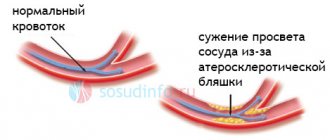
The reasons leading to impaired blood flow are as follows:
- The narrowing of the lumen of blood vessels as a result of the deposition of cholesterol plaques disrupts its elasticity, developing the atherosclerotic process. This often leads to myocardial infarction or stroke;
- Increased formation of blood clots can lead to their separation, migration through the bloodstream and closure of the lumen of the vessel (ischemic stroke).
- Previously suffered traumatic brain injuries, which seem to have ended successfully, can lead to an increase in intracranial pressure, which will also be expressed by manifestations of circulatory disorders.
REG of the brain can determine the presence or absence of a subdural hematoma resulting from traumatic brain injury. The hemorrhage formed in the brain tissue will naturally create an obstacle to the normal flow of blood.
If you don’t get too far ahead, but conduct a study when the symptoms are not clearly expressed and create discomfort from time to time, then the REG of the brain will not only determine the condition of the blood vessels, but will also help you choose tactics to prevent serious consequences that put a person’s life at risk.
In addition, REG shows not only the quality of blood flow through the great vessels, but will also evaluate collateral circulation (when the blood flow through the great vessels is obstructed and it is directed “bypassing”).
Reg and “non-serious” illnesses
There are conditions that, although not fatal, do not allow you to live normally. Now, neurocirculatory dystonia is present in many people, and therefore it is not particularly considered a disease, because “they don’t die from it.”
Or, for example, migraine (hemicrania), considered a whim of society ladies, has safely reached our days and does not leave many women alone.
Headache medications generally do not help unless the medication contains caffeine.
Considering a woman to be absolutely healthy (after all, there are no signs of any illness), those around her often brush it off. And she herself is slowly beginning to consider herself a malingerer, realizing, however, deep down that a head examination would not hurt. Meanwhile, unbearable headaches come monthly and are associated with the menstrual cycle.
A prescribed and performed REG of the head solves the problem in a matter of minutes, and the use of adequate medications relieves the patient of the fear of monthly physiological conditions. But this is a favorable course of the disease, but there is something else...
Few people know that migraine should not be considered frivolous, because not only women suffer from it, and not only at a young age. Men are also sometimes “lucky” in this regard. And the disease can manifest itself to such an extent that a person completely loses his ability to work and needs to be assigned a disability group.
When the need arises to do an REG, patients, as a rule, begin to worry. You can calm down here right away - the method is non-invasive, and therefore painless . The REG procedure does not cause harm to the body and can be performed even in early infancy.
An REG examination of the head is carried out using a 2-6 channel apparatus - a rheograph. Of course, the more channels the device has, the larger the study area will be covered. To solve large problems and record the work of several pools, polyreogreographs are used.
So, the step-by-step REG procedure looks like this:
- The patient is placed comfortably on a soft couch;
- Metal plates (electrodes) are placed on the head, which are previously treated with a special gel to prevent skin irritation;
- The electrodes are attached with a rubber band in the places where it is planned to assess the condition of the vessels.
- Electrodes are applied depending on which part of the brain is subject to REG examination:
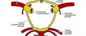
- If the doctor is interested in the internal carotid artery basin, then the electrodes will be placed on the bridge of the nose and mastoid process;
- If it concerns the external carotid artery, then the plates will be strengthened in front of the auditory canal and above the eyebrow from the outside (course of the temporal artery);
- Assessment of the functioning of the vessels of the vertebral artery basin involves the application of electrodes to the mastoid (mastoid) process and the occipital protuberances while simultaneously taking an electrocardiogram.
When examining the head with REG, the patient is advised to close his eyes so that external stimuli do not affect the final result. The data obtained by the device is recorded on paper tape.
The obtained REG results, the decoding of which requires additional skills, are sent to a doctor who has undergone special training in this area. However, the patient is very eager to find out what is going on in his vessels and what the graph on the tape means, because, as REG is done, he already has a good idea and can even reassure those waiting in the corridor.
In some cases, to obtain more complete information about the function of blood vessels, tests with drugs that act on the vascular wall (nitroglycerin, caffeine, papaverine, aminophylline, etc.) are used.
What do incomprehensible words mean: decoding REG
When a doctor begins to decipher the REG, he is first of all interested in the patient’s age, which must be taken into account to obtain adequate information.
Of course, the standards for tone and elasticity will be different for a young and an elderly person.
The essence of REG is to record waves that characterize the filling of certain areas of the brain with blood and the reaction of blood vessels to blood filling.
A brief description of the graphical representation of oscillations can be presented as follows:
- The ascending line of the wave (anacrotic) sharply tends upward, its top is slightly rounded;
- Descending (catacrota) goes down smoothly;
- An incisura located in the middle third, followed by a small dicrotic tooth, from where the descending wave descends and a new wave begins.
To decipher the REG, the doctor pays attention to:
- Are the waves regular?
- What is the top and how is it rounded;
- What the components look like (ascending and descending);
- Determines the location of the incisura, dicrotic tooth and the presence of additional waves.
Common types according to REG
After analyzing the rheoencephalography recording, the doctor records the deviation from the norm and makes a conclusion, which the patient strives to quickly read and interpret. The result of the study is to determine the type of vascular behavior:
- The dystonic type is characterized by a constant change in vascular tone, where hypotonicity with reduced pulse filling often predominates, which may be accompanied by difficulty in venous outflow;
- The angiodystonic type differs little from the dystonic type. It is also characterized by disturbances in vascular tone due to a defect in the structure of the vascular wall, leading to a decrease in the elasticity of blood vessels and complicating blood circulation in a certain pool;
- The hypertensive type according to REG is somewhat different in this regard; here there is a persistent increase in the tone of the afferent vessels with obstructed venous outflow.
Types of REG cannot be classified as separate diseases, because they only accompany another pathology and serve as a diagnostic guide for determining it.
The difference between REG and other brain studies
Often, when signing up for an REG head examination at medical centers, patients confuse it with other studies that contain the words “electro,” “graphy,” and “encephalo” in their names. This is understandable, all the designations are similar and sometimes it is difficult for people who are far from this terminology to understand.
Electroencephalography (EEG) is especially useful in this regard. That’s right, both study the head by applying electrodes and recording data on the work of some area of the head on a paper tape.
The differences between REG and EEG are that the first studies the state of blood flow, and the second reveals the activity of neurons in some part of the brain.
Vessels have an indirect effect during EEG, but long-term circulatory disorders will be reflected in the encephalogram. Increased convulsive readiness or other pathological focus on the EEG is clearly detected, which serves for the diagnosis of epilepsy and convulsive syndromes associated with trauma and neuroinfection.
Symptoms
The signs and characteristic manifestations of increased and decreased vascular tone are significantly different.
Hypotonicity
The main symptom of hypotonicity of the cerebral arteries is a dull, pressing, bursting headache, which is localized in the back of the head and temples.
The general condition is disturbed: a person is worried about weakness, malaise, which arise for no apparent reason. Mood and emotional state changes. A person may suffer from insomnia, and patients with hypotension often experience vomiting. Shortness of breath and arrhythmia develop even with minor physical activity.
Sweating increases, dizziness occurs, especially when getting up from a support.
Hypertonicity
In most cases, hypotension (hypertension) begins sluggishly, without obvious symptoms. A person attributes the first symptoms to banal overwork. Characteristic signs of hypotonicity at the initial stage include general malaise, the appearance of “goosebumps” before the eyes, and a low-intensity headache.
Additional clinical manifestations of hypertension include:
- increased sweating;
- redness of the skin on the face;
- swelling of the hands that occurs in the morning;
- memory impairment.
As hypertension progresses, painful headaches, dizziness, and chest pain occur. The risk of stroke increases and visual function is impaired.
In severe cases, if treatment measures are not followed, angina pectoris, heart failure, myocardial infarction, arrhythmia and other dangerous pathologies may develop.
How reg differs from other brain studies
Often, when signing up for an REG head examination at medical centers, patients confuse it with other studies that contain the words “electro,” “graphy,” and “encephalo” in their names. This is understandable, all the designations are similar and sometimes it is difficult for people who are far from this terminology to understand.
Electroencephalography (EEG) is especially useful in this regard. That’s right, both study the head by applying electrodes and recording data on the work of some area of the head on a paper tape.
The differences between REG and EEG are that the first studies the state of blood flow, and the second reveals the activity of neurons in some part of the brain.
Vessels have an indirect effect during EEG, but long-term circulatory disorders will be reflected in the encephalogram. Increased convulsive readiness or other pathological focus on the EEG is clearly detected, which serves for the diagnosis of epilepsy and convulsive syndromes associated with trauma and neuroinfection.
Help me understand the results of REG and RVG
Diabetes mellitus and its treatment
We were examined because we are going to a sanatorium
help me figure out what this all means
pulse arterial blood filling is normal // normal
tone of the main arteries—————-increased 2% // normal
total tone of regional arteries—increased 11% // increased 7%
tone region. arteries large caliber— —reduced 208%//reduced 196%
tone region. arteries average caliber— —reduced 58% // reduced 57%
dicrotic ind (tone of small-caliber arteries - normal//increased 21%
pulse arterial blood filling is normal // reduced 6%
tone of the main arteries——————norm // increased 16%
total tone of regional arteries———norm // increased 13%
tone region. arteries large caliber— —reduced 126% // reduced 72%
tone region. arteries average caliber——reduced 38% // norm
dicrotic.ind (tone of small-caliber arteries - reduced 60% // reduced 72%
These methods have very low diagnostic value and do not provide insight into the nature of blood flow.
Thank you very much, if it’s not too much trouble, please tell me what we need to pay attention to regarding our experience of illness. Dr.Anna
Pulse blood flow is slightly reduced on the right, moderately reduced on the left.
PC asymmetry is insignificant
The tone of arterioles and precapillaries is insignificant. elevated on the right, moderately elevated on the left.
We suggest you read: Normal hemoglobin levels during pregnancy
Hypotension of the venous network: no.
Venous outflow is preserved.
Blood circulation means. failure.
There is an increase in the time to reach the PV of the segment under study.
Pulse blood flow is normal on the right, moderately reduced on the left.
The tone of arterioles and precapillaries is moderate and increased.
Hypotension of the venous network - no.
Venous outflow is preserved on the right, preserved (the phenomena of venous stagnation are insignificant) on the left.
Even before undergoing RVG, Vitalik visited a vascular surgeon (before undergoing VTEK). He prescribed him the following medications: Tiklo: 250 * 2 times - 1 month
Mexicor 2.0 No. 10 intramuscular, then tablets 10 mg *2 rubles. 1 month
I didn’t see the doctor about the RVG results.
Judging by the conclusion of the RVG, is it still relevant to take these medications? And is it even worth taking them?
Treatment and therapy
To treat hypotension of cerebral vessels, an integrated approach and combination therapy are used. A course of treatment is usually prescribed to patients only after examination by a cardiologist and neurologist. After examining the patient and collecting anamnesis, a full clinical examination should be carried out, which includes the following tests:
- General and biochemical blood tests.
- Study of blood viscosity and coagulation rate.
- Dopplerography of cerebral vessels.
- Angiography of blood vessels.
Patients with hypotension should follow a diet, lead a healthy lifestyle, move more, and exercise. Among the medications used in the treatment of cerebral vascular hypotension, the following methods are used:
- Medications containing caffeine are used as first aid. They are used for a short period of time because they have only a symptomatic effect, but they are not used for permanent therapy.
- Nootropic drugs are used as maintenance therapy, which include aminalon, nootropil, phenibut and others.
- For depression and migraine-like pain, the drugs encephabol, stugeron, tanakan are prescribed, which accelerate blood flow and tone the walls of blood vessels.
- Herbal preparations have a good therapeutic effect for hypotension, of which ginseng root, Rhodiola rosea extract, Eleutherococcus tincture, liquid Leuzea extract and others are often used.
- Treatment with amino acids is used as nutrition and building material for blood vessels, many of which have the properties of neurotransmitters (substances that transmit signals between cells of the nervous system). Among the amino acids, glycine, glutamic and gamma-aminobutyric acids are used.
Problem in newborns
Pathologically altered vascular tone in infants is a dangerous phenomenon, which, if treatment is not started in a timely manner, can be fatal or cause the child to be assigned a disability group.
The most common consequence of insufficient oxygen supply to the brain with impaired vascular tone is cerebral ischemia. In this case, brain cells are inhibited due to insufficient blood supply, including oxygen. In approximately 40% of cases, the newborn dies.
To date, there is no specific treatment for cerebral ischemia in infants. Despite this, maintenance therapy can be started, which helps prevent complications and reduce the intensity of clinical manifestations.
Oxygen starvation can begin while the fetus is in the womb or during labor. During the prenatal period, this process is influenced by the following factors:
- alcohol and smoking abuse by a pregnant woman;
- development during pregnancy of an infectious disease, pathology of an endocrine nature, ARVI;
- age of the pregnant woman (before 18 years and after 35);
- high degree toxicosis in the third trimester.
Problems during labor also affect the condition of the baby’s brain. This:
- large fruit;
- use of drug stimulation of labor;
- premature birth;
- complications during childbirth, including birth injuries;
- entwining the fetus with the umbilical cord.
How is the functioning of the blood vessels in the head analyzed?
Indications for REG are determined by conditions in which the parameters of the tone and elasticity of the arteries and veins are disturbed or their asymmetry is observed in similar areas. With rheoencephalography, you can obtain indirect confirmation of increased intracranial pressure and assess the state of collateral (bypass) blood circulation (if the main vessel is blocked). Usually REG is prescribed when
- headaches, tinnitus, dizziness;
- suspected atherosclerosis of the vessels of the head;
- unstable and high blood pressure;
- strokes (to assess the collateral blood supply to the affected area of the brain);
- traumatic brain injuries;
- suspected increased intracranial pressure;
- osteochondrosis of the cervical spine
- migraine;
- memory disorders;
- sleep problems;
- hearing or vision impairment.
Rheoencephalography is possible for dynamic monitoring of the effectiveness of treatment and for the purpose of identifying pathology during preventive examinations of certain population groups.
As the disease progresses, it causes a decrease in the elasticity of the arteries, a narrowing of their lumen and a decrease in blood supply. On the REG there is a gradual smoothing of the top of the curve with the formation of a plateau. Over time, the shape becomes arched. The wave propagation time and its amplitude are shortened.
In the early stages, the dicrotic tooth moves toward the apex. The development of the disease leads to a rounding of the apex, and the dicrotic tooth may be located above the bend. The amplitude of the wave decreases. At a later stage, the shape of the curve is arched.
REG for migraine
Interhemispheric asymmetry in wave amplitude is recorded - it is increased on the affected side.
https://www.youtube.com/watch?v=upload
The course of the disease according to the hypertensive type on the REG is accompanied by a tendency to rounding the top of the wave, reducing the amplitude and smoothing out additional fluctuations.
If vascular tone is predominantly reduced, increased blood supply is observed. At the top of the REG curve, a plateau forms, the amplitude of the wave increases, and additional fluctuations become more noticeable.
- Hematoma. Causes significant asymmetry in the shape of the curve. On the side of the hematoma, the wave amplitude is reduced. Additional vibrations are almost invisible.
- Brain contusion. On the affected side, the amplitude of the wave is increased, the incisura becomes deeper and the angle of inclination of the ascending part of the REG curve increases.
- Concussion. Signs of vascular hypertonicity or hypotonicity without asymmetry are recorded.
Additional treatment
How to improve vascular tone? Treatment methods may differ depending on whether a person has a decrease or increase in tone. To determine the tone, appropriate studies are carried out (rheoencephalography, etc.), based on the results of which a final diagnosis is made (hypotension or hypertension).
Hypertonicity
It is recommended to start therapy for hypertension as early as possible, before the pathology has time to become chronic. To strengthen the walls of blood vessels, you need to follow some simple recommendations:
- giving up bad habits (smoking, drinking alcohol);
- taking preventive courses of cervical-collar massage;
- maintaining proper nutrition;
- timely cure of infections;
- maintaining an active lifestyle.
Physical activity should be moderate. You should not overload the body and lift weights, because this affects not only the blood vessels, but also all other organs and systems.
Among the medications to eliminate hypertension are Papazol, Eufillin, Revalgin, used intravenously. The medicine Papaverine is often used. Adaptogens, nootropics, and vasoactive calcium antagonists are prescribed. First aid medications for headaches - Spazgan, Nurofen, Spazmalgon.
Hypertonicity is also treated with cool baths, which help relax smooth muscles. The patient’s diet, which should be low in calories, is important.
REG and “non-serious” diseases
There are conditions that, although not fatal, do not allow you to live normally. Now, neurocirculatory dystonia is present in many people, and therefore it is not particularly considered a disease, because “they don’t die from it.” Or, for example, migraine (hemicrania), considered a whim of society ladies, has safely reached our days and does not leave many women alone. Headache medications generally do not help unless the medication contains caffeine.
Considering a woman to be absolutely healthy (after all, there are no signs of any illness), those around her often brush it off. And she herself is slowly beginning to consider herself a malingerer, realizing, however, deep down that a head examination would not hurt. Meanwhile, unbearable headaches come monthly and are associated with the menstrual cycle.
A prescribed and performed REG of the head solves the problem in a matter of minutes, and the use of adequate medications relieves the patient of the fear of monthly physiological conditions. But this is a favorable course of the disease, but there is something else...
Few people know that migraine should not be considered frivolous, because not only women suffer from it, and not only at a young age. Men are also sometimes “lucky” in this regard. And the disease can manifest itself to such an extent that a person completely loses his ability to work and needs to be assigned a disability group.