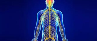
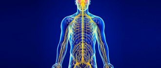
The peripheral nervous system connects the central nervous system to the organs and limbs. Unlike the central nervous system, the peripheral nervous system is not protected by bones and has no physiological barrier separating it from the circulatory system. Therefore, the peripheral nervous system may be susceptible to mechanical damage and is more easily affected by toxins.
Diseases of peripheral nerves are neuropathies. They are characterized by damage to the axons and myelin sheath of the nerves. Diseases of the nerve trunks and plexuses rank second in prevalence among the population after diseases of the spinal roots (“radiculitis”). Therefore, issues of prevention, early diagnosis and subsequent treatment of neuropathies remain pressing problems.
Types
- Demyelinating. The conduction of excitation through neurons is disrupted. Occurs in cases of lead poisoning, diphtheria, polyradiculoneuropathy, and diabetic polyneuropathy. The patient's health is restored within a few weeks if treatment is started in a timely manner.
- Axonopathies. In this case, the damage concerns the axons (the processes of nerve cells) - these are severe disorders of nerve function. As a result, muscle atrophy occurs. The cause of these disorders is the abuse of alcohol and other toxic substances.
Neuropathies of mixed origin are most common. Full recovery and restoration of nerve function depends in part on the severity of the damage.
If the recovery process is absent 3 months after the onset of the disease, then the prognosis is most often unfavorable.
Types of neuropathy
- Mononeuropathies. One nerve or a specific part of the nerve plexus is injured. The causes of damage can be trauma, compression of any level of the nerve trunk. Also, mononeuropathies are observed in diabetes mellitus, atherosclerosis, vascular damage, etc. Hypothermia and herpetic infections are not the least important factors in the functioning of one nerve.
- Multifocal neuropathies are a syndrome of partial damage to individual nerve trunks or their complete damage. Such neuropathies occur slowly and sequentially (from several days to several years). Causes: arthritis, vasculitis and a number of systemic connective tissue diseases.
- Polyneuropathy. Lesions of peripheral nerves are multiple. Moreover, the process is widespread and symmetrical. It occurs both acutely and chronically. It happens that the spinal roots are also affected.
The causes of diseases of the peripheral nerve trunks and plexuses can be:
- injuries received
- decrease in normal immune system function
- hereditary diseases
- intoxication with various substances
- infections
- lack of vitamins
- habitual intoxication (drug addiction and alcoholism)
- allergies
Names of certain nerves[edit]
Ten of the twelve cranial nerves arise from the brain stem and, with a few exceptions, primarily control the functions of the anatomical structures of the head. The nuclei of cranial nerves I and II lie in the forebrain and thalamus, respectively, so they cannot be considered truly cranial nerves. The tenth nerve viscerally receives sensory information from the chest and abdomen, and the 11th nerve is responsible for innervation of the sternocleidomastoid and trapezius muscles, neither of which are located entirely in the head.
Spinal nerves originate in the spinal cord and control the functions of the rest of the body. Humans have 31 pairs of spinal nerves: 8, 12 and 5, 5 sacral and 1 coccygeal. In the cervical region, the spinal nerves originate above the corresponding vertebra (that is, the nerve originating between the skull and the first cervical vertebra is called the first spinal nerve). From the thoracic region to the coccygeal nerves begin below the corresponding vertebrae
It is important to note that this method creates problems in the case of naming a spinal nerve that originates between the seventh superior and the first inferior (the so-called eighth spinal nerve). In the lumbar and sacral regions, the root ends of the nerves are located within the dural sac.
Symptoms
With neuropathies, symptoms can be different and depend on the affected areas. They cause a lot of discomfort to the patient:
- flaccid muscle paralysis
- pain in limbs
- change in skin sensitivity (there may be a contrast in sensations in one area of the skin compared to another)
- no sensation of pain and more
- amyotrophy
- speech disorder
- feeling of numbness in the face and limbs
- muscle weakness of the limbs
- impaired motor coordination
- dry skin
- focal blanching
- redness and bluish discoloration of the injured area
- Facial asymmetry may occur (if the facial nerves are damaged)
If you find yourself with at least one of the described symptoms, you should immediately seek help from a qualified neurologist.
Autonomic nervous system
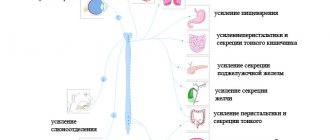
The ANS includes two divisions, the sympathetic (SNS) and the parasympathetic (PNS). The SNS is activated during a stressful situation. It increases the heart rate, constricts blood vessels, pupils, increases blood flow to the muscles and outflow from the gastrointestinal tract. The center of the SNS is located in the thoracic and lumbar regions of the spinal cord (Fig. 9). The PNS has the opposite effect. It is activated in a calm environment and leads to a rush of blood to the gastrointestinal tract, outflow from the muscles, a decrease in heart rate, pupil dilation, etc. PNS centers are located in the medulla oblongata, some nuclei of the cranial nerve and the sacral spinal cord.
The main difference between the autonomic reflex arc and the somatic one is the presence of another synaptic switch in the ganglion after the spinal cord. Thus, the autonomic reflex begins from the receptor, then the sensitive neuron from the ganglion transmits information to the neuron of the middle horns of the spinal cord (or another center of the ANS). The axon of the autonomic neuron exits through the anterior roots and goes to the ganglion, where it forms a synapse with the ganglion neuron, the process of which goes directly to the effector organ. The nerve fiber running from the spinal cord to the ganglion is called preganglionic. The nerve fiber running from the ganglion to the organ is called postganglionic. The ganglia of the SNS are located next to the spinal cord, so the preganglionic fiber is short and the postganglionic fiber is long. The ganglia of the PNS are located near or in the wall of the organ, so their preganglionic fiber is long and the postganglionic fiber is short. The effector neurotransmitter of the sympathetic nervous system is norepinephrine, and the parasympathetic nervous system is acetylcholine.
Rice. 9. Effects of the SNS and PNS.
Rice. 10. Comparison of the reflex arc of the somatic and autonomic reflex.
# Human anatomy
Treatment
In our clinic, traditional and alternative medicine methods are used to treat neuropathies. Complex treatment is selected only individually and depends entirely on the degree of damage to the nerve(s).
Initial treatment will be aimed at restoring the functions of the peripheral nerves, therefore, the cause will be eliminated.
An integrated approach is important in the treatment of neuropathies! Our specialists will take into account all the nuances of the damage and may prescribe:
- medications that improve metabolism, blood circulation and restoration processes in nervous tissue. Medicines can also be used in injection form, including intravenous drips in a cozy day hospital
- hormonal drugs (steroids) - in some cases
- therapeutic drug blockades
- certain types of physiotherapeutic treatment
- manual therapy (osteopathy)
- classic massage
Diagnostics
Before starting treatment for any disease, it is necessary to understand its cause. In the case of neuritis and neuralgia, the diagnosis is often made after a routine examination by a neurologist, since the symptoms of the disease of each nerve are often unique. To confirm the suspicion, the doctor may do some reflex tests.
But to identify the cause and extent of the disease, a certain examination will be needed:
- General tests to identify inflammatory processes and possible pathogens, metabolic disorders.
- Examination for circulatory pathologies.
- Ultrasound, x-ray, tomography to detect a physical cause that can cause nerve disease.
- Electromyography is a study of nerve conduction to determine the degree of destruction of its structure, as well as the specific location.
Recommendations and prevention
The main prevention of most diseases of the nervous system is maintaining a healthy and active lifestyle, giving up bad habits, timely and adequate treatment of infectious and non-infectious diseases.
If any neurological symptoms occur, do not delay contacting a doctor. Early diagnosis and timely treatment will help prevent the development of complications, prolongation of treatment and the consequences of uncontrolled use of medications. The most complete program of preventive measures is drawn up by a neurologist for each individual patient.
Stages of nervous system development
The diffuse nervous system is the most ancient, found in coelenterates (hydra). Such a nervous system is characterized by a multiplicity of connections between neighboring elements, which allows excitation to freely spread throughout the nervous network in all directions.
This type of nervous system provides wide interchangeability and thereby greater reliability of functioning, but these reactions are imprecise and vague.
The nodal type of nervous system is typical for worms, mollusks, and crustaceans.
It is characterized by the fact that the connections of nerve cells are organized in a certain way, excitation passes along strictly defined paths. This organization of the nervous system turns out to be more vulnerable. Damage to one node causes dysfunction of the entire organism as a whole, but its qualities are faster and more accurate.
The tubular nervous system is characteristic of chordates; it includes features of the diffuse and nodular types. The nervous system of higher animals took all the best: high reliability of the diffuse type, accuracy, locality, speed of organization of nodal type reactions.
FAQ
Three fingers on my right hand are numb and painful. What to do?
It is necessary to consult a neurologist to exclude carpal tunnel syndrome and treatment.
I had an attack of trigeminal neuralgia for a long time. Should I take something preventative to prevent another one?
It is imperative to see a neurologist. The doctor will select medications for you that need to be taken on a regular basis and prescribe preventive courses of injections (if necessary, drips in a day hospital) and tablet medications.
Severe burning pain appeared in the right half of the chest and some bubbles appeared. Which doctor should I see?
Similar manifestations, as you describe, can be observed with herpetic ganglioneuritis, the so-called. herpes zoster. This disease is treated by a neurologist.
After about 2 months, my legs began to go numb and weak. Why could this be?
Weakness and sensory disturbances in the limbs can be observed with polyneuropathies of various natures. For example, as a result of diabetes or intoxication. To establish the cause and select adequate therapy, you need to consult a neurologist. There may be a need for additional laboratory and instrumental examinations.
I developed weakness in my right hand after sleeping. Is it from the spine?
The cause of sudden weakness in the arm may also be diseases of the spine. However, a vascular cause or compression of the nerve trunk (nerve plexus) cannot be excluded. To find out the cause and prescribe treatment, you should consult a neurologist.
Can alcoholic neuropathy be cured?
Toxic and dysmetabolic polyneuropathies, incl. alcoholic diseases are most often chronic diseases. With adequate therapy, it is possible to achieve long-term and stable remission
AUTONOMIC NERVOUS SYSTEM
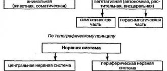
The autonomic, or autonomic, nervous system regulates the activity of involuntary muscles, the heart muscle, and various glands. Its structures are located both in the central nervous system and in the peripheral nervous system. The activity of the autonomic nervous system is aimed at maintaining homeostasis, i.e. a relatively stable state of the body's internal environment, such as a constant body temperature or blood pressure that meets the body's needs.
Signals from the central nervous system enter the working (effector) organs through pairs of sequentially connected neurons. The bodies of neurons of the first level are located in the CNS, and their axons end in the autonomic ganglia, which lie outside the CNS, and here they form synapses with the bodies of neurons of the second level, the axons of which are in direct contact with the effector organs. The first neurons are called preganglionic, the second - postganglionic.
In the part of the autonomic nervous system called the sympathetic nervous system, the cell bodies of preganglionic neurons are located in the gray matter of the thoracic (thoracic) and lumbar (lumbar) spinal cord. Therefore, the sympathetic system is also called the thoracolumbar system. The axons of its preganglionic neurons terminate and form synapses with postganglionic neurons in ganglia located in a chain along the spine. Axons of postganglionic neurons contact effector organs. The endings of postganglionic fibers secrete norepinephrine (a substance close to adrenaline) as a neurotransmitter, and therefore the sympathetic system is also defined as adrenergic.
The sympathetic system is complemented by the parasympathetic nervous system. The bodies of its preganglinar neurons are located in the brainstem (intracranial, i.e. inside the skull) and the sacral (sacral) part of the spinal cord. Therefore, the parasympathetic system is also called the craniosacral system. The axons of preganglionic parasympathetic neurons terminate and form synapses with postganglionic neurons in ganglia located near the working organs. The endings of postganglionic parasympathetic fibers release the neurotransmitter acetylcholine, on the basis of which the parasympathetic system is also called cholinergic.
As a rule, the sympathetic system stimulates those processes that are aimed at mobilizing the body's forces in extreme situations or under stress. The parasympathetic system contributes to the accumulation or restoration of the body's energy resources.
The reactions of the sympathetic system are accompanied by the consumption of energy resources, an increase in the frequency and strength of heart contractions, an increase in blood pressure and blood sugar, as well as an increase in blood flow to the skeletal muscles by reducing its flow to the internal organs and skin. All of these changes are characteristic of the “fear, flight or fight” response. The parasympathetic system, on the contrary, reduces the frequency and strength of heart contractions, lowers blood pressure, and stimulates the digestive system.
The sympathetic and parasympathetic systems act in a coordinated manner and cannot be viewed as antagonistic. They jointly support the functioning of internal organs and tissues at a level corresponding to the intensity of stress and the emotional state of a person. Both systems function continuously, but their activity levels fluctuate depending on the situation. See also HEART; EMOTION.
Treatment stories
Case No. 1
Patient Yu., 40 years old, noticed sudden facial asymmetry, lacrimation from the right eye and incomplete closure of the eyelids. I contacted a neurologist at the EXPERT Clinic. The patient was diagnosed with acute neuritis of the facial nerve and prescribed treatment and examination. The patient completed a course of intramuscular and intravenous drip injections in the day hospital of the EXPERT Clinic, courses of physical therapy and acupressure self-massage. Movements in the facial muscles have been completely restored. During the examination, the oncological nature of the disease was excluded, but its herpetic nature was revealed against the background of a secondary immunodeficiency state. After consulting an immunologist, patient Yu was prescribed a course of immunomodulatory therapy.
What is the central nervous system
The central nervous system (CNS) is the part of the vertebrate nervous system that coordinates sensory impulses and their corresponding responses in the body. The CNS includes the brain and spinal cord. The central nervous system can be roughly divided into gray and white matter. The outer cortex of the brain is made up of gray matter, while the inner region is made up of white matter. Gray matter consists of neurons, and white matter mainly consists of nerve axons. The retina, optic nerve, olfactory epithelium and olfactory nerves also belong to the central nervous system.
Figure 1: Central nervous system
Brain
The brain is made up of 100 billion nerve cells that are protected by the skull and protective membranes called meninges. The cells that support neurons in the brain are called glial cells or neuroglia. Astrocytes, oligodendrocytes, ependymal cells and radial glia are found in the CNS as glial cells. The brain can be divided into four lobes: frontal, occipital, parietal and temporal. The frontal lobes are responsible for voluntary body movements. The occipital lobes receive visual impulses from the eye. The parietal lobes receive sensory information such as temperature, touch, taste, and pain. The temporal lobes are responsible for memory and hearing. The brain initiates voluntary body movements.
Spinal cord
The spinal cord is protected by the vertebral column, which begins at the base of the brain. The main function of the spinal cord is to communicate with the brain and peripheral nerves. The spinal cord consists of eight cervical segments, twelve thoracic segments, five lumbar segments, five sacral segments and one coccygeal segment in humans.
