Sympathetic trunk
The sympathetic trunk (truncus sympathicus) is paired, formed by nodes connected by sympathetic fibers. The sympathetic trunk is located on the lateral surface of the spine along its entire length. Each node of the sympathetic trunk represents a cluster of autonomic neurons, with the help of which most of the preganglionic fibers are switched, emerging from the spinal cord and forming the white connecting branches (rr. communicantes albi). Preganglionic fibers contact vegetative cells in the corresponding node or are sent as part of internodal branches to the superior or inferior nodes of the sympathetic trunk. The white connecting branches are located in the thoracic and upper lumbar regions. There are no such connecting branches in the cervical, sacral and lower lumbar nodes. The nodes of the sympathetic trunk are also connected by special fibers to the spinal nerves - the gray connecting branches (rr. communicantes grisei), consisting mainly of postganglionic sympathetic fibers. The gray connecting branches extend from each node of the sympathetic trunk to each spinal nerve, within which they are directed to the periphery, reaching the innervated organs - striated muscles, smooth muscles and glands.
The sympathetic trunk is conventionally divided into cervical, thoracic, lumbar and sacral sections.
The cervical sympathetic trunk includes three nodes: superior, middle and inferior.
The upper node (gangl. cervicale superius) has a spindle-shaped shape measuring 5x20 mm. Located on the transverse processes of the II-III cervical vertebrae, covered with prevertebral fascia. Seven main branches depart from the node, containing postganglionic fibers to innervate the organs of the head and neck.
1. Gray connecting branches to the I, II, III cervical spinal nerves.
2. The jugular nerve (n. jugularis) is divided into two branches, the fibers of which join the vagus and glossopharyngeal nerves in the region of their lower nodes, and into a branch, the fibers of which join the hypoglossal nerve.
3. The internal carotid nerve (n. caroticus internus) penetrates the adventitia of the internal carotid artery, where its fibers form the plexus of the same name. From the plexus of this artery at the site of its entry into the carotid canal of the temporal bone, sympathetic fibers are separated, forming the deep petrosal nerve (n. petrosus profundus), passing into the pterygoid canal (canalis pterygoideus) of the sphenoid bone. Having left the canal, they pass through the pterygopalatine fossa, connecting to the postganglionic parasympathetic nerves of the pterygopalatine ganglion and the sensory nerves n. maxillaris, and diverge to the facial organs. Branches extend from the internal carotid plexus in the carotid canal, penetrating into the tympanic cavity, participating in the formation of the plexus of the tympanic cavity (plexus tympanicus). In the cranial cavity, the continuation of the internal carotid plexus is the cavernous one, the fibers of which are distributed along the branches of the cerebral vessels, forming the plexus of the anterior, middle cerebral arteries (plexus arteriae cerebri anterior et medius), as well as the plexus of the ophthalmic artery (plexus ophthalmicus). Branches extend from the cavernous plexus and pass into the ciliary parasympathetic ganglion (gangl. ciliare), connecting to its parasympathetic fibers to innervate the muscle that dilates the pupil (m. dilatator pupillae).
4. The external carotid nerve (n. caroticus externus) is thicker than the previous one. Around the artery of the same name, it forms an external plexus (plexus caroticus externus), from which the fibers are distributed to all its arterial branches, supplying blood to the facial part of the head, dura mater and neck organs.
5. The laryngopharyngeal branches (rr. laryngopharyngei) are distributed along the vessels of the pharyngeal wall, forming the pharyngeal plexus (plexus pharyngeus).
6. The superior cardiac nerve (n. cardiacus superior) is sometimes absent on the right and descends next to the cervical section of the sympathetic trunk. In the chest cavity, it participates in the formation of the superficial cardiac plexus, located under the aortic arch.
7. The branches that make up the phrenic nerve end in the pericardium, pleura, diaphragm, parietal peritoneum of the diaphragm, ligaments and liver capsule.
The middle node (gangl. cervicale medium), measuring 2×2 mm, is located at the level of the VI cervical vertebra at the intersection of the inferior thyroid and common carotid arteries; often absent. Four types of branches extend from this node:
1. Gray connecting branches to the V and VI cervical spinal nerves.
2. Middle cardiac nerve (n. cardiacus medius), located behind the common carotid artery. In the chest cavity, it takes part in the formation of the deep cardiac plexus, located between the aortic arch and the trachea.
3. Branches involved in the formation of the nerve plexus of the common carotid and subclavian arteries, as well as the plexus of the inferior thyroid artery. Autonomic plexuses are formed in these organs.
4. Internodular branch to the superior cervical sympathetic node.
The lower node (gangl. cervicale inferius) is located above the subclavian artery and behind the vertebral artery. Sometimes it connects with the first thoracic sympathetic node and is called the cervicothoracic (stellate) node (gangl. cervicothoracicum s. stellatum). 6 branches extend from the lower node.
1. Gray connecting branches to the VII and VIII cervical spinal nerves.
2. Branch to the plexus of the vertebral artery (plexus vertebralis), which extends into the skull, where it forms the basilar plexus and the plexus of the posterior cerebral artery.
3. Lower cardiac nerve (n. cardiacus inferior), located on the left behind the aorta, on the right - behind the brachiocephalic artery; takes part in the formation of the deep plexus of the heart.
4. Branches to the phrenic nerve do not form a plexus. Reach the pleura, pericardium and diaphragm.
5. Branches to the plexus of the common carotid artery (plexus caroticus communis).
6. Branches to the subclavian artery (plexus subclavius).
Thoracic nodes (ganglia thoracica) are located on the sides of the thoracic vertebrae on the necks of the ribs, covered with the parietal pleura and intrathoracic fascia (f. endothoracalis). The thoracic sympathetic ganglia have mainly six groups of branches:
1. The white connecting branches enter the nodes from the anterior roots of the intercostal nerves (Fig. 532).
2. Gray connecting branches extend from the nodes to the intercostal nerves.
3. Mediastinal branches (rr. mediastinales) start from the V superior sympathetic nodes and enter the region of the posterior mediastinum. They take part in the formation of the esophageal and bronchial plexuses.
4. Thoracic cardiac nerves (nn. cardiaci thoracici) start from the IV-V superior sympathetic nodes and are part of the deep cardiac plexus and the thoracic aortic plexus.
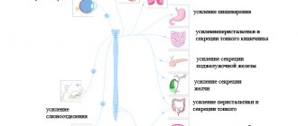
5. The great splanchnic nerve (n. splanchnicus major) is formed from the branches of the V-IX thoracic sympathetic nodes. The nerve is located under the intrathoracic fascia. Through the hole between the medial and intermediate crura of the diaphragm, the large splanchnic nerve penetrates the abdominal cavity, ending in the celiac plexus nodes. The nerve contains a large number of preganglionic fibers, which switch in the nodes of the celiac plexus to postganglionic fibers, and fewer postganglionic fibers, which have already switched in the thoracic nodes of the sympathetic trunk.
6. The small splanchnic nerve (n. splanchnicus minor) is formed from the branches of the X-XII nodes. It descends through the diaphragm lateral to the greater splanchnic nerve and reaches the celiac plexus. Preganglionic fibers switch to postganglionic fibers in the sympathetic ganglia, and another group of preganglionic fibers, switched in the thoracic ganglia, are sent to the organs.
The lumbar nodes (ganglia, lumbalia) of the sympathetic trunk are a continuation of the chain of nodes of the thoracic part, located between the lateral and intermediate legs of the diaphragm. They include 3-4 nodes located on the sides of the spine on the medial edge of m. psoas major. On the right, the nodes are visible lateral to the inferior vena cava, and on the left, lateral to the aorta. Branches of the lumbar sympathetic ganglia:
1. White connecting branches approach only the I, II nodes from the I and II lumbar spinal nerves.
2. The gray communicating rami connects the lumbar ganglia with all lumbar spinal nerves.
3. Lumbar splanchnic nerves (nn. splanchnici lumbales) from all nodes are connected to the celiac (plexus celiacus), renal (plexus renalis), superior mesenteric (plexus mesetericus superior), abdominal aortic (plexus aorticus) and superior hypogastric (plexus hypogastricus superior) , plexuses.
The sacral nodes (ganglia sacralia) of the sympathetic trunk include 3-4 paired sacral and 1 unpaired coccygeal nodes, which are located medial to the anterior sacral foramina.
1. Gray communicating branches go to the spinal and sacral nerves.
2. The splanchnic nerves (nn. splanchnici sacrales) participate in the formation of the autonomic plexuses of the pelvis. The visceral branches form the inferior hypogastric plexus (plexus hypogastricus inferior), located on the branches of the internal iliac artery; along its branches, the sympathetic nerves reach the pelvic organs.
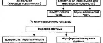
Specifics of the structure of the SNS
The sympathetic nervous system is formed by the sympathetic trunk - a paired row of nerve nodes that are located to the left and right of the spine. The nodes that form the SNS are interconnected by nerve bridges, due to which a kind of network is formed. At the bottom of the spine, 2 rows join to form the coccygeal node (Table 1).
| SNA departments | Number of nerve ganglia |
| Cervical | 3 |
| Chest | 9-12 |
| Lumbar | 2-7 |
| Sacral | 4 |
| Coccygeal | 1 |
The described departments form the central SNS. From them depart peripheral nerve fibers responsible for the activity of internal organs and various physiological functions. Thus, the SNS has a segmental type of structure.
Video about the sympathetic nervous system:
Content
- 1 Morphology 1.1 Central and peripheral sections 1.1.1 Central section
- 1.1.2 Peripheral department
- 2.1 General significance of autonomic regulation
Main functions of SNA
The sympathetic nervous system is responsible for a large number of processes occurring in the human body. With the participation of the SNS, vital forms of activity, especially reflex, are maintained in the body.
The main functions include the following:
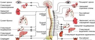
- Increased blood flow to muscle tissue with increasing loads
- Reduced secretory and absorption processes in the gastrointestinal tract
- An increase in the concentration of sugar or cholesterol in the blood
- Regulation of metabolic processes
- Activation of the heart muscle in response to lack of oxygen in tissues
- Transmission of nerve impulses to the spinal cord
- Reaction of the eye pupils
- Release of neurotransmitters into the blood, including under the influence of stress factors
- Activation of the process of processing nutrients into energy
- Control of the secretory functions of the salivary glands
- Changes in the size of the lumen of blood vessels
- Bronchial dilatation
Cerebral palsy ICD 10: classification of cerebral palsy
In addition to the processes described above, the SNS is also responsible for sweating and the expansion of skin pores. The sympathetic department is involved in the breakdown of complex carbohydrates in the liver and provokes relaxation of smooth muscles. Due to the work of the SNS, involuntary urination is prevented, as the urinary sphincter contracts.
During the period of activation, the SNS performs trophic functions. Due to this, the muscle tissues that ensure human mobility receive more oxygen. This allows you to increase your performance, and in the event of extreme situations or exposure to stress, a person does not experience physical fatigue. The function of the SNS, due to which the body quickly adapts to unfavorable factors, is called adaptive.