The human body is a multi-stage structure, each organ and system of which is closely interconnected with each other and with the environment. And so that this connection is not interrupted even for a split second, a nervous system is provided - a complex network that permeates the entire human body and is responsible for self-regulation and the ability to adequately respond to external and internal stimuli. Thanks to the well-coordinated work of the nervous system, a person can adapt to the factors of the external world: any, even minor, change in the environment causes nerve cells to transmit hundreds of impulses at an incredibly high speed so that the body can instantly adapt to new conditions. Internal self-regulation works in a similar way, in which the activity of cells is coordinated in accordance with current needs.
The functions of the nervous system affect the most important processes of life, without which the normal existence of the body is unthinkable. These include:
- regulation of the work of internal organs in accordance with external and internal impulses;
- coordination of all units of the body, from the smallest cells to organ systems;
- harmonious interaction between humans and the environment;
- the basis of higher psychophysiological processes characteristic of humans.
How does this complex mechanism work? What cells, tissues and organs make up the human nervous system and what is each of its sections responsible for? A brief excursion into the basic anatomy and physiology of the human body will help you find answers to these questions.
Organization of the human nervous system
Nerve cells cover the entire body, forming an extensive network of fibers and endings. This system, on the one hand, unites every cell of the body, forcing it to work in one direction, and on the other hand, it integrates a particular person into the environment, balancing his needs with external factors. The nervous system ensures the normal processes of digestion, respiration, blood circulation, the formation of immunity, metabolism, etc. - in a word, everything without which normal life activity is unthinkable.
The effectiveness of the nervous system depends on the correct formation of the reflex - the body's response to irritation. Any impact, be it external changes or internal imbalance, triggers a chain of impulses that instantly affect the body, and it, in turn, forms a response. Thus, the human nervous system forms the unity of the tissues, organs and systems of the human body with each other and with the outside world.
The entire nervous system consists of millions of nerve cells - neurons, or neurocytes, each of which has a body and several processes.
The classification of neuron processes depends on what function it performs:
- the axon sends a nerve impulse from the body of the neuron to another nerve cell or the final target of the chain - a tissue or organ that must perform a certain action;
- The dendrite receives the sent impulse and leads it to the body of the neuron.
Due to the fact that each nerve cell is polarized, the chain of nerve impulses never changes direction, falling into the right direction. In this way, each nerve impulse moves forward, initiating the work of muscles, internal organs and systems.
Types of nerve cells
Before considering the nervous system as a whole, it is necessary to understand what functional units it consists of. The NS includes:
- Sensory neurons. Located in nerve ganglia that receive information directly from receptors.
- Interneurons are an intermediate link, thanks to which the received impulse is transmitted from sensitive neurons further along the chain.
- Motor neurons. They act as initiators of a response to a stimulus, transmitting a signal from the brain to the muscles or glands, which normally should perform their assigned function.
It is according to this scheme that any response of the human body to an external or internal stimulus signal, which acts as an impetus for a specific action, is built. As a rule, the passage of a nerve impulse takes a few fractions of a second, but if this time is delayed or the chain is interrupted, this indicates the presence of a pathology of the nervous system and requires serious diagnosis.
Abstract
The central regulatory systems include: nervous, humoral (endocrine) and immune. The nervous system is a set of various structures of nervous tissue that regulate the activities of all organs and systems of the body, communicate between organs and the body as a whole with the external environment.
The nervous system carries out nervous regulation of body functions , participating in maintaining homeostasis; provides mental processes (learning, speech, memory, thinking, etc.) that allow not only learning, but also changing the external environment.
Nerve cell. Nerve fibers
The main structural and functional element of the nervous system is the neuron ( nerve cell ). Consists of a body and processes extending from it: dendrites and axons , which form nerve fibers (see.
"Animal tissue") Dendrites (short tree-like branching processes) provide perception of irritation and transmission of excitation to the neuron body.
An axon is the most powerful and longest (up to 1 m) non-branching process that conducts nerve excitation from the neuron body to other nerve cells or various organs.
Neurons are divided into:
- sensitive (receptor, afferent), which transmit excitation from receptors in the central nervous system (CNS);
- intercalary (intermediate), which transmit excitations within the central nervous system, connect nerve cells to each other, and make up the bulk of the central nervous system;
- motor (effector, efferent), which transmit impulses to the working organs.
Clusters of neuron cell bodies outside the CNS are called ganglia . Nerves are bundles of nerve fibers that extend beyond the central nervous system and give numerous branches to all organs, enclosed in a common connective tissue sheath. They are divided into sensitive, motor and mixed .
Nerve endings are the terminal (end) parts of nerve fibers.
Sensitive nerve fibers end in organs with receptors that perceive stimuli from the external or internal environment of the body, convert them into nervous excitation, and then transmit it to the central nervous system. Motor nerve fibers end in effectors that transmit excitation to the working organ (muscle, gland).
Synapses are specialized contacts between excitable cells that serve to transmit and transform nerve impulses. The synapse is divided into a presynaptic part (axon terminal), a synaptic cleft, and a postsynaptic part (a section of a neuron, muscle or secretory cell).
Signal transmission is carried out by an electrical (electrical synapses), humoral (chemical synapses, more common) mechanism, or a combination of both.
In chemical synapses, the presynaptic part contains synaptic vesicles with mediators (norepinephrine, acetylcholine, serotonin, gamma-aminobutyric acid, etc.). An incoming nerve impulse excites the presynaptic membrane and causes the release of a transmitter.
The transmitter entering the synaptic cleft reaches the postsynaptic membrane and causes the formation of an impulse in it. Through synapses, nerve impulses are transmitted in only one direction.
Scheme. Human nervous system. Basic principles
The regulatory activity of the nervous system is based on reflexes
Reflex is the body’s response to irritation, carried out and controlled by the nervous system.
Reflexes are important for maintaining the functional integrity of the body and the constancy of its internal environment ( homeostasis ); they also ensure the effective interaction of the body with the external environment.
Reflexes are divided into unconditioned (innate, genetically fixed) and conditioned (individually acquired).
The implementation of the reflex is associated with the reflex arc - the path along which excitation passes during the reflex.
The reflex arc consists of a receptor (nerve ending that perceives irritation); sensitive nerve fiber (transmits impulses from receptors to the central nervous system); the nerve center located in the central nervous system (a set of interneurons that ensure switching of excitation from sensory neurons to motor ones); motor nerve fiber motor neurons (transmit impulses from the central nervous system to the working organs); working organ (muscle, gland, etc.) A prerequisite for the implementation of a reflex is the integrity of all parts of the reflex arc.
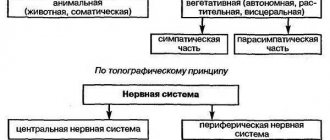
Structure of the nervous system
Anatomically, the nervous system is divided into
Functionally, the nervous system is divided into:
- somatic , innervating striated muscles and sensory organs;
- vegetative (autonomous), innervating internal organs.
This is a summary of the topic "The Nervous System: Basic Principles" . Choose what to do next:
Source: https://uchitel.pro/%D0%BD%D0%B5%D1%80%D0%B2%D0%BD%D0%B0%D1%8F-%D1%81%D0%B8%D1% 81%D1%82%D0%B5%D0%BC%D0%B0/
Structure and types of the nervous system: structural classification
To simplify the structure of the nervous system, in medicine there are several classification options depending on the structure and functions performed. Thus, anatomically, the human nervous system can be divided into 2 broad groups:
- central (CNS), formed by the brain and spinal cord;
- peripheral (PNS), represented by nerve ganglia, endings and nerves themselves.
The basis of this classification is extremely simple: the central nervous system is a kind of connecting link in which the analysis of the incoming impulse and further regulation of the activity of organs and systems is carried out. And the PNS serves to transport the received signal from the receptors to the CNS and the subsequent activator, but from the CNS to the cells and tissues that will perform a specific action.
central nervous system
The central nervous system is a key component of the nervous system, because it is here that the main reflexes are formed. It consists of the spinal cord and brain, each of which is reliably protected from external influences by bone structures. Such thoughtful protection is necessary, since each part of the central nervous system performs vital functions, without which it is impossible to maintain health.
Spinal cord
This structure is contained within the spinal column. It is responsible for the simplest reflexes and involuntary reactions of the body to stimuli.
In addition, spinal cord neurons coordinate the activity of muscle tissue, which regulates defense mechanisms. For example, upon feeling an extremely hot temperature, a person involuntarily withdraws his palm, thereby protecting himself from a thermal burn. This is a typical reaction controlled by the spinal cord.
Brain
The human brain consists of several sections, each of which performs a number of physiological and psychological functions:
- The medulla oblongata is responsible for the vital functions of the body - digestion, respiration, blood movement through the vessels, etc. In addition, the nucleus of the vagus nerve is located here, which regulates the autonomic balance and psycho-emotional reaction. If the nucleus of the vagus nerve sends active impulses, a person’s vitality decreases, he becomes apathetic, melancholic and depressed. If the activity of impulses emanating from the core decreases, the psychological perception of the world changes to a more active and positive one.
- The cerebellum regulates the precision and coordination of movements.
- The midbrain is the main coordinator of muscle reflexes and tone. In addition, neurons regulated by this part of the central nervous system contribute to the adaptation of the sensory organs to external stimuli (for example, pupil accommodation at dusk).
- The diencephalon is formed by the thalamus and hypothalamus. The thalamus is the most important organ that analyzes incoming information. The hypothalamus regulates the emotional background and metabolic processes; there are centers responsible for the sensation of hunger, thirst, fatigue, thermoregulation, and sexual activity. Thanks to this, not only physiological processes are coordinated, but also many human habits, for example, the tendency to overeat, the perception of cold, etc.
- Cerebral cortex. The cerebral cortex is a key link in mental functions, including consciousness, speech, perception of information and its subsequent comprehension. The frontal lobe regulates motor activity, the parietal lobe is responsible for bodily sensations, the temporal lobe controls hearing, speech and other higher functions, and the occipital lobe contains the centers of visual perception.
Peripheral nervous system
The PNS provides interconnection between organs, tissues, cells and the central nervous system. Structurally, it is represented by the following morphofunctional units:
- Nerve fibers, which, depending on the functions performed, are motor, sensory and mixed. Motor nerves transmit information from the central nervous system to muscle fibers, sensitive nerves, on the contrary, help to perceive information received through the senses and transmit it to the central nervous system, and mixed nerves are involved to one degree or another in both processes.
- Nerve endings, which are also motor and sensory. Their function is no different from fiber structures with the only nuance - the nerve endings begin or, conversely, end the chain of impulses from the organs to the central nervous system and back.
- Nerve ganglia, or ganglia, are clusters of neurons outside the central nervous system. The spinal ganglia are responsible for transmitting information received from the external environment, and the autonomic ganglia are responsible for transmitting information about the state and activity of the internal organs and resources of the body.
In addition, all peripheral nerves are classified depending on their anatomical features. Based on this characteristic, there are 12 pairs of cranial nerves that coordinate the activity of the head and neck, and 31 pairs of spinal nerves that are responsible for the torso, upper and lower extremities, as well as internal organs located in the abdominal and thoracic cavities.
Cranial nerves originate from the brain. The basis of their activity is the perception of sensory impulses, as well as partial participation in respiratory, digestive and cardiac activity. The function of each pair of cranial nerves is presented in more detail in the table.
| No. | Name | Function |
| I | Olfactory | Responsible for the perception of various odors, transmitting nerve impulses from the olfactory organ to the corresponding center of the brain. |
| II | Visual | Regulates the perception of visual data by delivering impulses from the retina. |
| III | Oculomotor | Coordinates the movement of the eyeballs. |
| IV | Block | Along with the oculomotor pair of nerves, it takes part in the coordinated movement of the eyes. |
| V | Trigeminal | Responsible for sensory perception of the facial area, and also participates in the act of chewing food in the oral cavity. |
| VI | Abductor | Another nerve that regulates the movements of the eyeballs. |
| VII | Facial | The nerve that coordinates facial contractions of the facial muscles. In addition, this pair is also responsible for taste perception, transmitting signals from the papillae of the tongue to the brain center. |
| VIII | vestibulocochlear | This pair is responsible for the perception of sounds and the ability to maintain balance. |
| IX | Glossopharyngeal | Regulates the normal activity of the pharyngeal muscles and partially transmits taste sensations to the brain center. |
| X | Wandering | One of the most significant cranial nerves, the functionality of which determines the activity of internal organs located in the neck, chest and abdominal wall. These include the pharynx, larynx, lungs, heart muscle and digestive tract organs. |
| XI | Dorsal | Responsible for contractions of muscle fibers of the cervical and shoulder regions. |
| XII | Sublingual | Coordinates the activity of the tongue and partially forms speech skills. |
The activity of the spinal nerves is classified much more simply - each specific pair or complex of pairs is responsible for its assigned area of the body with the same name:
- cervical - 8 pairs,
- infants - 12 pairs,
- lumbar and sacral - 5 pairs respectively,
- coccygeal - 1 pair.
Each representative of this group belongs to mixed nerves, formed by two roots: sensory and motor. That is why the spinal nerves can both perceive an irritating effect, transmitting an impulse along the chain, and intensify activity in response to a message from the central nervous system.
Morphofunctional division of the nervous system
There is also a functional classification of parts of the nervous system, which includes:
- The somatic nervous system regulates the functions of skeletal muscles. It is controlled by the cerebral cortex, and therefore is completely subordinate to the conscious decisions of a person.
- The autonomic nervous system, responsible for the activity of internal organs. Its centers are located in the brain stem, and therefore it is not consciously regulated in any way.
In addition, the autonomic system is divided into 2 more significant functional departments:
- Sympathetic. Activated when energy is consumed;
- Parasympathetic. Responsible for the recovery period of the body.
Somatic nervous system
Somatics is a department of the nervous system that is responsible for the delivery of motor and sensory impulses from receptors to the organs of the central nervous system and back. Most of the nerve fibers of the somatic system are concentrated in the skin, muscle frame and organs responsible for sensory perception. It is the somatic nervous system that coordinates almost 100% of the conscious part of the activity of the human body and the processing of information received from the receptors of the sensory organs.
The main elements of somatics are 2 types of neurons:
- sensory or afferent. Regulate the delivery of information to the cells of the central nervous system;
- motor or efferent. They work in the opposite direction, transporting nerve impulses from the central nervous system to cells and tissues.
Both neurons stretch from parts of the central nervous system directly to the final target of impulses, that is, to muscle and receptor cells, and the body in most cases is located directly in the central part of the nervous system, and the processes reach the necessary localization.
In addition to conscious activity, somatics also includes some reflexes controlled unconsciously. With the help of such reactions, the muscular system comes into an active state without waiting for an impulse from the brain, which allows it to act instinctively. This process is possible if the nerve fiber paths pass directly through the spinal cord. An example of such actions is the jerking of the hand when feeling a high temperature or the knee-jerk reflex when hitting a tendon with a hammer.
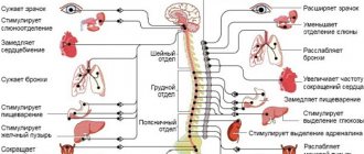
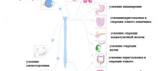
Autonomic nervous system
The autonomic nervous system, or autonomic nervous system, is a department that coordinates the activity of primarily internal organs. Since the basic processes of life - breathing, metabolism, heartbeat, blood flow, etc. - are not subject to consciousness, autonomic nerve fibers react primarily to changes occurring in the internal environment of the body, remaining indifferent to conscious impulses. Thanks to this, optimal conditions are maintained in the body to provide the energy resources necessary in a particular situation.
The peculiarities of autonomic nervous activity imply that the main fibers are concentrated not only in the organs of the central nervous system, but also in other tissues of the human body. Numerous nodes are scattered throughout the body, forming an autonomous nervous system outside the central nervous system, between the brain centers and organs. Such a network can regulate the simplest functions, but more complex mechanisms still remain under the direct control of the central nervous system.
The key role of vegetatives is to maintain relatively constant homeostasis by self-adjusting the activity of internal organs depending on the needs of the body. Thus, vegetative fibers optimize the secretion of hormones, the speed and intensity of blood supply to tissues, the intensity and frequency of respiration and heart rate and other key mechanisms that must respond to changes in the external environment (for example, with intense physical activity, increased temperature or humidity, atmospheric pressure and etc.). Thanks to these processes, compensatory and adaptive reactions are ensured that keep the body in optimal shape under any circumstances. Since the unconscious activity of internal organs can be regulated in two directions (activation and suppression), autonomics can also be divided into 2 sections - parasympathetic and sympathetic.
Sympathetic nervous system
The sympathetic division of the autonomic system is directly connected to the spinal substance, located from the first thoracic to the third lumbar vertebra. It is here that the activity of internal organs is stimulated, which is necessary during times of increased energy consumption - during physical exertion, during stress, intense work or emotional shock. Such mechanisms make it possible to support the body by providing it with the resources necessary to overcome unfavorable conditions.
Under the influence of sympathy, breathing and pulsation of blood vessels become more frequent, due to which the tissues are better supplied with oxygen and energy is released from the cells more quickly. Thanks to this, a person can work more actively, coping with increased loads in unfavorable conditions. However, these resources cannot be infinite: sooner or later the amount of energy reserves decreases, and the body can no longer function “at high speed” without a break. Then the parasympathetic department of the vegetative system comes into play.
SENSITIVE NERVE ENDINGS (RECEPTORS)
They represent the terminal branches of the dendrites of a neurocyte. CLASSIFICATION. There are several principles for classifying receptor nerve endings.
1. At the place of perception of the stimulus. Receptor nerve endings are divided into three groups: exteroceptors, which perceive irritation from the external environment; interoreceptors, which serve to perceive stimuli from the internal environment of the body; proprioceptors that perceive information from the musculoskeletal system.
2. Depending on the specificity of the irritation perceived by the receptor. There are: mechanoreceptors that perceive mechanical stimuli, movements of body parts; chemoreceptors perceive chemical stimuli; thermoreceptors detect changes in temperature, and nociceptors perceive the feeling of pain.
3. According to the method of perception of the stimulus , contact receptors are distinguished which come into a state of excitation when directly exposed to a part of the body, and distant receptors, which perceive the stimulus remote from the body (receptor cells of the retina, hearing, smell).
4. Morphological classification. Depending on their structure, all receptors are divided into free and non-free. Free receptor nerve endings consist only of the terminal branches of the dendrite of a sensitive neuron, and non-free nerve endings, in addition to the terminals of the nerve process, also have oligodendroglia cells (lemmocytes), which surround the dendrite terminals and are involved in the perception of irritation. In turn, non-free nerve endings are divided into non-encapsulated (not surrounded at the periphery by a connective tissue capsule) and encapsulated, which have such a capsule. Free nerve endings primarily perceive pain stimuli. Most non-free nerve endings are mechanoreceptors. Recently, however, a groundless point of view has been put forward that there is no separation of receptors depending on the type of perceived stimulation, all receptors are capable of perceiving stimuli of any modality, and the nature of the sensation depends on the strength of the stimulus.
MORPHOLOGY OF RECEPTORS. 1. Free nerve endings. They are most abundant in the skin. These are mechanoreceptors on hair follicles, nociceptive (perceiving pain stimuli) nerve endings in the epidermis (Fig. 14.3). There are also many of them in the stratified squamous non-keratinizing epithelium and serous membrane. In the epidermis they are represented by tree-like branching of the dendrites of pseudounipolar neurons of the spinal ganglia.
2. Non-free, non-encapsulated nerve endings are represented by Merkel tactile well as nerve endings of connective tissue. There are especially many of them in the dermis. Merkel's tactile discs (Fig. 14.3 b)
consist of a process of a nerve cell, which ends in an extension in the form of a P. disc. This disc forms a synapse with the Merkel cell, which lies in the epidermis. In the cytoplasm of the Merkel cell there are secretory granules with a neurotransmitter. Mechanical irritation causes the release of granules from Merkel cells, their contents lead to depolarization of the neurocyte process.
Non-free, non-encapsulated endings in connective tissue are constructed as follows. The axial cylinder is freed from myelin and is surrounded at a considerable distance by glial cells, in close contact with them. Very often, a cross-section shows the bilateral symmetry of such endings.
3. Non-free encapsulated nerve endings are built according to a general principle. These endings include nerve endings in connective and muscle tissues. There are the following varieties of these endings: lamellar corpuscles of Vater-Pacini, tactile corpuscles of Meissner, terminal flasks of Krause, genital corpuscles of Dogel, corpuscles of Ruffini, neuromuscular and neurotendon spindles , etc.
The most common are Vater-Pacini lamellar bodies. They are found in the skin, mammary gland, mesentery, internal organs, near blood vessels, near joints. These are large formations with a diameter of 1 to 5 mm (Fig. 14.4, 14.5). They are oval in shape and consist of a connective tissue capsule, terminals of the dendrite of a pseudounipolar neuron and neurolemmocytes (oligodendroglia). When approaching the capsule, the dendrite loses its myelin sheath and is surrounded on all sides by neurolemocytes. They form the so-called inner flask. This flask is covered on the outside with a connective tissue capsule, which is often called the outer flask. The capsule consists of! from layer-by-layer parallel collagen fibers (forming from 10 to 60 layers) and fibrocyte cells. Blood vessels are found in the outer capsule. Between the outer and inner flasks there are specialized branched oligodendrogliocytes in contact with the axial cylinder. When pressure is applied to the body, the mechanical effect is greatly enhanced by the layers of the outer bulb, which makes this receptor very sensitive. The pressure moves the outer flask relative to the inner one. In this case, process oligodendrocytes are irritated, transmitting excitation to the dendrite.
Meissner's tactile corpuscles are found in the papillary dermis They are mechanoreceptors and are smaller in size than Father-Pacini bodies (50-140 µm). They have an oval shape (Fig. 14.3 and 14.6). Outside there is a very thin layered capsule - the outer flask. The dendrite of a pseudounipolar neuron loses its myelin sheath, branches, and its branches enter the capsule in a spiral. Perpendicular to them lie glial cells, which, together with the terminals of the dendrites, form an internal flask. A slight deformation of the capsule is transmitted to gliocytes, which have a synaptic connection with the dendrite.
Krause end flasks are baroreceptors and thermoreceptors. They lie in the dermis of the skin and mucous membranes. They have small (40-150 microns) sizes. They also consist of an outer capsule and an inner flask. The inner flask is formed by flat gliocytes, between which thin branches of the dendrite pass, forming a kind of glomerulus. The outer capsule is very thin.
Genital Dogel bodies are located in particularly sensitive areas of the skin, primarily in the area of the external genitalia and the skin of the mammary glands. They are similar in structure to Krause's flasks, but unlike them, the body contains several processes from several neurocytes. Therefore, irritation of the genital body causes a strong irradiation of excitement.
Ruffini corpuscles are found in the connective tissue of the skin and in the capsules of the joints. Perceive a feeling of pressure. They look like versiform formations up to 2 mm long. The axial cylinder in the inner flask branches to form a large number of branches with club-shaped thickenings at the end. The capsule is well defined.
In smooth muscle tissue, sensory nerve endings are also encapsulated; they contact a group of smooth myocytes.
In skeletal muscle tissue, the sensory nerve endings are called neuromuscular spindles. They are encapsulated nerve endings (Fig. 14.7, 14.8). The outer connective tissue capsule of the neuromuscular spindle surrounds several thin so-called intrafusal muscle fibers. Unlike ordinary muscle fibers lying outside and called extrafusal, intrafusal fibers are thin, contain few myofibrils and have light cytoplasm. There are two types of intrafusal muscle fibers (Fig. 14.8). 1. YaS fibers. The nuclei of these fibers lie in the central part of the muscle fiber, forming a cluster in the form of a nuclear bursa (abbreviated as BU). At the location of the nuclei, the fiber expands sharply.
2. JC fibers. These fibers have a uniform thickness, and the nuclei lie along the entire length of the fiber in its center, forming a nuclear chain.
Around these two types of intrafusal fibers in their central part, specific synapses of the dendrites of sensory neurons are formed in the form of:
1) annulospiral (ring-spiral) endings, in which the processes of nerve cells are twisted around the central part of the intrafusal fiber in a spiral and enter into a synaptic connection with it over a long distance; annulospiral endings are present on both JS and JC fibers. 2) cluster-shaped endings, which are found only on NC fibers. Moreover, they are formed not in the central part, but on the periphery of the fiber.
The intrafusal fibers also contain motor nerve endings, which are represented by the axons of the γ-motoneurons of the anterior horns of the spinal cord. They regulate the length of intrafusal fibers and maintain their tone. All free space between muscle fibers is filled with fluid and limited by a thin capsule. Any change in muscle tone leads to a change in fluid pressure in the capsule cavity. In this case, pressure is transferred to the dendrites. Annulospiral endings respond to changes in the length of the muscle fiber and the speed of this change, and pampiniform endings respond only to changes in length. Thanks to neuromuscular spindles, the body
constantly receives information about the degree of muscle contraction, which forms an idea of the position of the body in space.
INTERNEURAL SYNAPSES
This is a special type of nerve endings, when the branches of the processes of some nerve cells end on other nerve cells. With the help of synapses, excitation is transmitted from one nerve cell to another.
Classification of synapses. There are several approaches to classifying synapses.
i. According to the mechanism of nerve impulse transmission. Synapses are divided into chemical, electrical and mixed. In chemical synapses, excitation a neurotransmitter. These synapses are the most common in the nervous system of higher animals. At electrical synapses, an action potential is transmitted directly from the membrane of one neuron to another. Mixed synapses are a combination of features of both chemical and electrical synapses.
2. Morphological classification of synapses. Takes into account the characteristics of the contacting areas of neuerocytes. There are axo-somatic, axo-dendritic, axo-axonal, dendro-dendritic, somato-somatic synapses.
3. Physiological classification. According to the effect they cause on a nerve cell, synapses are divided into excitatory and inhibitory.
4. Mediator classification of synapses. cholinergic, aminergic (adrenergic, serotonergic, dopaminergic) synapses purinergic, amino acid (mediators are amino acids: GABA, glycine, glutamate, aspartate, etc. ), peptidergic (see mediator classification of neurocytes).
STRUCTURE OF SYNAPSES. Any synapse consists of three parts: a pre-synaptic pole with a presynaptic membrane, a synaptic cleft and a postsynaptic pole with a postsynaptic membrane.
Electrical synapses. These synapses are built like nexuses: two membranes (pre- and postsynaptic) of neighboring neurons come close to each other up to a distance of 2 nm, and this place of contact is penetrated by numerous connexons. Consequently, there is practically no synaptic cleft in the electrical synapse. Connexons are a kind of pore across both membranes, which is bounded at the edges by special protein molecules called connexins. Connexons allow passage not only of alkali metal ions, which play an important role in the formation of electrical potentials, but also of molecules with MW of 1000–2000. Therefore, in addition to electrical coupling, connexons enable neurons to exchange metabolites. Unlike chemical synapses, in which the signal conduction is somewhat delayed, in electrical synapses the impulse is transmitted almost without delay and in both directions. The significance of electrical synapses is unknown. It is believed that it is associated with the need for rapid coupling of nerve cells.
Chemical synapses. Unlike electrical synapses, chemical synapses transmit nerve impulses only in one direction and with a delay (synaptic delay). This is the most common type of synapse in mammals. Chemical synapses have all three distinct components: non-resynaptic and nost-synaptic poles and a synaptic cleft (Fig. 14.9) (in a light microscope, synapses are visible in the form of button-shaped thickenings on neurocytes, see Fig. 13.1).
At the presynaptic pole there are presynaptic vesicles with a transmitter, mitochondria, agranular EPS, neurotubules and neurofilaments. Synaptic vesicles have different structures depending on the mediator they contain. Thus, acetylcholine bubbles are small and electronically transparent. Synaptic vesicles containing norepinephrine are larger and have an electron-dense part in the center. The vesicles containing peptides are large in size, have a dense core, and are surrounded by a peripheral light rim.