The morphology of the nervous system is one of the complex sections of anatomy.
The nervous system ensures the interaction of all human organs and systems, exchanges information, and determines the adaptive relationship of the body with the environment.
Correct mastery of the anatomy of the nervous system determines the materialistic formation of the concept of the body as an integral system and creates the basis for the study of theoretical and clinical disciplines (neuropathology, neurosurgery, psychiatry, etc.).
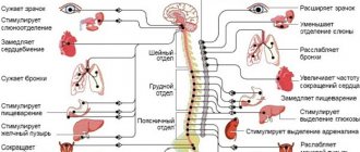
The nervous system consists of central and peripheral. The peripheral nervous system is formed by 12 pairs of cranial nerves and 31 pairs of spinal nerves.
The study of the peripheral nervous system is of great practical and diagnostic importance. In clinical practice, diseases of the spinal nerves such as radiculitis, meningoradiculitis, radiculoneuritis, funiculitis, neurodermatitis, neuritis, neuromyositis, herpes zoster, etc. are often encountered.
This information is in addition to existing anatomical manuals and textbooks and does not replace them.
Nervous system function
- complex and diverse. The nervous system (system nervosum) exchanges information between the body and the external environment, regulates and coordinates the functions of all organs, ensures the functional unity and integrity of the body, and determines the adaptive behavior of the body in the environment.
Conventionally, the nervous system is divided according to functional and topographic principles as follows (Fig. 5.1).
Rice. 5.1. Scheme of division of the nervous system according to functional and topographic principles
Propagation of nerve impulses
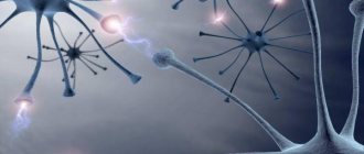
As a result of the evolution of the nervous system of humans and other animals, complex information networks arose, the processes of which are based on chemical reactions. The most important element of the nervous system are specialized cells called neurons.
.
Neurons consist of a compact cell body containing a nucleus and other organelles. Several branched processes extend from this body. Most of these projections, called dendrites
, serve as contact points for receiving signals from other neurons.
One process, usually the longest, is called an axon
and transmits signals to other neurons. The end of an axon can branch multiple times, and each of these smaller branches is able to connect to the next neuron.
The outer layer of the axon contains a complex structure formed by many molecules that act as channels through which ions can flow both into and out of the cell. One end of these molecules, deflecting, attaches to the target atom. Energy from other parts of the cell is then used to push that atom out of the cell, while the process in the opposite direction brings another molecule into the cell. The most important is the molecular pump, which removes sodium ions from the cell and introduces potassium ions into it (sodium-potassium pump).
When a cell is at rest and not conducting nerve impulses, the sodium-potassium pump moves potassium ions into the cell and removes sodium ions out (imagine a cell containing fresh water and surrounded by salt water). Because of this imbalance, the potential difference across the axon membrane reaches 70 millivolts (approximately 5% of the voltage of a regular AA battery).
However, when the state of the cell changes and the axon is stimulated by an electrical impulse, the equilibrium on the membrane is disturbed, and the sodium-potassium pump begins to work in the opposite direction for a short time. Positively charged sodium ions enter the axon, and potassium ions are pumped out. For a moment, the internal environment of the axon acquires a positive charge. In this case, the channels of the sodium-potassium pump are deformed, blocking further sodium influx, and potassium ions continue to flow out, and the original potential difference is restored. Meanwhile, sodium ions spread inside the axon, changing the membrane at the bottom of the axon. At the same time, the state of the pumps located below changes, promoting further propagation of the impulse. A sudden change in voltage caused by the rapid movement of sodium and potassium ions is called an action potential
. When an action potential passes through a certain point on the axon, the pumps turn on and restore the resting state.
The action potential travels quite slowly—no more than a fraction of an inch per second. In order to increase the speed of impulse transmission (since, after all, it is not good for a signal sent from the brain to take a minute to reach the hand), the axons are surrounded by a sheath of myelin, which prevents the influx and outflow of potassium and sodium. The myelin sheath is not continuous - at certain intervals there are breaks in it, and the nerve impulse jumps from one “window” to another, due to this the speed of impulse transmission increases.
When the impulse reaches the end of the main part of the axon body, it must be transmitted either to the next underlying neuron or, in the case of neurons in the brain, through numerous branches to many other neurons. For such transmission, a completely different process is used than for transmitting an impulse along the axon. Each neuron is separated from its neighbor by a small gap called a synapse
.
The action potential cannot jump across this gap, so some other way must be found to transmit the impulse to the next neuron. At the end of each process there are tiny sacs called ( presynaptic
)
vesicles
, each of which contains special compounds -
neurotransmitters
. When an action potential occurs, these vesicles release neurotransmitter molecules that cross the synapse and bind to specific molecular receptors on the membrane of underlying neurons. When a neurotransmitter attaches, the balance on the neuron membrane is disrupted. Now we will consider whether a new action potential arises with such an imbalance (neuroscientists continue to search for the answer to this important question to this day).
After neurotransmitters transmit a nerve impulse from one neuron to the next, they can simply diffuse out, undergo chemical breakdown, or return back to their vesicles (this process is awkwardly called reuptake
). At the end of the 20th century, an astonishing scientific discovery was made - it turns out that drugs that affect the release and reuptake of neurotransmitters can radically change a person’s mental state. Prozac* and similar antidepressants block the reuptake of the neurotransmitter serotonin. It appears that Parkinson's disease is associated with a deficiency of the neurotransmitter dopamine in the brain. Researchers studying borderline states in psychiatry are trying to understand how these compounds affect human reasoning.
There is still no answer to the fundamental question of what causes a neuron to initiate an action potential - in the professional language of neurophysiologists, the mechanism of “firing” of a neuron is unclear. Particularly interesting in this regard are neurons in the brain, which can receive neurotransmitters sent by a thousand neighbors. Almost nothing is known about the processing and integration of these impulses, although many research groups are working on this problem. We only know that the neuron carries out the process of integrating incoming impulses and makes a decision whether or not to initiate an action potential and transmit the impulse further. This fundamental process controls the functioning of the entire brain. It is not surprising that this greatest mystery of nature remains, at least today, a mystery for science!
Biochemistry of nerve impulse transmission. Main components and stages.
A synapse is a functional contact between specialized areas of the plasma membranes of two excitable cells. A synapse consists of a presynaptic membrane, a synaptic cleft, and a postynaptic membrane. The cell membranes at the point of contact have thickenings in the form of plaques - nerve endings. A nerve impulse that reaches a nerve ending is unable to overcome the obstacle that has arisen in front of it - the synaptic cleft. After this, the electrical signal is converted into a chemical one. The presynaptic membrane contains special channel proteins similar to the proteins that form the sodium channel in the axon membrane. They also respond to the membrane potential by changing their conformation and forming a channel. As a result, Ca2+ ions pass through the presynaptic membrane along a concentration gradient into the nerve ending. The Ca2+ concentration gradient is created by Ca2+-dependent work.
ATPase is a calcium pump. An increase in Ca2+ concentration inside the nerve ending causes the 200-300 acetylcholine-filled vesicles present there to merge with the plasma membrane. Next, acetylcholine is secreted into the synaptic cleft by exocytosis and binds to receptor proteins located on the surface of the postsynaptic membrane.
The acetylcholine receptor is a transmembrane oligomeric glycoprotein complex consisting of 6 subunits: 2-, 2-beta, 1-gamma and 1-delta. The density of receptor proteins in the postsynaptic membrane is very high - about 20,000 molecules per 1 µm2. The spatial structure of the receptor strictly corresponds to the conformation of the mediator. When interacting with acetylcholine, the receptor protein changes its conformation so that a sodium channel is formed inside it. The cation selectivity of the channel is ensured by the fact that the gate of the channel is formed by negatively charged amino acids. Thus, the permeability of the postsynaptic membrane to sodium increases and a new impulse (or contraction of the muscle fiber) occurs. Depolarization of the postsynaptic membrane causes dissociation of the acetylcholine-protein-receptor complex and acetylcholine is released into the synaptic cleft. Once acetylcholine is in the synaptic cleft, it undergoes rapid hydrolysis within 40 μs by the enzyme acetylcholinesterase.
During the hydrolysis of acetylcholine, an intermediate enzyme-substrate complex is formed, in which acetylcholine is bound to the active center of the enzyme through serine.
Irreversible inhibition of cholinesterase causes death. Cholinesterase inhibitors are organophosphorus compounds (chlorophos, dichlorvos, tabun, sarin, soman, binary poisons). These substances bind covalently to serine in the active site of the enzyme. Some of them are synthesized as insecticides, and some as chemical warfare agents (nerve poisons). Death occurs as a result of respiratory arrest. Reversible cholinesterase inhibitors are used as therapeutic drugs. For example, in the treatment of glaucoma and intestinal atony.
Catecholamines: norepinephrine and dopamine. Adrenergic synapses are found in postganglionic fibers, in fibers of the sympathetic nervous system, in various parts of the brain. Catecholamines in nervous tissue are synthesized according to a general mechanism from tyrosine. The key enzyme in the synthesis is tyrosine hydroxylase, which is inhibited by the end products.
Catecholamines, like acetylcholine, accumulate in synaptic vesicles and are also released into the synaptic cleft upon receipt of a nerve impulse. But regulation in the adrenergic receptor occurs differently. In the presynaptic membrane there is a special regulatory protein - -achromogranin (Mm = 77 kDa), which, in response to an increase in the concentration of the transmitter in the synaptic cleft, binds the already released transmitter and stops its further exocytosis. There is no enzyme that destroys the transmitter in adrenergic synapses. After transmitting the impulse, the transmitter molecule is pumped by a special transport system through active transport with the participation of ATP back through the presynaptic membrane and is reincorporated into the vesicles. In the presynaptic nerve ending, excess transmitter can be inactivated by monoamine oxidase, as well as catecholamine-O-methyltransferase by methylation at the hydroxy group. Cocaine inhibits the active transport of catecholamines.
Binding of the transmitter to the postsynaptic receptor almost instantly causes an increase in the concentration of c-AMP, which leads to rapid phosphorylation of proteins of the postsynaptic membrane. As a result, the generation of nerve impulses by the postsynaptic membrane changes (inhibits). In some cases, the immediate cause of this is an increase in the permeability of the postsynaptic membrane for potassium, or a decrease in conductivity for sodium (these events lead to hyperpolarization).
GABA is an inhibitory neurotransmitter. Increases the permeability of postsynaptic membranes for potassium ions. This leads to a change in membrane potential.
Glycine is an inhibitory neurotransmitter, similar in effects to GABA.
Formation of neurotransmitters - acetylcholine, adrenaline, dopamine, serotonin.
The information that allows neurons to establish only certain connections with certain neurons is encoded in the structure of the polysaccharide branches of membrane glycoproteins. The formation of such connections, which were not laid down during embryonic development, is the result of the experience of an individual organism and constitutes the material basis for storing information that determines the behavioral characteristics of a given organism.
Neurotransmitters are substances that are characterized by the following characteristics. They accumulate in the presynaptic structure in sufficient concentration. Released upon transmission of impulse. After binding to the postsynaptic membrane, they cause a change in the rate of metabolic processes and the occurrence of an electrical impulse. They have a system for inactivation or a transport system for removal from the synapse, which have a high affinity for them. Thus, neurotransmitters play an important role in the functioning of nervous tissue, providing synaptic transmission of nerve impulses. Their synthesis occurs in the body of neurons, and accumulation occurs in special vesicles, which gradually move with the participation of systems of neurofilaments and neurotubules to the tips of axons.
Chemical classification of neurotransmitters: Amino acids (and their derivatives) - These include taurine, norepinephrine, DOPAminGABA, glycine, acetylcholine, homocysteine and some others (adrenaline, serotonin, histamine, serotonin).
Taurine is formed from the amino acid cysteine. First, sulfur in the SH group is oxidized to a sulfuric acid residue (the process occurs in several stages), and then decarboxylation occurs. Taurine is an unusual acid that does not have a carboxyl group, but rather a sulfuric acid residue. Taurine takes part in the conduction of nerve impulses in the process of visual perception.
Acetylcholine - The amino acids serine and methionine are required for the synthesis of choline. Ethanolamine can also be used in finished form. But, as a rule, ready-made choline enters the nervous tissue from the blood. The second precursor of this neurotransmitter, Acetyl-CoA, is synthesized in nerve endings. The product of this reaction, acetylcholine, is involved in the synaptic transmission of nerve impulses. It accumulates in synaptic vesicles, forming complexes with the negatively charged protein vesiculin. The transfer of excitation from one cell to another is carried out using a special synaptic mechanism.
Norepinephrine is a transmitter in postganglionic fibers of the sympathetic and in various parts of the central nervous system.
Dopamine is a transmitter of pathways, the bodies of neurons of which are located in the part of the brain that is responsible for the control of voluntary movements. Therefore, when dopaminergic transmission is disrupted, the disease parkinsonism occurs.