Late neurosyphilis
Late neurosyphilis occurs long after the initial infection (from 5 to 25 years). Basically, the disease takes the following forms:
- meningovascular neurosyphilis;
- tabes dorsalis;
- gummous lesion of the central nervous system.
Meningovascular neurosyphilis occurs with damage to both the meninges and blood vessels of the brain. As a result of changes in the vascular bed, the disease leads to arterial thrombosis and circulatory disorders, which is clinically expressed in repeated strokes.
Tabes dorsalis is a form of late neurosyphilis with predominant damage to motor neurons and dorsal roots of the spinal cord. As a result, the leading symptom of the disease is progressive paralysis.
Gummy lesions of the central nervous system are accompanied by the formation of gummas - giant tumor-like nodes in the brain or spinal cord. Symptoms resemble those of benign tumors.
How the disease develops
Treponema pallidum penetrates the nervous system by hematogenous and lymphogenous routes in the early stages of untreated syphilis. They affect the membranes, vessels and membranes of the roots and peripheral nerves. Over time, these structures lose the ability to hold treponema pallidum and neutralize them, and then the bacteria penetrate the substance (parenchyma) of the brain and spinal cord, causing the development of a number of diseases.
In the first years from the onset of infection, the patient may develop a latent (asymptomatic) form of neurosyphilis, when the patient does not have any neurological disorders, but lymphocytic pleocytosis and increased protein content are noted in the cerebrospinal fluid.
In the primary (rarely) and secondary (more often) periods of syphilis, the development of syphilitic meningitis is recorded. The main symptom complex called neurosyphilis develops in the tertiary period of syphilis.
- In the first five years of the disease, early syphilis of the nervous system develops, which is characterized by the development of inflammatory changes in the mesenchyme - blood vessels and meninges.
- Late neurosyphilis develops in later stages of the disease - 10 - 25 or more years from the moment of primary infection. Following the mesenchyme, the parenchyma begins to be affected - nerve cells, fibers and glia.
Modern neurosyphilis occurs with minimal severity of symptoms and is characterized by a milder course and less change in the cerebrospinal fluid. Complaints that come to the fore include weakness, lethargy, insomnia, and decreased performance. The longer the infectious process, the more often the symptoms and clinical manifestations of neurosyphilis are recorded.
Rice. 2. The photo shows manifestations of tertiary syphilis - gumma. During this period, late neurosyphilis develops.
Symptoms of early neurosyphilis
The following symptoms are characteristic of early latent neurosyphilis:
- fatigue, muscle weakness;
- severe and moderate headaches, “heaviness in the head”;
- tinnitus, hearing loss, dizziness;
- memory impairment.
Acute syphilitic meningitis is accompanied by severe symptoms:
- severe headaches;
- dizziness, tinnitus;
- severe vomiting without nausea, not associated with food intake or dietary errors;
- fever, high temperature;
- pathological reflexes;
- convulsions;
- deterioration of vision, narrowing of visual fields, facial asymmetry, drooping eyelids, deviation of the tongue to the side when cranial nerves are involved in the inflammatory process.
When brain tissue is involved in the process, strokes, paralysis, and paresis of the lower and upper extremities are possible.
Symptoms of late syphilis
The meningovascular form of neurosyphilis is disguised as vascular disorders. Symptoms include:
- moderate headaches;
- dizziness, weakness;
- paralysis, paresis, speech disorders;
- symptoms of ischemic stroke - loss of consciousness, coma, impaired brain function.
Tabes dorsalis manifests itself as progressive paralysis with pain in areas corresponding to the level of damage to the spinal cord. Possible:
- dagger pains simulating attacks of angina, renal or hepatic colic;
- numbness, a feeling of “goosebumps” on the skin, tingling in the soles, areas corresponding to the level of the lesion;
- dysfunction of the pelvic organs - impotence, urinary and fecal incontinence;
- impairment of tactile and pain sensitivity in areas corresponding to the level of the lesion;
- unsteadiness of gait, hearing loss, progressive decrease in visual acuity;
- arthrosis, arthritis, osteoporosis are possible due to tissue nutritional disorders;
- because of them, ulcers also appear on the skin of the feet and legs, teeth, hair, nails fall out, sweat glands die off;
- paralysis of the lower and upper limbs occurs 10-20 years after infection with syphilis.
Gumma of the brain is a tumor-like formation of scarred and necrotically altered nervous tissue. When it occurs, it can grow, leading to symptoms similar to those of a brain tumor. The changes are irreversible; usually, surgical treatment is performed for partial improvement. Since gummas most often form at the base of the brain, paralysis, dysfunction of the cranial nerves, and possibly intracranial pressure due to impaired circulation of the cerebrospinal fluid are possible. The outcome of progressive gumma is complete paralysis.
After 10-20 years, the disease turns a person into a weak-minded freak, unable to perform basic actions, take care of himself, a bedridden and hopeless patient. This is what advanced, untreated neurosyphilis looks like; symptoms, progressing, make the diagnosis obvious. Such a person will live, but not for long and very badly.
Fortunately, thanks to the widespread introduction of tests for syphilis, such forms of the disease are extremely rare. According to the rules, screening for this infection is carried out annually during a clinical examination at the place of residence or upon hiring. This is especially relevant, since there are known cases of syphilis infection through everyday life, when seeking the services of a cosmetologist or stylist. A single scratch made by an inattentive hairdresser on an infected instrument can cause illness. 1-2 spirochetes are enough for infection.
Why spinal cord puncture for syphilis?
… In parallel with the increase in the overall incidence of syphilis, there is an increase in the proportion of cases of neurosyphilis.
According to the instructions for the treatment and prevention of syphilis, a study of cerebrospinal fluid must be carried out before treatment in patients with early and late latent syphilis in the presence of clinical lesions of the nervous system, as well as in latent and late forms of neurosyphilis.
Liquorological examinations are carried out upon deregistration:
( 1 ) patients whose treatment was started for early and late neurosyphilis;
( 2 ) persons who, during clinical and serological control, developed any clinical manifestations of a specific lesion of the nervous system (acute syphilitic meningitis, meningovascular syphilis, syphilitic meningomyelitis, tabes dorsalis, syphilitic gumma, syphilitic neuritis and polyneuritis, syphilitic damage to the optic nerves, syphilitic damage to the auditory nerves, syphilitic meningomyelitis, progressive paralysis);
( 3 ) persons with serological resistance that persists at the end of the clinical and serological observation period.
Cerebrospinal fluid for research is obtained by lumbar puncture between the III and IV or IV and V lumbar vertebrae using long thin puncture needles (diameter from 0.4 to 0.8 mm and length 10-12 cm). 3-4 ml of liquor is collected in two sterile tubes (no more than 8-10 ml). It is necessary to ensure that the liquid flows out and the cannulas drop in drops (20-40 drops per minute). If liquid flows out in a stream after removing the mandrin from the needle, then you must immediately insert the mandrin back to a shallow depth in order to adjust the rate of fluid flow.
If the first drops of cerebrospinal fluid are stained with blood, you should change the position of the needle (push it deeper or pull it out slightly). Then the clear liquid is collected into another test tube (a portion of the cerebrospinal fluid mixed with blood is not examined).
After puncture, the puncture site is treated with 3-5% iodine tincture and a sterile bandage is applied. To prevent complications, the patient is placed on the bed with his stomach down (the foot end of the bed is raised by 20-30 cm). After 3-4 hours he is allowed to turn on his side. Bed rest is observed for 24-48 hours. The patient is recommended to drink plenty of fluids and fast for 6-8 hours after the puncture.
One portion of cerebrospinal fluid (3-4 ml) is sent to a clinical-biochemical laboratory to study cytosis, protein content, and perform globulin (Pandey, Nonne-Appelt, Weichbrodt or Takata-Ara) and colloid (Lange or paraffin) reactions.
Nonne-Apelt reaction . Using a saturated solution of ammonium sulfate, a protein ring is formed at the interface of the reagent with the cerebrospinal fluid. After shaking off, and therefore mixing, the liquids, opalescence or cloudiness of varying intensity is obtained. The Nonne-Apelt reaction is assessed using a four-point system: weakly positive (+) - opalescence is faintly noticeable, moderately positive (++) - slight turbidity, positive (+++) - pronounced turbidity and sharply positive (++++) - intense turbidity . Ross-Jones slightly modified this reaction. He suggested not mixing the cerebrospinal fluid with a saturated solution of ammonium sulfate, but carefully pouring it dropwise from a pipette onto a more concentrated reagent. Moreover, cerebrospinal fluid is prepared in various degrees of dilution. After 3 minutes, the degree of turbidity of the ring formed at the interface of the liquids and the ammonium sulfate solution is determined. An opalescence ring is formed when the protein content in the cerebrospinal fluid is at a concentration of 3 g/l.
The Pandey reaction precipitates not only globulins, but also all proteins of the cerebrospinal fluid. It consists of applying 10-15 drops of a 10% carbolic acid solution to a watch glass, which is placed against a dark background, then one drop of cerebrospinal fluid. The reaction is highly sensitive and is determined by the intensity of the turbidity formed at the site of contact between the liquid and the reagent after 3 minutes.
The Weinbrocht reaction is performed in such a way that 3 parts of a 0.1% silver nitrate solution are added to 7 parts of cerebrospinal fluid and then shaken. In this case, turbidity of varying degrees occurs, which is used to judge the content of protein substances (not only globulins).
Takata-Ara reaction . Colloidal reaction. When solutions of sublimate and fuchsin are added to alkalized liquor, a purple mixture is normally observed. When the fluid clears and sediment appears, the reaction is designated as metasyphilitic (progressive paralysis); when stained red, it is called meningitis.
The Lange reaction is based on the ability of pathologically altered cerebrospinal fluid, when mixed with colloidal solutions, to change the dispersion of the solution and thereby its color; with normal cerebrospinal fluid, the purple-red color of the solution does not change. In pathology, there are several types of color change curve. The first type is paralytic: with gradually increasing degrees of cerebrospinal fluid breakdown in test tubes, discoloration occurs only in the first 5 test tubes with a rather steep return to normal. This course of reaction is characteristic of progressive paralysis. For other forms of neurosyphilis, a syphilitic tooth is more characteristic - a moderate change in color in the 2-5th tube. The second type of curve is meningitic (acute meningitis): a color change is noted from the 2-3rd tube, reaches a maximum in the 6th and 7th tubes, after which it abruptly returns to normal. In other diseases of the central nervous system, the Lange reaction is quite typical and cannot serve as an auxiliary diagnostic tool.
The second portion of cerebrospinal fluid (3-4 ml) is sent to the serological laboratory to carry out the Wasserman reaction with cardiolipin and treponemal antigens, RIF and RIBT.
The Wasserman reaction (complement fixation reaction, RSK) with treponemal and cardiolipin antigens is used to confirm the diagnosis of syphilis in the presence of active manifestations of the disease, to examine persons who have been in direct contact with a patient with syphilis, to identify latent (latent) syphilis, and the effectiveness of therapy. When examining patients in psychiatric and neurological hospitals. Donors and pregnant women, including persons referred for abortion.
Blood for research is taken in an amount of 5-7 ml from the ulnar vein with a sterile needle. In infants, blood is taken from the temporal vein or from incisions in the heel. Blood collection is performed strictly on an empty stomach. And leave it in clean, dry test tubes for 2-3 hours at room temperature for coagulation. Testing of DCS and specific reactions is carried out in serological laboratories of dermatovenerological institutions.
The principle of RSC is that reagins found in the blood serum of patients with syphilis have the property of combining with various antigens. The resulting complexes sort the complement introduced into the reaction. To indicate the reagin–antigen–complement complex, the hemolytic system (a mixture of sheep erythrocytes with hemolytic serum) is used. In the presence of the complex, red blood cells precipitate. Which is noticeable to the naked eye. The severity of hemolysis is indicated by the doctor using the following keys: strongly positive 4+, positive 3+, weakly positive 2+ or 1+ and negative. In addition to a qualitative assessment of these reactions, there is also a quantitative assessment, which is important in the diagnosis of certain stages of syphilis and in monitoring the effectiveness of therapy.
Currently, it is recommended to use cordylipyl antigen (an extract from bovine heart enriched with cholesterol and lecithin) and triponemal antigen (an ultrasound-treated suspension of apathogenic cultural treponemes) as antigens. The reaction of complement fixation with cardiolipin and treponemal antigens becomes positive after 2-4 weeks, gradually increases and reaches a maximum (1:160 - 1:320 and above) with secondary fresh syphilis. Then the reagin titer gradually falls and in case of secondary recurrent syphilis it usually does not exceed 1:180 - 1:120. Among patients with tertiary syphilis, these reactions give a positive result in only 70%.
It should be emphasized that CSRs are not strictly specific for syphilis and in some cases can give false-positive (nonspecific) results; they can be obtained due to technical errors (complete hemolysis, unsterile blood collection, insufficient qualifications of laboratory technicians). False reactions are observed in patients with leprosy, malaria, sometimes with situational diseases, neoplasms, tuberculosis, liver diseases, when taking medications, and also during pregnancy. During menstruation, etc. It is not recommended to test blood during the first week after vaccination, injuries, surgical interventions, in febrile conditions during the first 2 weeks after birth, in newborns in the first 10 days of life, because physicochemical changes in the blood serum in these conditions may be similar to those observed in patients with syphilis.
RIF is based on an indirect method for determining fluorescent antibodies. The antigen in this reaction is a suspension of killed cultural Treponema pallidum, fixed to glass slides on which the test and anti-species fluorescent serum is applied. The results of RIF are determined under a fluorescent microscope by assessing the glow of the treponemes in the preparation. With a positive result, the treponema has a yellowish-green glow, the degree of which is indicated by pluses from 1+ to 4+; with negative results, the treponema does not glow. RIF is currently installed in several modifications (RIF - abs, RIF - 200).
RIBT , proposed in 1949 by R. Nelson and M. Meyer, is based on the phenomenon of immobilization of Treponema pallidum by antigens from the patient’s blood serum in the presence of complement. A suspension of pale live treponemes obtained from rabbits infected with syphilis is used as an antigen for RIBT. Counting treponemes that have lost mobility (immobilized) is carried out under a microscope. The results of reactions are assessed as percentages from 0 to 20% - negative, from 21 to 31% - doubtful, from 31 to 50% - weakly positive, from 51 to 100% - positive. RIBT becomes positive at the end of the primary period of syphilis and remains so throughout all periods of this disease, and sometimes even after full anti-syphilis treatment. RIBT can give false-positive results if the test serum contains treponematous substances (antibiotics - penicillin, tetracycline) that cause nonspecific immobilization of Treponema pallidum. Therefore, blood cannot be tested for this reaction earlier than 2 weeks after finishing taking antibiotics.
To carry out specific serological reactions (RIF and RIBT), blood is taken from the ulnar vein on an empty stomach in an amount of 5-10 ml. After sterilization, the syringe and needle are washed with isotonic sodium chloride solution, and the blood is poured into a dry test tube for testing for RIF and into a sterile test tube for testing for RIBT. Specific serological reactions to syphilis are performed in specialized laboratories of dermatovenerological institutions.
The results of a cerebrospinal fluid examination are assessed using the Robustov scale , supplemented by the latest data on RIF and RIBT with cerebrospinal fluid. According to this classification, there are four degrees of changes in the cerebrospinal fluid in patients with syphilis.
I degree – minor isolated or combined changes in the cerebrospinal fluid.
Changes in the cerebrospinal fluid are considered insignificant when the following are determined: cytosis more than 8 cells in 1 mm 3, protein more than 0.33%, Nonne-Apelt reaction (++), Pandi reaction (+++), Lange reaction more than twos or one three, PB is weakly positive, RIF is positive.
If one of these changes is detected, they speak of isolated changes in the cerebrospinal fluid, but if two of them are detected, they are said to be combined. most often they are detected before treatment in patients with primary and secondary syphilis, vascular neurosyphilis, in some patients who have recently completed treatment for neurosyphilis, as well as in individual subjects after a control period of observation.
II degree – significant changes in the cerebrospinal fluid with negative results of RT and RIBT.
Protein-cell dissociation is noted (cytosis is within normal limits or slightly increased, the amount of protein is sharply increased, globulin colloid reactions are positive) or cell-protein dissociation (pleocytosis and a slight increase in protein, Pandi and Nonne-Appelt reactions are weakly positive or negative).
Such changes are more often found in patients with various clinical forms of syphilitic meningitis and meningovascular syphilis.
III degree – sharp changes in the cerebrospinal fluid (cell-protein or protein-cell dissociation, globulin reactions are often positive) and positive results of RT, RIBT and RIF.
It is determined in patients with advanced early latent or acute early syphilitic meningitis, meningovascular syphilis, late mesenchymal neurosyphilis and in some patients with tabes dorsalis.
IV degree – paralytic type of changes in the cerebrospinal fluid: high pleocytosis or high protein content, globulin reactions, RV, RIF, RIBT are positive, paralytic type Lange curve.
Such changes in the cerebrospinal fluid are most typical for patients with progressive paralysis, taboparalysis, to a lesser extent for patients with tabes dorsalis and to an even lesser extent for patients with late mesenchymal neurosyphilis.
Based on materials from doctorspb.ru
Early meningovascular syphilis. In the acute form, communication between the ventricles of the brain and the subarachnoid spaces stops. Syphilis affects all parts of the central and peripheral nervous system. Neuritis and polyneuritis in patients with syphilis. The classification of syphilis of the nervous system is based on anatomical and clinical principles.
Typically, a lumbar puncture is performed before an additional course of treatment. The possibility of damage to the nervous system in the earliest stages of syphilis has been fully proven: various researchers note its changes in 26-96% of patients with infectious forms of syphilis. The disease is based on damage to the blood vessels and membranes of the brain and spinal cord, as well as nerve trunks.
Some patients exhibit hyperemia of the optic nerve head and papillitis. The patient's condition improves after spinal puncture. Congestive optic discs develop, detected ophthalmologically, and protein-cell dissociation in the cerebrospinal fluid.
Damage to the optic nerves. With syphilis, optic neuritis occurs (usually bilateral), more often observed with basal meningitis. In the literature there are descriptions of isolated cases of deafness occurring in patients with early syphilis. This method provides irreplaceable information in the diagnosis of nervous system disorders, the presence of infections and many systemic diseases.
This pathology occurs when epithelial cells are transferred to the membranes of the spinal cord, which leads to dilation and displacement of intracranial vessels. Syphilitic damage to the nervous system is a chronic progressive disease caused by Treponema pallidum. Treponema pallidum penetrates the vessels, membranes and substance of the nervous system by hematogenous and lymphogenous routes; the last path is the main one.
The inflammatory process spreads to the entire mesenchymal apparatus of the nervous system: vessels, perineurium and endoneurium of roots and peripheral nerves. Over time, the mesenchyme loses the ability to retain pale treponema, disinfecting them, and they penetrate the parenchyma of the central nervous system, causing degenerative changes in it.
3 courses of treatment and that I am no longer eligible for treatment? How does the doctor explain the need for a puncture after three full courses of treatment for this disease? If the process affects the membranes and vessels of the brain or spinal cord, neurosyphilis is called mesenchymal; if brain matter is involved in the process, they speak of parenchymal neurosyphilis.
Modern neurosyphilis is characterized by less severe subjective and objective symptoms, a milder course, and less changes in the cerebrospinal fluid than before. The degree and frequency of clinical manifestations and changes in the cerebrospinal fluid increase as the infectious process lengthens. It is expressed by various clinical variants of specific meningitis, vascular neurosyphilis and mono-polyneuritis.
In most patients, clear clinical symptoms of meningitis cannot be detected. Acute generalized syphilitic meningitis. Lately it is very rare. It is observed mainly during the period of recurrent syphilis.
Occurs in 10-20% of cases of early neurosyphilis. There are acute and latent hydrocephalus. The process develops acutely over 3-5 days and is manifested by severe increasing headache, dizziness, uncontrollable vomiting, confusion, stupor, and sometimes delirious syndrome.
An ophthalmological examination reveals congestive papillae of the optic nerves. This is an inflammation of the soft meninges with specific endatheritis of the spinal cord. Accounts for 0.5% of early neurosyphilis. The course can be acute or chronic, and its distribution can be diffuse or limited. There are two clinical forms: meningoradiculitis and meningomyelitis. Positive reactions with cerebrospinal fluid: RIBT, RIF and Wasserman. It is characterized by moderate involvement of the meninges in the pathological process and damage to blood vessels.
The disease develops 3-5 years after infection; the ratio of early and late meningovascular syphilis is 1:30 and 1:40. Signs of meningitis in patients are not pronounced. It occurs in 10-15% of patients with primary and in 20-50% of patients with secondary fresh, recurrent and early latent syphilis.
Based on materials from zdravbaza.ru
Often syphilis of the brain and spinal cord occupies a person. The main danger of the disease is its sometimes irreversible consequences. If treatment is not taken in a timely manner, it can lead to mental disorders. At risk are people who do not neglect fleeting intimate relationships.
Neurosyphilis is a chronic infectious process in the head or back area that causes irreparable harm to organs and the nervous system. This form of pathology develops when the central nervous system is infected with Treponema pallidum, which is the causative agent of the disease.
There are three periods:
If treatment is not started in time, the pathology leads to serious internal damage to the vessels of the brain nucleus and its membranes - the appearance of nodes in the hard shell, proliferation of lymphoid cells, chronic vascular disease and tissue necrosis.
In the last phase, it occupies the cerebrospinal fluid. First, the vessels are affected, and if treatment is not started, the substance and membranes are affected. Damage to the brain substance almost always provokes the appearance of meningitis. When vessels are affected, their walls are almost completely destroyed, which leads to hemorrhage.
Often this disease manifests itself too late. And in the advanced phase, treatment will take quite a long time. In the most severe cases, this pathology leads to death. You should know that before the discovery of antibacterial therapy, this disease was considered incurable and leading to death.
Syphilis of the brain and spinal gray matter occurs for a number of reasons :
- sex without protection;
- blood transfusions that do not comply with safety standards;
- household items - towel, dishes, etc.;
- from mother to child through the birth canal;
- the most innocent kiss if there is damage in the oral cavity.
After infection occurs, the infection does not immediately manifest itself, gradually passing through all its stages.
In advanced forms of syphilis of the brain, the membranes of the nucleus become inflamed and the following symptoms :
- unbearable headache;
- problems in the gastrointestinal tract;
- tinnitus and dizziness;
- increased pressure inside the skull.
As soon as the head disease reaches the blood vessels, the condition worsens. The patient feels worse and is plagued by migraine pain and lack of sleep. Later, deterioration of tactile sensitivity and dysfunction of the pelvic organs appear.
Syphilis of the spinal cord can be perceived as the result of an untreated pathology of the head.
- First, it manifests itself by increasing tactile sensitivity and decreasing the sensitivity of muscles and joints.
- Later, the person begins to experience loss of coordination of movements, constriction of the pupils and lack of reaction to light.
- The disease is often accompanied by non-healing wounds on the legs and dystrophy of the leg muscles.
- But the most obvious symptom of the disease is unbearable back pain.
If left untreated, the disease develops in the tissues and destroys them. This leads to a complication such as progressive paralysis. This condition is characterized by some personality disorders and changes in mental state :
- hallucinations - the infected person hears insults and abuse directed at himself, which soon convinces him that he is being pursued by murderers or robbers;
- episodes of impaired consciousness - delirium that has ordinary content;
- attacks of unmotivated aggression - the patient begins to perform actions unusual for him before, becomes rude;
- a state of euphoria with delusions of grandeur;
- epileptic seizures;
- strokes;
- there is a disturbance in gait and articulation;
- decreased memory and intelligence;
- general absurdity.
For accurate diagnosis and subsequent treatment, it is necessary to pass all the required tests. Treatment is carried out as follows:
- Penicillin plays the main role in treatment. This drug enters the body through a vein, in very large dosages. If the patient is allergic to penicillin, it is replaced with ceftriaxone.
- Sometimes anti-inflammatory drugs are prescribed to protect a person from complications such as: hyperthermia, tachycardia, increased headache, hypotension, muscle pain and increased general symptoms.
During treatment, the patient's condition improves.
Analysis of cerebrospinal fluid obtained as a result of research helps to most accurately determine the presence of spinal cord disease and prescribe optimal treatment. This method is indispensable in the diagnosis of neurosyphilis with its asymptomatic manifestations. The process involves taking fluid from the spinal cord using a special needle.
To protect yourself from spinal and head syphilis, do not neglect such a means of protection as a condom . But even if an illness of the brain or spinal region has manifested its symptoms, you need to undergo an examination as soon as possible and begin treatment for this dangerous disease. If treatment is not undertaken, death will be the logical consequence of a disregard for oneself.
Based on materials from rus-urologiya.ru
Spinal cord puncture. Such a terrible phrase can often be heard at a doctor’s appointment, and it becomes even scarier when this procedure concerns you specifically. Why do doctors puncture the spinal cord? Is such manipulation dangerous? What information can be obtained from this study?
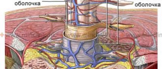
The first thing you need to understand when it comes to spinal cord puncture (which is what patients most often call this procedure), it does not mean a puncture of the tissue of the central nervous system organ itself, but only the collection of a small amount of cerebrospinal fluid, which washes the spinal cord and brain . Such manipulation in medicine is called a spinal, or lumbar, puncture.
Why is a spinal cord puncture performed? There can be three purposes for such manipulation – diagnostic, analgesic and therapeutic. In most cases, a lumbar puncture of the spine is done to determine the composition of the cerebrospinal fluid and the pressure inside the spinal canal, which indirectly reflects the pathological processes occurring in the brain and spinal cord. But specialists can perform a spinal cord puncture for therapeutic purposes, for example, to administer medications into the subarachnoid space to quickly reduce spinal pressure. Also, one should not forget about such an anesthesia method as spinal anesthesia, when anesthetics are injected into the spinal canal. This makes it possible to perform a large number of surgical interventions without the use of general anesthesia.
Considering that in most cases, spinal cord puncture is prescribed for diagnostic purposes, this type of research will be discussed in this article.
A lumbar puncture is taken to examine the cerebrospinal fluid, which can help diagnose some diseases of the brain and spinal cord. Most often, such manipulation is prescribed for suspected:
- infections of the central nervous system (meningitis, encephalitis, myelitis, arachnoiditis) of a viral, bacterial or fungal nature;
- syphilitic, tuberculous lesions of the brain and spinal cord;
- subarachnoid bleeding;
- abscess of the central nervous system;
- ischemic, hemorrhagic stroke;
- traumatic brain injury;
- demyelinating lesions of the nervous system, such as multiple sclerosis;
- benign and malignant tumors of the brain and spinal cord, their membranes;
- Guienne-Barré syndrome;
- other neurological diseases.
It is prohibited to take a lumbar puncture for space-occupying formations of the posterior cranial fossa or temporal lobe of the brain. In such situations, taking even a small amount of cerebrospinal fluid can cause dislocation of brain structures and cause strangulation of the brain stem in the foramen magnum, which entails immediate death.
It is also prohibited to perform a lumbar puncture if the patient has purulent-inflammatory lesions of the skin, soft tissues, or spine at the puncture site.
Relative contraindications are severe spinal deformities (scoliosis, kyphoscoliosis, etc.), since this increases the risk of complications.
With caution, puncture is prescribed to patients with bleeding disorders, those who take drugs that affect blood rheology (anticoagulants, antiplatelet agents, non-steroidal anti-inflammatory drugs).
The lumbar puncture procedure requires preliminary preparation. First of all, the patient is prescribed general clinical and biochemical blood and urine tests, and the state of the blood coagulation system is necessarily determined. The lumbar spine is examined and palpated. To identify possible deformations that may interfere with the puncture.
You need to tell your doctor about all the medications you are currently taking or have recently taken. Particular attention should be paid to drugs that affect blood clotting (aspirin, warfarin, clopidogrel, heparin and other antiplatelet agents and anticoagulants, non-steroidal anti-inflammatory drugs).
You also need to inform the doctor about possible allergies to medications, including anesthetics and contrast agents, recent acute illnesses, or the presence of chronic illnesses, as some of them may be a contraindication to the study. All women of childbearing age should tell their doctor if they may be pregnant.
It is forbidden to eat for 12 hours before the procedure and drink for 4 hours before the puncture.
The procedure is performed with the patient lying on his side. In this case, you need to bend your legs as much as possible at the knee and hip joints, bringing them to the stomach. The head should be bent forward as much as possible and close to the chest. It is in this position that the intervertebral spaces widen well and it will be easier for the specialist to get the needle into the right place. In some cases, the puncture is performed with the patient sitting with the back as rounded as possible.
The specialist selects the puncture site by palpating the spine so as not to damage the nerve tissue. The spinal cord in an adult ends at the level of the 2nd lumbar vertebra, but in short people, as well as in children (including newborns), it is slightly longer. Therefore, the needle is inserted into the intervertebral space between the 3rd and 4th lumbar vertebrae or between the 4th and 5th. This reduces the risk of complications after puncture.
After treating the skin with antiseptic solutions, local infiltration anesthesia of soft tissues is carried out with a solution of novocaine or lidocaine using a regular syringe with a needle. After this, a lumbar puncture is performed directly with a special large needle with a mandrel.
The puncture is made at the selected point, the doctor directs the needle sagittally and slightly upward. At approximately a depth of 5 cm, resistance is felt, after which a peculiar dip of the needle follows. This means that the end of the needle has entered the subarachnoid space and you can begin collecting cerebrospinal fluid. To do this, the doctor removes the mandrin (the inner part that makes the instrument airtight) from the needle and cerebrospinal fluid begins to drip from it. If this does not happen, you need to make sure that the puncture is performed correctly and that the needle enters the subarachnoid space.
After collecting the cerebrospinal fluid into a sterile tube, the needle is carefully removed and the puncture site is sealed with a sterile bandage. For 3-4 hours after the puncture, the patient should lie on his back or side.
The first step in cerebrospinal fluid analysis is to assess its pressure. Normal values in a sitting position are 300 mm. water Art., in a lying position – 100-200 mm. water Art. As a rule, pressure is assessed indirectly - by the number of drops per minute. 60 drops per minute corresponds to the normal value of cerebrospinal fluid pressure in the spinal canal. Pressure increases during inflammatory processes of the central nervous system, with tumor formations, with venous stagnation, hydrocephalus and other diseases.
Next, the cerebrospinal fluid is collected into two 5 ml tubes. They are then used to carry out the necessary list of studies - physicochemical, bacterioscopic, bacteriological, immunological, PCR diagnostics, etc.
In the vast majority of cases, the procedure takes place without any consequences. Naturally, the puncture itself is painful, but pain is present only at the stage of inserting the needle.
Some patients may develop the following complications.
It is generally accepted that after a puncture a certain amount of cerebrospinal fluid flows out of the hole, as a result of which intracranial pressure decreases and a headache occurs. This pain resembles a tension headache, has a constant aching or squeezing character, and decreases after rest and sleep. It can be observed for 1 week after the puncture; if cephalgia persists after 7 days, this is a reason to consult a doctor.
Sometimes traumatic complications of puncture can occur, when the needle can damage spinal nerve roots and intervertebral discs. This is manifested by back pain, which does not occur after a correctly performed puncture.
If large blood vessels are damaged during the puncture, bleeding and hematoma formation may occur. This is a dangerous complication that requires active medical intervention.
Occurs when there is a sharp drop in cerebrospinal fluid pressure. This is possible in the presence of space-occupying formations in the posterior cranial fossa. To avoid such a risk, before taking a puncture, it is necessary to perform a study for signs of dislocation of the midline structures of the brain (EEG, REG).
They may occur due to violation of the rules of asepsis and antisepsis during puncture. The patient may develop inflammation of the meninges and even form abscesses. Such consequences of puncture are life-threatening and require the prescription of powerful antibacterial therapy.
Thus, spinal cord puncture is a very informative technique for diagnosing a large number of diseases of the brain and spinal cord. Naturally, complications during and after the manipulation are possible, but they are very rare, and the benefits of puncture far outweigh the risk of developing negative consequences.
Based on materials from moyaspina.ru
Diagnosis of neurosyphilis
Early neurosyphilis is diagnosed based on characteristic symptoms with positive tests for infection. To identify the disease, the Wasserman reaction, Treponema pallidum immobilization test (TPI), enzyme-linked immunosorbent assay (ELISA), and polymerase chain reaction (PCR) are widely used.
Late neurosyphilis is more difficult to determine, since in more than half of the cases the pathogen is absent in free form in the patient’s blood. Waserman and RIBT reactions can be negative. MRI and MSCT data record nonspecific changes in the brain. However, a qualified specialist is able to distinguish them from other similar symptoms. Positive tests for syphilis in the past can help in diagnosis.
Most forms of neurosyphilis are diagnosed by analyzing the cerebrospinal fluid. There are large numbers of leukocytes and lymphocytes in it and the absence of pronounced symptoms of meningitis is a characteristic symptom of neurosyphilis.
Asymptomatic meningitis
Asymptomatic (latent) meningitis is registered in 10 - 15% of cases in patients with primary syphilis, in 20 - 50% in patients with secondary and latent early syphilis. In most cases, symptoms of meningitis cannot be identified. Previously, latent meningitis was called “syphilitic neurasthenia”, since the symptoms of neurasthenia came to the fore - severe fatigue, exhaustion, decreased mood, absent-mindedness, forgetfulness, indifference, irritability, decreased performance. Sometimes patients are bothered by persistent headaches, attacks of dizziness, a feeling of stupefaction, and difficulty concentrating. Meningeal symptoms are rare. Serological reactions of the cerebrospinal fluid (Wassermann reaction and RIF) are positive, pleocytosis (increased lymphocytes and polynuclear cells) of more than 5 cells per 1 mm3 and an increased amount of protein are noted - more than 0.46 g/l.
In early forms of syphilis, asymptomatic meningitis is one of its manifestations, like chancre or secondary syphilides. But in late forms of syphilis, asymptomatic meningitis requires active treatment, as neurosyphilis develops against its background.
Only with neurosyphilis are changes in the cerebrospinal fluid observed in the absence of clinical symptoms.
Rice. 4. Damage to the oculomotor nerve (photo on the left) and pupillary disorders (anisocoria) in the photo on the right with neurosyphilis.
Treatment of neurosyphilis
The basis of treatment for any form of syphilis is aggressive antibacterial therapy. For this purpose, penicillin and cephalosporin drugs are used. Treatment of early neurosyphilis is often combined, with the simultaneous administration of several drugs. The usual regimen is penicillin, ceftriaxone and probenecid. Method of administration: intravenously. Penicillin is also injected into the spinal canal. Treatment is continued for two weeks, in adequate doses. With proper inpatient treatment, the disease recedes.
Late neurosyphilis cannot be treated with antibiotics, since the pathogen itself may not be in the body, and the disease “rolls” along the rails laid by treponema pallidum. For this reason, patients with this form of the disease are usually unable to infect other people. In addition, due to severe damage to the nervous system, sexual relations are the last thing on the patient’s mind.
To treat late neurosyphilis, bismuth and arsenic preparations are used, which are highly toxic and therefore used only in specialized hospitals.