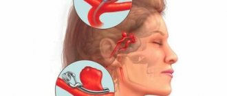
Causes of cerebral arachnoiditis
Children and people under 40 years of age are patients diagnosed with arachnoiditis. Weakness of the body contributes to serous inflammation of the arachnoid membrane of the brain.
Working in low temperatures, in chemical plants with toxic substances, lack of vitamins and sunlight, and alcohol dependence predispose to the disease. A combination of factors of various origins influences the development of the pathological process.
Pathogenesis of arachnoiditis
Classification of the causes of arachnoiditis:
- allergic;
- infectious;
- traumatic;
- oncological
In addition, a distinction is made between true and residual (in the form of a complication).
Bacterial infection from chronic lesions near the brain (tonsillitis, otitis, periodontitis, chronic sphenoiditis), complications from previous infectious diseases of the membranes cause inflammation of the connective tissue.
Bruises and concussions disrupt the structure of the arachnoid part and provoke a pathological process. Neoplasms (benign and malignant) destroy brain cells, which manifests itself in the form of impaired circulation of cerebrospinal fluid.
The cause of true arachnoiditis is an allergic reaction of the body to the transport of cerebrospinal fluid. An autoimmune attack is accompanied by a response: thickening and adhesion of the membranes. The frequency of manifestations does not exceed a few percent.
All other causes cause a residual form of the pathological process.
Violation of the circulation function of the membranes leads to the accumulation of cerebrospinal fluid in the ventricle and the formation of cysts. Such phenomena cause an increase in intracranial pressure and corresponding symptoms:
- headaches with nausea and vomiting;
- vegetative-vascular disorders;
- dysfunction of the optic nerve;
- fatigue;
- dizziness;
- convulsions.
Violation of liquor flow does not appear immediately, with a delay in time, for example:
- after a viral infection – several months;
- after TBI – a year and a half later.
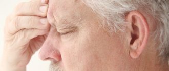
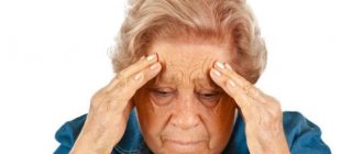
In arachnoiditis, the symptoms in the early stages are similar to asthenia and neurasthenia. Common to all patients is the complaint of severe morning headache. Attacks can range from 1-2 times a month to weekly or more often. The duration of the jump in intracranial pressure can be from several minutes (mild) to several days (severe).
Depending on the location of the source of pathology in the cerebral cortex, the manifestations of the disease have special features:
- impaired sensitivity and mobility in the limbs;
- convulsive seizures, including epileptic seizures;
- inflammation of the visual, auditory, facial nerves;
- weakening of memory;
- deterioration in coordination of movements.
Swelling of brain tissue can override the body's neurosympathetic regulation, causing breathing and heartbeat to stop.
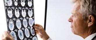
A diagnosis of suspected damage to the arachnoid membrane is made in a hospital setting, using radiography, CT, MRI, and EEG.
Diagnostic signs of cerebral arachnoiditis
During the examination, attention is paid to the relationship between previous infectious diseases (influenza, measles), inflammation of the meninges, head and spinal cord injury and neurological signs.
Manifestations of the disease are progressive, long-term in nature with periods of exacerbation and rest, affecting the functional state of the head structures, musculoskeletal system, mental and neurological reactions.
Diagnosis of symptoms of arachanoiditis determines:
- presence of intracranial pressure (X-ray);
- the amount of intracranial pressure (cerebrospinal fluid sampling);
- presence of cysts and adhesions (CT and MRI);
- hydrocephalus (Echoelectrography).
The increased content of protein, cells and serotonin in the cerebrospinal fluid makes it possible to distinguish this pathology from other neurological diseases.
Differential symptoms of the disease
Foci of aranchoidal inflammation have their own symptoms, which can be identified during examination.
Convextal arachnoiditis (based on EEG):
- increased excitability of the cerebral cortex;
- epilepsy attacks.
A narrowing of the visual field is typical for patients with damage to the basal layer. Basal arachnoiditis is diagnosed after examination by an ophthalmologist, who reveals swelling and compression of brain tissue in the area of the optic nerve.
The otolaryngologist determines the degree of damage to the auditory nerve (hearing loss, tinnitus), which is typical for pathology of the posterior cranial fossa.
Symptoms of different stages
With true arachnoiditis, the damage to the meninges is diffuse and therefore does not have obvious manifestations. The consequences of neuroinfection, trauma, and oncology that are localized occur in a more severe form.
The development of the disease can occur in one of three ways:
- in acute form;
- subacute;
- chronic.
Signs of acute course:
- vomit;
- Strong headache;
- temperature.
Subacute course:
- weakness;
- insomnia;
- decreased hearing and vision;
- lack of coordination;
- dizziness;
- impaired skin sensitivity in the extremities.
The chronic course is expressed in an increase in all symptoms:
- the appearance of convulsions and seizures;
- deafness;
- blindness;
- weakening of mental abilities;
- paralysis and paresis.
Most often, the disease occurs in a subacute form with transition to chronic. Headache has different symptoms: morning, worse with tension, occurring when jumping with a hard landing (on the heels). In addition, dizziness, weakening of memory, attention, insomnia, irritability and weakness are also symptomatic.
Types of arachnoiditis and their signs
Based on the location of the inflammatory focus, arachnoiditis is divided into several types.
- Cerebral
Cerebral arachnoiditis is an inflammation of the arachnoid membrane and the cortex of the cerebral hemispheres. Depending on the location, it can be convextal or basal. It is characterized by a sharp increase in intracranial pressure, especially after mental fatigue, physical exertion, and cold exposure. Accompanied by epileptic seizures and memory impairment.
- Optical-chiasmatic
Post-traumatic cerebral arachnoiditis causes the formation of adhesions and cysts in the basal layer. Compression and disruption of the nutrition of the optic and auditory nerves causes their atrophy, which leads to a decrease in acuity and a narrowing of the field of vision, and the development of hearing loss. Sinusitis, tonsillitis, syphilis can cause opto-chiasmatic arachnoiditis.
Chronic sphenoiditis (inflammation of the mucous membrane of the nasal sinus) is a source of infection located next to the optic nerve. This disease is difficult to diagnose and often causes inflammation of the membranes of the brain.
Associated symptoms: pain in the back of the head, nausea, vomiting. The process develops slowly, first affecting one eye and then the other.
- Spinal
Traumatic damage to the spine, as well as purulent foci (furunculosis, abscess) lead to inflammation of the arachnoid membrane of the spinal cord. The affected areas are the thoracic, lumbar, and sacral regions. Compression of the nerve processes is accompanied by pain, decreased conductivity, and impaired circulation in the extremities.
- Adhesive
Adhesive arachnoiditis means the occurrence of numerous adhesions due to purulent inflammation of the brain tissue. The circulation of cerebrospinal fluid is impaired, and hydrocephalus develops. Headaches upon awakening with nausea and vomiting, depression of visual function, constant drowsiness, apathy are characteristic signs of the adhesive process.
- Cystic
Cystic arachnoiditis is the formation of cavities filled with cerebrospinal fluid, changing the structure of the brain due to compression of nearby tissues. Constant pressure on the hard shell of the brain causes persistent, bursting headaches. The most common cause of cystic formations is a concussion. The consequences manifest themselves in the form of convulsive seizures without loss of consciousness, unsteady gait, nystagmus (involuntary eye movements).
- Cystic-adhesive
Cystic adhesive arachnoiditis is characterized by the formation of cystic areas in the adhesive membrane. As a result of a constant destructive process, the following are observed:
- headaches when concentrating;
- dizziness;
- fainting;
- weather sensitivity;
- metabolic disorders;
- changes in skin sensitivity;
- epileptic seizures.
As a result, nervous exhaustion and depression develop.
Diagnostics
In some cases, it takes some time to make a correct diagnosis. First of all, it is imperative to exclude a brain tumor, since the same symptoms are observed with this disease. It is necessary to conduct a survey craniogram, the results of which most often make the correct diagnosis and prescribe treatment.
When examining the spinal fluid, a slight increase in the number of lymphocytes is detected. During puncture, fluid flows out under high pressure and this is another sign of arachnoiditis. However, computed tomography and magnetic resonance imaging are of decisive importance in diagnosis.
It is also necessary to conduct a survey of the patient himself, because inflammation of the arachnoid membrane most often occurs after an infection, and this in most cases is otitis media, sinusitis and other purulent diseases of the sinuses.
Complications and consequences of arachnoiditis
The pathological process leads to the development of cerebral hydrocele and increased intracranial pressure. As a result, the vegetative-vascular system, vestibular apparatus, visual and auditory nerves suffer, and epilepsy develops.
Vegetative-vascular disorders:
- changes in blood pressure;
- tingling and burning in the fingertips;
- skin hypersensitivity.
Vestibular apparatus:
- intermittent claudication;
- unsteadiness on one leg;
- falling when landing on the heel;
- inability to connect the fingers with the tip of the nose.
Nystagmus, decreased vision to the point of blindness, hearing loss are complications of arachnoiditis.
Convulsions and convulsive seizures over time can develop into status epilepticus (a seizure lasting more than half an hour or a series of short-term, incessant seizures). A disorder of consciousness and the development of mental disorders occur.
Decreased ability to work is the main consequence of cerebral arachnoiditis. Depending on the severity of the disease, the patient becomes either partially limited in ability to work or completely disabled. High ICP levels at a constant level can lead to the death of the patient.
Correct therapy
Arachnoiditis should never be treated with folk remedies. This is a very serious disease, and only a specialist can help here to prevent complications from developing. But if the disease is left unattended, then after a while the patient may become disabled.
In the treatment of the disease, antiallergic drugs are used, such as suprastin, tavegil, pipolfen. Biogenic stimulants are also used to resolve adhesions. This is aloe, Phibs, lidase. When intracranial pressure increases, diuretics are added to treatment. If the prescribed therapy does not produce any result, surgical treatment is performed.
Even after treatment, patients often receive group III disability. If a person experiences frequent attacks of epilepsy, he receives group II. But group I is given to those whose disease has led to blindness.
Treatment of arachnoiditis
Treatment of cerebral arachnoiditis is carried out comprehensively:
- therapy for the cause of inflammation;
- dissolution of adhesions;
- decreased intracranial pressure;
- suppression of convulsive excitability;
- treatment of mental and nervous disorders.
To suppress foci of infection, including neuroinfections, therapeutic agents in the form of antibacterial drugs are used. In the diffuse form, antiallergic drugs and glucocorticoids are prescribed.
Absorbable drugs help normalize the cerebrospinal fluid balance in the brain and spinal cord. Diuretics are used to reduce blood pressure.
Anticonvulsant treatment is aimed at inhibiting motor centers using medication. To restore nerve conduction, neuroprotectors are prescribed.
In general, it is necessary to restore the body's strength through diet, sleep and rest.
All types of arachnoiditis require long-term treatment.
Surgical intervention is used if blindness and the patient’s life are threatened. It is aimed at ensuring the outflow of cerebrospinal fluid. For this purpose, dissection of adhesions, shunting (discharge of cerebrospinal fluid beyond the skull), and removal of cysts are used.
Physiotherapy
Patients with arachnoiditis may be prescribed magnetic therapy.
Treatment with physical factors complements medication and is prescribed to improve cerebral liquor dynamics, microcirculation and metabolism of nervous tissue, as well as to restore the normal functioning of the nervous system.
The main physical methods used to treat arachnoiditis:
- drug electrophoresis of neurostimulants, vasodilators and metabolic stimulants;
- low-frequency magnetic therapy (improves metabolic processes, stimulates neuroendocrine processes);
- low-intensity DMV therapy (reduces intracranial pressure by increasing renal blood flow and diuretic effect, normalizes the functioning of the nervous system);
- transcerebral UHF therapy (increases blood and lymph circulation, metabolic processes, reduces inflammation);
- sodium chloride baths (have a diuretic effect as a result of reducing the reabsorption of sodium ions from primary urine, normalizes the activity of the sympathetic-adrenal system);
- fresh baths (increases blood flow in organs and tissues, glomerular filtration and diuresis);
- aerotherapy (increases the body’s nonspecific reactivity, improves the psycho-emotional state, activates metabolism);
- Thalassotherapy (improves microcirculation, trophism and tissue metabolism);
- peloidotherapy (increases metabolism, improves the functioning of the autonomic nervous system).
Prevention of arachnoiditis
Timely diagnosis of arachnoiditis at the first symptoms of neurological abnormalities will prevent the development of the disease. Examination after infectious diseases or brain injuries must be carried out if headaches appear over time. Foci of infection, especially purulent ones, must be treated until complete recovery, preventing them from becoming chronic.