What's happened
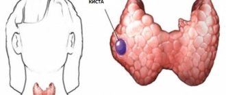
All cystic defects consist of a cavity filled with liquid contents. A subependymal cyst in a newborn child is formed due to exposure to provoking factors.
Minor neoplasms are not dangerous to the health of the baby and are prone to spontaneous resorption. Large tumors, on the contrary, require constant monitoring by a specialist, as they can cause serious consequences, including death.
Subependymal cystic formations are considered common among newborns who were born with various problems. The defect is formed due to injuries to the walls of small blood vessels, after which a small outpouring of blood occurs. Special cells process blood, and the resulting cavity is filled with cerebrospinal fluid.
Such minor neoplasms subside after a few months, do not cause neurological disorders, and therefore do not require drug therapy. However, this does not apply to extensive defects that threaten the child’s health.
Subependymal cyst in a newborn baby: symptoms, treatment, consequences
The main source of the problem is oxygen starvation of the fetal brain. Insufficient blood supply leads to many pathologies, including the formation of a cerebral cyst. According to statistics, 10 out of 100 women are diagnosed with hypoxia during childbirth. That is why expectant mothers are strongly recommended to carry out the necessary examinations in time and rule out an abnormality in the baby’s development.
- multiple pregnancy;
- difficult childbirth, birth injuries;
- anemia;
- infectious diseases during pregnancy;
- iron deficiency;
- placental insufficiency.
As for the treatment of this pathology, removal of a brain cyst occurs only in cases of increased risk to the child’s life, for example, with a progressive increase in the volume of the tumor. In such a situation, tissue displacement in the brain occurs and immediate surgical intervention is required.
If the baby’s health is favorable, the child’s subependymal cyst may completely resolve or its growth may stop. In such cases, there is nothing to worry about; you just need to monitor and maintain the health of the little patient. Young parents need to seek help from competent specialists: a therapist and a neurologist.
What does Komarovsky say about the cyst?
Cystic formations of the brain occur in all age groups.
The reasons for their appearance are not fully understood; it is believed that most often they arise due to injuries, including birth injuries, and circulatory disorders.
Every tenth newborn experiences intrauterine hypoxia due to insufficient blood supply to the brain. Therefore, subependymal cysts in infants are diagnosed quite often.
What kind of education is this? How does the pathology manifest itself? Does the child need medical or surgical treatment? Parents need to know the answers to these questions precisely because of the prevalence of the problem.
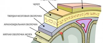
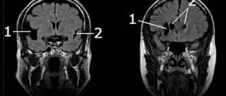
Ependyma is a neuroepithelium that lines the inside of the cavity of each cerebral ventricle and the central canal of the spinal cord. It can serve as a source of formation of intracerebral cysts.
In this case, cell hyperplasia is activated and their differentiation is slowed down. The neoplasm can be located either on top of the ependyma, that is, grow in the cavity of the ventricles, or under the ependymal layer.
Various pathological influences lead to disruption of blood supply to certain areas of the brain. Due to a lack of oxygen and nutrients, an area of necrosis forms in the brain tissue, located subependymal. As a result of tissue necrosis, a cavity filled with liquid is formed.
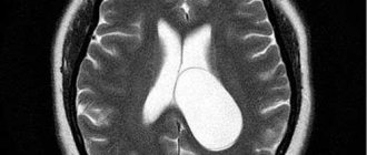
According to medical statistics, intracerebral cysts in most cases are located in the 4th ventricle, sometimes the lateral ventricles are affected.
Etiology
The most common cause of the appearance of this neoplasm is intrauterine hypoxia. Birth trauma, complicated by hemorrhage into the ventricles or into the brain tissue, also contributes to the occurrence of cystic cavities. As a result, the intrauterine development of the fetus is disrupted, and congenital abnormalities of the brain structure may form.
Most often, oxygen deficiency develops in the following pathological conditions of the expectant mother:
- anemia;
- multiple pregnancy;
- late toxicosis;
- viral and bacterial infectious diseases.
Also, a subepindymal neoplasm can appear during Rh-conflict pregnancy or placental circulatory disorders.
Provoking factors include drug and alcohol intoxication of a woman, stress, poor ecology and exposure to ionizing radiation during pregnancy.
It is believed that at any age, the impetus for the occurrence of cystic formations can be a brain injury, inflammatory diseases of the central nervous system (meningitis, encephalitis). Heredity plays an important role in the etiology of benign neoplasms of the central nervous system.
In articles on medical topics, you can often find the expression cerebral pseudocyst in newborns. Unlike cysts, it is not a pathological phenomenon and has a different mechanism of formation.
After the birth of a child, cerebrospinal fluid enters the choroid plexuses of the ventricles of the brain, forming pseudocysts.
They are small, round, do not grow, and are involved in the production of cerebrospinal fluid necessary for the normal functioning of the brain.
Subependymal pseudocyst is usually determined only using instrumental examination methods. As a rule, it does not give any clinical symptoms. Pseudocysts resolve on their own in children.
A cyst of subepinedymal localization is distinguished by the fact that it is mainly benign histologically, but there is a possibility of rapid growth and malignancy.
Therefore, all children with a history of intrauterine hypoxia or birth trauma require careful dynamic monitoring of the condition and size of this formation.
They are at risk.
Based on the growth pattern and histological structure, brain tumors are conventionally divided into benign and malignant. But with rapid growth, they are all malignant.
After all, tumors develop in the confined space of the cranium, put pressure on the surrounding brain structures, block the circulation of cerebrospinal fluid and venous outflow, causing a clear clinical picture of malignancy.
Clinic
Subependymal cysts of the brain in newborns can be asymptomatic and resolve within the first year of life, even without treatment. Especially if it does not exceed 5 mm in diameter. But often a cyst, not detected during the first month, after about 6 months, and more often in the second year of life, gives a certain clinical picture.
Symptoms and dynamics of the disease in infants depend on the type, size, and location of the cyst.
A small intracerebral cyst is not dangerous. A growing benign tumor can cause compression and displacement of surrounding brain tissue. A clinical picture similar to brain cancer arises, so an increase in size always alarms specialists.
The localization of intracerebral cysts is determined clinically by focal signs.
The symptom of pressure of a large intracerebral cyst on the occipital lobe is visual impairment. The child has poor vision and at a conscious age complains of blurred vision and double contours of objects.
An intracerebral cyst of the left hemisphere, affecting the cerebellar tracts, causes muscle hypotonia, hearing loss and coordination, more pronounced on the left.
Any mass formation gives cerebral and focal symptoms.
General cerebral signs include hypertensive-hydrocephalic syndrome. It is caused by the development of intracranial hypertension and cerebral edema, manifested by a number of cerebral symptoms:
- headache (monotonous crying, causeless anxiety);
- vomiting (constant regurgitation);
- expansion of superficial veins on the head;
- increase in head size;
- bulging and pulsation of the fontanel;
- seizures;
- dizziness, loss of coordination;
- mental changes (mental retardation);
- forced head position;
- hearing impairment.
Complications of cyst development: formation of a brain abscess, rupture of the cyst membrane, transformation into a cancerous tumor. The consequence of a rapid increase in size of an intracerebral cyst with compression of vital centers is coma with possible death.
Diagnostics
In the presence of intrauterine hypoxia, birth trauma, birth by cesarean section, the baby must undergo neurosonography (ultrasound through the fontanel).
Treatment
Small cysts are tried to be treated conservatively. The choice of medications is determined by the cause of the development of the pathology. Antibiotics, antivirals, immunostimulants, and absorbent drugs are prescribed.
With progressive growth and the appearance of neurological symptoms, treatment with surgical intervention is necessary.
Surgical treatment can be performed in the following ways:
- Bypass surgery - only fluid is removed from the cystic formation.
- Endoscopic intervention - the anomaly is removed using a gentle method.
- Neurosurgical operation - craniotomy.
After the operation, courses of immunostimulating, anti-inflammatory and restorative therapy with control examinations of the brain are required
The prognosis in most cases can be considered favorable. Often intracerebral cysts resolve on their own without any consequences.
But dynamic observation by a neurologist with periodic ultrasound, MRI of the brain and course treatment are necessary in any case.
Often in adolescence, small intracerebral cysts begin to grow rapidly, causing numerous complaints.
Subependymal cyst is a serious pathology that requires early diagnosis and constant dynamic monitoring by specialists. Treatment must be timely to avoid complications, often life-threatening. The joint efforts of doctors and parents will help the baby grow up healthy.
Total: 45
A subependymal cyst is a benign formation whose cavity is filled with fluid. A tumor forms in brain structures during fetal development or in newborns.
Classification
Symptoms and selection of treatment depend on the characteristics of the subependymal cyst in the newborn. Depending on the size, formations are divided into small and large.
The first include defects with a diameter of up to 3 cm. Most often, such cavities resolve on their own, do not threaten the child and do not require any medical intervention. However, it is better to carry out routine inspections periodically.
With large formations, the risk of developing serious complications increases significantly. To protect the baby from dangerous problems, the doctor is obliged to prescribe the correct treatment, including surgery.
Based on the tendency to increase, tumors can be enlarged or non-enlarged. The first type includes cystic formations that rapidly progress, compress the tissue structures of the gray matter, and disrupt the functioning of the brain. In this case, immediate surgical intervention is required.
Non-enlarging defects do not reach large sizes and the patient’s condition does not worsen. In such a situation, the prognosis is considered favorable.
Depending on the number of chambers, cysts are divided into single-chamber and multi-chamber neoplasms. The first type is not so dangerous and can be effectively treated.
On this topic
- central nervous system
How does a headache with a brain tumor hurt?
- Natalya Gennadievna Butsyk
- December 3, 2021
Multi-chamber cavities cause symptoms similar to those of many diseases, so diagnosing this pathology is difficult. In addition, the second type is difficult to eliminate.
It is equally important to differentiate the subependymal capsule from other cystic diseases of the brain in newborns. The development of an arachnoid cyst is affected by damage and infection. This disease progresses rapidly and requires immediate treatment.
Sometimes a vascular plexus cyst is also diagnosed. This defect is formed during the prenatal period if the expectant mother is infected with the herpes virus. the disease often goes away on its own.
Types and forms
Based on its size, the subependymal cyst is classified into:
- small. They are less than 3 cm. Usually such neoplasms do not threaten the child. They often resolve on their own and only rarely require treatment. It is quite enough to be examined regularly to monitor the development of such formations;
- big. The larger the tumor, the higher the risks for the baby. Such cysts require active treatment, and in some cases, surgery.
These tumors can also be:
- not increasing. If they are small and do not grow, then the chance of independent recovery is very high;
- increasing. In such cases, intensive treatment and even surgery may be necessary.
Also, such formations differ in the number of chambers:
- single-chamber. They pose less danger and are easier to treat than others;
- multi-chamber. These cysts are more difficult to diagnose (symptoms may resemble other diseases) and much more difficult to treat.
Causes
Cystic pathology occurs due to the influence of the following provoking factors:
- Hypoxia. This condition occurs with anemia, diabetes, problems with the kidneys, heart, lungs, and smoking.
- Birth injuries. Head injuries during childbirth significantly increase the likelihood of developing the disease. This often happens if the birth canal is narrow, the fetal head is too large, or the pelvis is deformed.
- Toxicosis.
- Lack of microelements and vitamins. Deficiency occurs if the expectant mother is hungry or malnourished.
- Anemia. A decrease in the level of hemoglobin in the blood leads to a lack of oxygen, developmental delays, and vulnerability to infectious pathologies.
- Hemolytic disease. Due to different Rhesus levels in the mother and fetus, the immune system attacks the embryo. This condition requires immediate medical attention, as it threatens the child’s life.
- Infections. Even a common cold can seriously harm the fetus.
On this topic
- central nervous system
What are the dangers of a porencephalic cyst of the brain?
- Olga Vladimirovna Khazova
- May 27, 2021
Multiple pregnancies significantly increase the risk of complications. In addition, in this case, children are often born with low birth weight.
Excessive or deficient amount of amniotic fluid also affects the child’s health. Polyhydramnios leads to brain pathologies, and a lack of fluid leads to a lack of protection for the embryo from external factors.
Qualitative approach to therapy
Treatment of a cerebral cyst is aimed at preventing its growth. To do this, you need to find and eliminate the main cause that provokes oxygen starvation. During certain life stages of a child's growth, medical care will vary.
First aid
After birth, a neonatologist performs resuscitation care.
Removes fluid from the respiratory tract, stimulates artificial respiration; in severe cases, an oxygen mask is used to supply pure oxygen.
Article on the topic: Meningioma of the brain: treatment without surgery
The first three days after birth...
A neurologist conducts a daily examination and prescribes a restorative course of medication and rehabilitation therapy. Massage, physical therapy and sedatives are prescribed.
In childhood, the doctor observes the dynamics of the child’s development rate.
Medicines are used to stimulate speech functions and normalize the psycho-emotional state. At this stage, you may need the help of specialists such as a speech therapist and a psychologist.
...and beyond
When a child reaches adolescence, vitamin medications are prescribed to stimulate brain function and normalize metabolism. It is also necessary to take hormone replacement medications.
All physiotherapeutic and medicinal methods are prescribed only after the results of the examination and identification of deviations in the child’s development.
Clinical picture
Subependymal cystic formations affect both the left and right hemispheres of the gray matter. Most often, the ventricles filled with cerebrospinal fluid are affected.
Clinical manifestations depend on the location, size and rate of growth of the cyst. Different parts of the brain perform specific functions, so compression of them leads to specific symptoms.
Damage to the occipital zone leads to deterioration of visual acuity, up to complete blindness. When the temporal region is affected, hearing is impaired. A tumor of the cerebellum causes deterioration in motor coordination.
On this topic
- central nervous system
Everything you need to know about pituitary adenoma
- Olga Vladimirovna Khazova
- February 28, 2021
When the pituitary gland is damaged, developmental delays occur, and sometimes the child suffers from dwarfism. If the pathogenic process affects the frontal part, then speech formation is disrupted.
In addition to the signs described above, the baby may experience general symptoms such as worsening sleep, anxiety, weight loss, pathological muscle tone, and frequent crying for no reason.
A newborn child with a subependymal cyst often refuses to drink breast milk, spits up heavily, moves excessively, periodically loses consciousness, and suffers from attacks of epilepsy and tremors in the limbs.
Localization in the brain and symptoms
A subependymal cyst is localized at the site of impaired blood supply to brain tissue, most often in the ventricular region. Symptoms of the disease depend on the location and size of the cyst. The presence of a pathogenic focus in a certain area of the brain leads to disruption of the body functions for which it is responsible.
A cyst in the occipital region affects the functioning of the visual system, visual acuity decreases, double vision, etc. A cyst in the temporal lobe leads to hearing impairment, in the cerebellum – gait and coordination of movements are impaired, in the pituitary gland – hormonal imbalances.
The small size of the cyst may not manifest itself in any way on the child’s behavior. But larger ones lead to convulsions and strokes, which can result in paralysis of the body.
The main symptoms observed in the presence of a cerebral cyst in the head of a newborn child:
- sleep disturbance;
- anxious behavior;
- breast refusal;
- poor weight gain or loss;
- long, causeless crying;
- hyper- or hypotonicity of muscles;
- tremor of the chin, twitching of the limbs;
- hearing impairment;
- blurred vision, sometimes complete loss;
- regurgitation “fountain”;
- loss of consciousness, coma;
- pulsation and swelling of the fontanelle;
- convulsions.
The consequences of growth and lack of treatment of the cyst lead to mental and physical retardation in the development of the child.
Diagnostics
The main method for diagnosing subependymal cysts is ultrasound neurosonography. This procedure allows you to achieve an accurate result. Moreover, the manipulation is absolutely safe and painless for the child.
Ultrasound examination of the brain is prescribed for infants whose fontanelles have not yet ossified. The planned procedure should be carried out when the baby is one month old. However, if there are pathologies during pregnancy or after a difficult birth, manipulation is prescribed earlier.
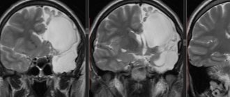
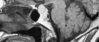
Additional methods are sometimes used to clarify the diagnosis. Computed tomography or magnetic resonance imaging allows you to accurately study the characteristics and location of the tumor. Due to exposure to radiation, CT scans are done no more than twice a year.
Preventive measures
If the mother cannot protect herself or the child from birth or intrauterine trauma, then it will be possible to exclude the causes leading to hypoxia if:
- give up smoking, alcohol, drugs;
- lead a healthy lifestyle;
- diversify your diet, filling it with vitamins and minerals;
- during pregnancy, do not start treatment for infection;
- Avoid contact with toxic substances: detergents, hair dyes, alkaline hygiene products.
Early detection, observation, and immediate treatment (if necessary) give a favorable prognosis. Comprehensive measures for the prevention and treatment of SEC can reduce the number of diseases and the negative consequences of the disease.
Treatment
The basis of therapy is the elimination of the factor that provoked the development of a subependymal cyst in a newborn. Most often it is necessary to treat hypoxia. The treatment process occurs in several stages.
Immediately after birth, in the presence of hypoxia, resuscitation procedures are immediately performed to prevent the development of formation. To do this, the trachea, oropharynx and nasopharynx are cleared of fluid, and artificial respiration is used.
After this, the baby’s condition is monitored by a neurologist for three days, who selects treatment tactics. Special medications are prescribed, courses of therapeutic massage and physiotherapeutic procedures are prescribed to improve the condition and eliminate the lack of oxygen.
If the cyst was diagnosed in childhood, then drugs are used to normalize metabolic processes in the gray matter, and vitamin complexes. In case of rapid increase and large size, surgery is performed.
Using an endoscope, the cavity is cleared of liquid contents. Sometimes open manipulation of the brain is required.
Treatment methods
Therapy for a subependymal cyst, namely the selection of a technique, will depend on the characteristics of the pathology. The doctor may prescribe resuscitation procedures if disturbances in the functioning of important organs are observed. If children are born in hypoxic conditions, they may need;
- artificial ventilation;
- correction of biological constants through the use of infusion treatment;
- detoxification procedures carried out in intensive care conditions.
If no threats to the life of the newborn are observed, but there are symptoms indicating damage to the brain, the specialist may resort to drug treatment, which includes the use of:
- nootropic drugs (nicergoline, piracetam, etc.), the work of which is aimed at improving metabolism in tissues;
- minerals and vitamins;
- diuretics if there is a risk of brain swelling or intracranial hypertension;
- anticonvulsants in the presence of seizures (Depakine, carbamazepine, etc.).
Treatment for infection involves the use of immunotherapy using immunoglobulin medications (pentaglobin and a number of others). In addition, antiviral pharmacological products are prescribed, which provide positive results in almost all cases.
Observation of an asymptomatic subependymal cyst is usually not accompanied by treatment. It is enough to carry out regular examinations, visiting a neurologist, and also undergo prescribed diagnostic measures (MRI or ultrasound). In some cases, if there is an asymptomatic cyst, specialists may prescribe the use of medications (nootropics and vitamins).
Complications
If diagnosis is delayed, the cystic neoplasm will progress, grow and compress the surrounding tissue structures. Focal manifestations will become more pronounced as the defect increases, and the child’s visual acuity will significantly decrease.
On this topic
- central nervous system
What threatens a cyst in the head?
- Olga Vladimirovna Khazova
- February 28, 2021
Often, a subependymal cyst causes an increase in intracranial pressure, as a result of which the newborn will begin to suffer from severe headaches, apathy, constant weakness, fatigue, dizziness, nausea with vomiting, and diplopia. Frequent loss of consciousness will also accompany an infant with this disease.
In the absence of timely treatment, extensive formation disrupts the development of the skull bones, causing physical and mental development to deteriorate. In advanced situations, the pathology leads to death. Sometimes cystic defects cause stroke.
How to treat a subependymal cyst in a child
For the normal development of the fetus in the womb, the supply of sufficient oxygen is very important. Otherwise, the risk of developing serious pathologies that will negatively affect the future life of newborns increases.
One of the most dangerous among them is a subependymal cyst in a newborn baby. Its location is always in the brain.
Pathology occurs against the background of improper blood circulation, prolonged absence of air and insufficient quantities of nutrients entering the baby’s body.
A cerebral cyst is classified as a benign formation in the ventricle of the brain. There is liquid inside it. Education is formed due to the lack of oxygen entering the body in sufficient quantities.
If hypoxia was diagnosed during the period of gestation, then the likelihood of developing pathology increases several times at once. In this case, the cyst forms against the backdrop of the body’s response to atrophic changes in tissue.
Hypoxia during pregnancy should be treated without fail. Otherwise, the cyst begins to actively increase in size. Against this background, the baby may periodically experience seizures, mood disorders or psycho-emotional states.
Cysts in a newborn are diagnosed in case of oxygen starvation of the fetus during gestation. That is why, when making this diagnosis, a woman must undergo the prescribed course of treatment. Additionally, it should be noted that the pathology in mothers occurs in acute and moderate forms.
Subependymal cysts are formed under the influence of the following negative external and internal factors:
- Ischemic disease was previously diagnosed in the brain. Quite often, cysts are formed under the influence of this particular factor. Ischemia brings about negative changes in the blood supply to certain areas of the brain. The formation provokes the appearance of empty areas, which over time are filled with a special liquid. If the formation is small in size, then no serious changes in the functioning of the child’s brain are observed. Otherwise, it is advisable to resort to drug treatment. Convulsions, vomiting, bulging of the fontanel outward and inhibition of development appear if the cyst continues to increase in size.
- Quite often, a neoplasm forms against the background of a cerebral hemorrhage. This situation is especially dangerous if it occurs in the womb. Hemorrhage occurs when the fetus becomes infected with infections, acute hypoxia, or receives a serious birth injury.
- Against the background of acute and moderate hypoxia, the fetus experiences a negative change in the functioning of the body. This situation has a bad effect on the woman and the placenta itself. Hypoxia develops against the background of anemia, toxicosis, multiple births and Rh conflict. Additional negative factors include polyhydramnios and infection with various infectious diseases.
A subependymal cyst is located in the brain, and therefore interferes with the necessary flow of blood to its tissues. Symptoms of the disease directly depend on the location of the formation. The pathogenic focus negatively affects the functioning of the body. The disorder occurs in the system for which the specific affected area of the brain is responsible.
If the cyst was formed in the occipital part, then infants experience deterioration in vision. With a detailed examination, it can be established that they are beginning to see double.
Damage to the temporal lobe is dangerous and can lead to serious hearing impairment. When the cyst is localized in the cerebellum, the child’s gait and coordination change negatively.
Children with the disease are often nervous and irritable
A small cyst cannot have a negative impact on the child’s behavior and well-being. However, large sizes are very dangerous. They can lead to seizures, stroke and even partial paralysis of the body. That is why a child with a disease must be constantly registered in the hospital. The location of the formation - left or right - is of no small importance.
The appearance of a hollow formation is a natural replacement reaction of the body to the death of brain tissue caused by hypoxia.
The presence of a small subependymal cyst in a newborn cannot be determined by any pathological signs or behavior. Symptoms characteristic of brain disorders begin to appear if the cyst grows and, due to its large size, puts pressure on the nerve centers and brain tissue. In such cases, the following changes in behavior and well-being can be observed in the child:
- sleep disturbance;
- excessive excitability or lethargy, accompanied by changes in muscle tone;
- causeless anxiety and tearfulness during wakefulness;
- lack of weight gain or weight loss;
- decreased visual and auditory functions;
- decreased sucking reflex and breast refusal as a consequence;
- twitching of limbs, trembling of the chin;
- profuse regurgitation;
- swelling of the fontanel and its pronounced pulsation;
- convulsions, epileptic attacks;
- loss of consciousness, which in severe cases can progress to the coma stage.
After a year of life, the presence of a subependymal cyst can be detected by signs of psychomotor and physical development. In such cases, the child experiences speech delay, decreased memory and concentration on the environment.
The location and size of the subependymal cyst determines the nature of the symptoms that appear. The volumetric formation, localized in the occipital part, affects the visual centers. A cyst in the temporal part negatively affects hearing, in the cerebellum - on the functions of the motor apparatus and coordination center, and in the pituitary gland - on the functioning of the hormonal system.
Since the cyst almost always develops on one side of the head (for example, in the left lobe of the temple or the right occipital part), the corresponding symptoms manifest themselves to a greater extent on this side (right or left). For example, with a tumor located on the left, visual, auditory and motor disorders affect the left side more.
Very rarely, when the tumor reaches a large size and its presence poses a threat to the health and life of the child, doctors may decide to perform an operation. In other cases, a conservative approach is used to treat subepindeminal cysts.
Depending on age and life stage, the following measures for the treatment of brain cysts are distinguished:
- Providing assistance to the baby in the first minutes after birth. Resuscitation care is provided by a neonatologist: he quickly removes fluid from the respiratory system; if necessary, performs artificial respiration; provides breathing with the help of an oxygen mask if the baby has a pronounced impairment of respiratory functions.
- Drug therapy and observation in the first three days. A neurologist examines the newborn daily. For obvious symptoms of oxygen starvation, he prescribes a complex of drugs that reduce fluid pressure on the brain and normalize blood supply to the brain. Depending on the indications, the baby may be recommended a course of physiotherapy and massages.
- Observation of the child by a neurologist in subsequent years. Regular examination by a neurologist allows you to track the dynamics of the baby’s physical and psychomotor development. To support brain activity, the doctor may prescribe a course of medications, refer to a psychologist or specialized specialists if the presence of a subependymal cyst causes inhibition in development.
- Therapeutic activities in adolescence. During this period, observation by a neurologist is mandatory. According to the indications, the teenager is prescribed vitamin complexes and medications that help improve brain function. Almost always, when a child is diagnosed with a subependymal cyst, hormone replacement therapy is prescribed.
Medications and courses of physiotherapy should be prescribed by a pediatric neurologist with the direct participation of a family pediatrician.
Localized in the structures of the brain, a small subependymal cyst does not pose any risks or threats to the child and does not reduce the full value of his mental development and growth.
Severe consequences can be caused by an increase in education provoked by infections, brain hypoxia and other pathological factors.
The growth of a subependymal cyst, depending on its location, causes delayed consequences that appear not in infancy, but 2-3 years after birth. In such situations, the child can observe:
- developmental delay (memory disorders, speech delay);
- psycho-emotional disorders (mood changes, irritability, tearfulness);
- anemia;
- cardiac and vascular pathologies;
- disturbances in the functioning of the bronchopulmonary system.
When a small cyst forms, doctors give a positive prognosis for recovery. With intensive growth of the subependymal cyst, the child may develop acute attacks of hypoxia, as a result of which the prognosis for healthy development and growth is significantly worsened, and the risk of death increases.
To reduce the likelihood of a subependymal cyst and irreversible consequences resulting from its growth, the expectant mother must take preventive measures while carrying the baby. During this period she needs:
- eat well;
- prevent the development of inflammatory processes and infections;
- avoid contact with toxic substances;
- Regularly visit an obstetrician-gynecologist and undergo appropriate diagnostic tests.
If a child is diagnosed with a subependymal cyst, then preventive measures to prevent its growth should be taken by a neurologist after a detailed examination.
When treating cystic formations, conservative therapy methods are used. The main task of medical personnel is to prevent further enlargement of the cavity. To do this, it is necessary to eliminate the cause-catalyst causing hypoxia.
Forecast
The prognosis of a subependymal cyst in a newborn is influenced by the size of the pathological cavity, location, timeliness of diagnosis and rate of increase. Minor neoplasms that do not increase in diameter do not cause serious consequences, so the prognosis in such situations is favorable.
Large formations that constantly grow threaten not only the health of the baby, but also his life. Sometimes even timely surgical intervention is not able to eliminate the problems that have arisen, as a result of which the child becomes disabled.
Therefore, it is important to detect a tumor at an early stage, when the defect does not compress neighboring tissues.
Etiological factors in the development of brain cysts in infants
In newborn babies, brain cysts are divided into 3 types:
- subependymal;
- choroid plexus cysts;
- arachnoid.
The following reasons can provoke fetal hypoxia, which is the main provocateur of the appearance of a cyst during intrauterine development:
- lack of iron in the blood;
- severe toxicosis, especially in the last trimester;
- anemia;
- Rh conflict between mother and child;
- multiple pregnancy;
- placental insufficiency;
- infectious lesions of the body.
The baby may experience oxygen starvation during labor. For this reason, a brain cyst can be a consequence of birth trauma.
Oxygen starvation can affect the further development of the child, so if there is a suspicion of hypoxia in the newborn, the doctor will prescribe an additional examination that will help determine whether there is a pathology. The child undergoes ultrasound diagnostics (echography) and additionally undergoes laboratory testing of the baby’s blood and amniotic fluid.
A subependymal cyst of the brain is located in that part of the brain that is filled with fluid and is called cerebrosinal.
When the blood circulation process in the brain tissue is disrupted, brain cells begin to die and a cavity forms in their place, which will soon fill with fluid. Impaired blood supply can lead to hemorrhage in the baby’s head, which will trigger the formation of a cyst.
The consequences of a subependymal cyst do not affect the physical and psychological health of the child. Problems begin when the cyst grows in size over time and puts pressure on the surrounding brain structures. As a rule, the symptoms of the pathology begin to manifest themselves in the second year of the baby’s life. The child has speech problems, poor concentration, lethargy and increased excitability.
In severe cases, the disease is accompanied by convulsions, neurological disorders, general deterioration of health and can cause death.
The reasons for the formation of a cyst of the nervous system in newborns in most cases are related to the mechanisms of its formation and various pathological factors (viruses, toxins, drugs) affecting the fetal brain cells in the prenatal period; a hereditary predisposition to the occurrence of neoplasms is also important.
1) a choroid plexus cyst, which appears due to infection of the fetus with the herpes virus, requires surgical treatment;
2) a subependymal (intracerebral) cyst develops as a result of oxygen starvation of brain tissue, which causes the death of neurons, and a cystic neoplasm forms in their place. Without timely surgical intervention, this type of cyst can cause significant disturbances in the development of the child (mental retardation, speech delay, visual impairment, vestibular disorders);
3) arachnoid cyst - this type of tumor is localized between the cerebral spaces and can develop in any part of the fetal brain. Treatment of arachnoid cysts is carried out using various surgical methods (endoscopic surgery, craniotomy or shunt surgery). In the absence of surgical intervention, the baby develops significant disorders in the psychoneurological sphere;
4) traumatic (acquired) cyst - formed as a result of birth trauma, compression or bruise during childbirth, with the development of intracranial bleeding and contributes to the development of various types of brain tumors.
A choroid plexus cyst in newborns and infants is a pathological neoplasm that appears in the prenatal period due to cystic growths of cerebral vessels as a result of the negative influence of pathogens of intrauterine infections (usually when infected with the herpes virus or toxoplasmosis) during pregnancy.
The choroid plexuses are structures that do not have nerve endings and play a huge role in the blood supply to the fetal brain and its maturation; their active development begins from the sixth week of the baby’s development. When a child becomes infected early and a choroid plexus cyst forms, these formations often resolve on their own before 25-38 weeks of pregnancy - experts associate this with the active growth and development of the fetal nervous system.
Also, these neoplasms do not affect the development of the child. Choroid plexus cysts of medium and large sizes are determined using ultrasound at 17-20 weeks of fetal development. But these pathological neoplasms of the choroid plexuses of the brain can appear in a newborn after birth with massive infection of the fetus in late pregnancy or during childbirth with the gradual implementation of intrauterine infection.
Choroid plexus cysts in newborns are classified as “soft markers” that are absolutely harmless and do not affect the functions and development of the brain, but can increase the possibility of developing other diseases or cause disturbances in the functional systems of the body. In most cases, these neoplasms disappear without a trace by the first year of a child’s life.
Due to the risk of developing various diseases of other organs, when diagnosing a choroid plexus cyst, mandatory ultrasound monitoring of the presence, location and concomitant pathologies is necessary. The child is re-examined at three months of age, then at six months and when he reaches one year. In the absence of positive dynamics for independent resorption of the cyst, the attending physician, based on the results of research and the child’s development, makes a decision on further observation or treatment of the baby individually.
A subependymal cyst is considered a serious pathology that forms in the brain tissue of a fetus or newborn due to significant oxygen deprivation of brain tissue or as a result of hemorrhages in the ventricles of the brain due to birth injuries. Often this type of cystic neoplasm resolves on its own, but mandatory monitoring (ultrasound of the brain) and a special course of treatment are required.
In most cases, this type of cyst does not increase in size and does not affect the baby's development. But when large, a subependymal cyst can cause displacement of brain tissue, which leads to the appearance and progression of neurological symptoms, requiring immediate surgical treatment.
A choroidal cyst in a newborn is a cystic neoplasm of the choroidal plexus of the brain. This type of cyst can develop due to the introduction and progression of an infectious process in the body or traumatic brain injury to the fetus during pregnancy or as a result of birth trauma. Choroidal cysts must be removed due to the fact that the probability of spontaneous resorption of this type of cyst is 45%.
Signs of a choroidal cyst in a newborn are:
- muscle twitching and/or convulsive reactions;
- constant restlessness of the child or, on the contrary, severe drowsiness;
- constant screaming due to severe headaches;
- constant regurgitation and vomiting;
- impaired coordination of movements.
Also, this type of cyst can significantly slow down the development and formation of the baby. Diagnosis of this cystic formation is carried out by ultrasound examination (neurosonography of the brain through the large fontanel). Treatment is prescribed individually and, in most cases, surgically in combination with drug therapy.
An arachnoid cyst in a newborn is considered a rare brain anomaly, occurring in 3% of infants.
This type of cyst is a thin-walled intracranial formation between the arachnoid membrane and the surface of the brain.
There are two types of arachnoid cysts:
- primary (congenital neoplasms), which is diagnosed in late pregnancy or in the first hours of the baby’s life;
- secondary (acquired) develop as a result of an inflammatory process or surgical intervention (the formation of a cyst occurs when another type of tumor is removed or hematomas are removed).
Most often, this type of cyst develops in newborn boys.
Symptoms of an arachnoid cyst in a newborn are: headaches, vomiting, tremors of the limbs, convulsions.
An arachnoid cyst in most cases has a positive prognosis and, with timely treatment, does not affect the development of the baby.
A periventricular cyst is formed as a result of damage to the white matter of the brain due to the formation of foci of necrosis and is one of the types of hypoxic-ischemic brain damage, infectious diseases, abnormalities in brain development in utero and during childbirth, as well as the most common cause of paralysis in infants.
Treatment of periventricular cysts is very complex and is determined individually, combining drug therapy and surgery. This type of cyst rarely resolves on its own.
A subependymal cyst in a newborn develops due to circulatory failure in the ventricles of the brain, which causes the death of cells and tissues, and in their place a cavity forms and a cystic neoplasm forms.
This type of cyst may be asymptomatic and not affect the development of the baby, but it can cause the development of other pathological processes in the brain. Treatment of subependymal cysts involves drug therapy, surgery, and follow-up with a neurologist.