General information
Facial nerve neuropathy (synonym: facial nerve neuritis, Bell's palsy) is paralysis/paresis of the facial nerve, accompanied by sensory, motor and autonomic disturbances in the area of innervation of the facial muscles and facial asymmetry.
Facial nerve neuropathy (FN) is one of the most common and pressing problems in neurology. Specific lesions of the facial nerve, according to various authors, range from 9.3 to 12.8% of all diseases of the peripheral nervous system. Facial nerve neuritis code according to ICD-10: G51.0 - Bell's palsy. In modern terminology, the term “Bell's palsy” is usually used only to refer to damage to the facial nerve of unknown etiology (idiopathic origin), while neuropathy of the facial nerve is a broader concept that includes a whole variety of forms.
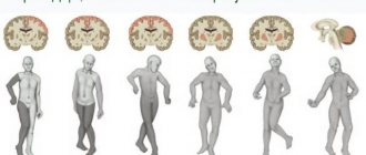
First of all, it should be noted that NLN always develops only when the nerve fiber is damaged from the motor nucleus of the facial nerve to its exit from the stylomastoid foramen (peripheral paresis) and always on the same side, in contrast to central paresis, which occurs mainly during a stroke and is often combined with paresis of the limbs developing on the side opposite to the lesion (Fig. below).
The facial nerve (FN) refers primarily to the motor nerves that provide facial expressions, the processes of blinking, chewing, swallowing, and frowning. However, the trunk of the facial nerve also contains components of the intermediate nerve - parasympathetic (secretory) and sensory (taste) fibers innervating the salivary glands, as well as the taste sensitivity of the tongue.
The relatively high incidence of damage to the facial nerve is largely due to its inherent anatomical and topographical features - the nerve has a complex and long course in the narrow bony canal of the temporal bone. The most vulnerable segment of the FN (in which it is pinched/compressed) is the segment located in a narrow convoluted canal where, in cases of edema development due to various reasons (for example, inflammation), it is compressed.
Among different localizations of damage to the peripheral part of the FN, Bell's palsy is the most common pathology (16-25 cases / 100,000 population) and is caused by the development of edema and its subsequent compression in the bone canal (tunnel syndrome). The high vulnerability of the FN in the fallopian canal is explained by its prevalence in the cross section of the canal, where it occupies 40%-70% of the total area. Moreover, despite the fact that the canal narrows in some places, the thickness of the nerve trunk itself remains unchanged.
In the vast majority of cases, peripheral paresis of the facial nerve is manifested by unilateral damage to the facial nerve. The right/left facial side is affected with equal frequency. Bilateral neuropathy of the FN accounts for only 6.2% of all its lesions. The average age of onset is about 40 years, but can occur at any age. The lowest incidence rate is observed in children under 10 years of age, increases in persons in the age group 10–29 years, stable rates are typical for persons 30–69 years of age, and reaches maximum rates in the population of patients after 70 years of age.
The disease is characterized by a high frequency of complications (7–18% of cases); recurrent FN neuropathies are observed in 24.5% of cases. Recurrent neuropathies, compared to primary ones, are more severe, more difficult to treat, and extremely rarely result in complete recovery. Neuropathy of the facial nerve, as many patients who visit a special forum write, is an extremely psychotraumatic situation for patients and has an extremely negative impact on the psycho-emotional sphere and physical condition of patients, up to the development of neurosis. FN palsy is a common cause of long-term disability and significantly reduces quality of life.
Diagnostics
Since facial neuritis has a clear clinical picture, the diagnosis is usually based on the patient's examination and medical history.
During the examination, the doctor asks the patient to frown, puff out his cheeks, close his eyes and perform other similar actions to determine the degree of damage to the facial muscles. Neuritis of the facial nerve is accompanied by a sail symptom (when exhaling, there is a passive swelling of the cheek on the affected half), when closing the eyes, Bell's symptom is revealed, and weakness of the entire affected half of the face is observed (with a stroke and brain tumor, weakness of the lower part of the face is observed mainly).
In order to assess the degree of damage to the facial nerve, in case of a recent disease (up to 3 months), the K. Rosier scale is often used, which consists of 4 degrees of severity of paralysis.
The F.M. method is also used. Farber, taking into account changes in the degree of eyebrow raising and reduction, lip extension, eye closing, the presence of the brow reflex and the corneal reflex before and after treatment. This method allows you to assess the severity of the disease and the effectiveness of treatment for neuritis of any age.
In 1985, the Committee on Facial Nerve Disorders approved the six-level House-Brackmann Facial Nerve Grading Scale, which is used in cases of incomplete facial nerve repair to assess:
- degree of muscle weakness;
- symmetry;
- the presence of synkinesis;
- the presence of facial contractures.
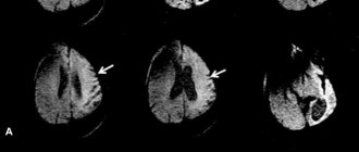
Since similar symptoms are observed in other diseases (supranuclear lesions of the facial nerve, fractures), radiography, CT and MRI are performed to exclude such pathologies.
In Bell's palsy, according to radiography performed according to Schüller-Mayer, in 84% of patients a pneumatic (with a large number of cells) type of structure of the mastoid process is detected. In half of the cases, this type of structure extends to the top of the petrous part of the temporal bone and causes a local narrowing of the lumen of the fallopian canal due to the protruding walls of individual cavities. The same structure can be identified by tomography performed according to Stenvers.
For differential diagnosis, laboratory tests are also used, which make it possible to detect a slight increase in the amount of protein in the cerebrospinal fluid (CSF) in 1/3 of cases.
The functions of the facial nerve are assessed using electroneuromyography (EMG), which, when conducting research in the acute period, makes it possible to find out:
- whether the facial nerve paresis is central or peripheral;
- affects individual branches of the nerve or its trunk;
- what type of lesion is observed (axonopathy, demyelination, mixed process);
- prognosis for recovery of the facial nerve.
The first EMG (examination of the facial nerve and blink reflex on both sides) is recommended to be carried out in the first 4 days of the disease, the second - 10-15 days after the moment of paralysis, the third - after 1.5 - 2 months. If necessary, additional studies are carried out on an individual basis.
During the EMG study, distal latency (the speed with which the impulse is conducted from the angle of the lower jaw), the amplitude of the M-response (depending on the synchrony and amount of activation of the muscle motor units caused) and the speed with which the impulse is conducted along the nerve are assessed.
If on days 5-7 from the onset of the disease the first two indicators are within normal limits, the prognosis is favorable for damage of any severity.
The increased latency indicates the process of demyelination, but the observed preservation of the normal M-response amplitude (or the presence of 30% compared to the healthy side) indicates the possibility of recovery within 2 months.
An M-response amplitude of 10 to 30% indicates a fairly good, but longer recovery (from 2 to 8 months).
The amplitude of the M-response, which is less than 10% compared to the healthy side, with the speed of impulse conduction along the facial nerve differing by 40% from the indicators of the healthy side, indicates an incomplete and prolonged restoration of the functions of the facial muscles.
The fibrillation potential detected at 2-3 weeks indicates the presence of a process of axonal degeneration. In this case, the prognosis is unfavorable - there is a high probability of developing contractures.
Facial neuritis must be distinguished from infections of the middle ear or mastoid, chronic meningeal infections, Ramsay Hunt syndrome, Guillain-Baré syndrome, Lyme disease and multiple sclerosis.
Pathogenesis
The triggering factor for FLN is irritation of the vessels of the craniocervical region, which contributes to the development of vasospasm of the vertebral and branches of the external carotid artery, which leads to primary ischemia of the FLN root. Increasing disturbances of microcirculation in the structures of the FN lead to anoxic swelling of the nerve . This, in turn, leads to compression (squeezing) of the nervous tissue in the facial (fallopian) canal of the temporal bone, disruption of neuromuscular conduction caused by blockade of the process of release of acetylcholine and a disorder in the interaction of acetylcholine with receptors located on the postsynaptic membrane. As disorders increase in the nervous tissue, secondary ischemia of the FN develops.
Classification
There are primary lesions of the FN, caused by hypothermia, and secondary, as a complication of other diseases.
Based on etiology, they are distinguished:
- Bell's palsy (idiopathic neuropathy).
- Otogenic neuritis (with inflammation of the middle ear/mastoid process of the temporal bone).
- Infectious neuritis ( influenza , herpes , mumps , polio , etc.).
- Traumatic neuritis (damage to the facial nerve).
- Ischemic (in cases of impaired blood supply to the nerve).
Treatment of Bell's palsy
This is an inflammatory disease, so glucocorticoid hormones are used to relieve swelling: Prednisolone, Dexamethasone. Potassium supplements (iodide) and diuretics are also needed to treat Bell's palsy.
For viral infections, interferons and Acyclovir are used. If one of the seventh pair of cranial fibers is torn by injury, it must be sutured.
To prevent drying of the cornea, artificial tears are used. Wet compresses on the eye help maintain the normal condition of the cornea.
The innervation of facial muscles is improved by the drugs Prozerin, vitamins Milgamma, Neuromultivit. Facial massage and gymnastics are needed: various grimaces, exercises for the tongue, lips, eyes, eyebrows.
It is useful to know how multiple sclerosis manifests itself in children and pregnant women: signs of pathology.
It is interesting to read what vestibular neuritis is: causes of development, symptoms, diagnosis, treatment.
All about inflammation of the occipital nerve: causes, signs, treatment.
Treatment of Bell's palsy with folk remedies includes warming compresses with herbal infusions (linden flowers), facial massage with fir oil. To warm up, use a compress of heated salt in a cotton bag and a boiled egg.
In the first period, diuretic herbs are used for swelling: Orthosiphon collection, corn silk decoction. Physiotherapy is used: electrophoresis with vitamin B1, Lidase, aloe extract, vitreous. They also provide sanatorium-resort treatment and vitamin therapy.
Causes of neuritis of the facial nerve
With the peripheral nature of the FN lesion, it is quite difficult to establish the causes of the disease in most cases. It is generally accepted that the causes of FN neuropathy are polyetiological (ischemic, otogenic, idiopathic, traumatic, infectious and other origins). As already indicated, Bell's palsy develops as a result of compression of the nerve in the narrow convoluted canal of the temporal bone, which occurs for various reasons (inflammation, hereditary predisposition in the form of congenital narrowness of the facial nerve canal).
Provoking factors for FN neuropathy include hypothermia, infections, nerve compression by a tumor ( neurinoma ), trauma to the bones of the base of the skull/face with mechanical damage/rupture of nerve fibers, and poisoning. Also, neuropathy can develop as a complication of otitis , mumps , mesotympanitis , neurotropic viral infection ( poliomyelitis , herpes ), inflammatory processes in the brain.
Making a diagnosis and conducting research
Typically, otolaryngologists, psychoneurologists, psychiatrists, neurosurgeons and pulmonologists are involved in the diagnosis of plegia. When identifying the causes of the syndrome, a detailed history taking and identifying each patient’s propensity for such psychogenic reactions is important.
When collecting anamnesis, the patient must answer several questions:
- duration of decrease in muscle strength;
- approximate reasons that caused the complaints (fever, diarrhea, headache, ingestion of canned food);
- whether harmful substances are used in the work;
- Do relatives have such phenomena?
A neurological examination is also carried out with an assessment of muscle strength using 5 points and a search for other manifestations (loss of reflexes, facial asymmetry, muscle atrophy).
The patient is prescribed a blood test to determine the erythrocyte sedimentation rate and the number of leukocytes. A toxicological analysis is carried out to identify poisoning.
Additional studies include:
- electroneuromyography (to assess the electrical activity of muscles, the time of nerve impulse conduction);
- test with Proserin (to determine myasthenia gravis);
- magnetic resonance angiography (to assess the integrity and patency of arteries in the skull, detect brain tumors);
- MRI, computed tomography of the spinal cord and head to study their structure, identify tissue disorders, tumors, hemorrhages, ulcers, etc.;
- electroencephalography to assess the electrical activity of areas of the brain.
A neurosurgeon may be needed.
Symptoms
Symptoms of facial neuritis are determined by the level of its damage. Let us consider only the symptoms of compression-ischemic damage to the FN (Bell's palsy). Most often, compression-ischemic neuropathy manifests itself as acutely developed paresis/paralysis of facial muscles in the form of:
- Marked smoothness of all skin folds of the face on the affected side, the eyelid and corner of the mouth are drooping.
- Bloating of the cheek when exhaling/speaking when pronouncing consonants (sail sign).
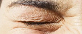
- “Hare eye” - when you close your eyes, the eye does not close on the affected side, while the eyeball turns slightly outward/upward.
- Inability to raise an eyebrow, close an eye, wrinkle a forehead, whistle, the mouth slit is pulled to the healthy side when grinning.
- Getting solid food between the gum and cheek when chewing and pouring liquid over the edge of the mouth on the affected side.
- Problems with diction (not always).
- Pain behind the ear.
Facial asymmetry is extremely specific and difficult to miss (photo below). In the acute stage of the disease, the patient’s face is asymmetrical and at rest, while the “healthy” side of the face pulls on the paretic muscles, thereby causing additional discomfort.
Facial paralysis usually begins suddenly. At the onset of the disease, some patients develop moderate/mild pain and paresthesia in the mastoid/ear area 1-2 days before the onset of movement disorders or simultaneously.
Traditional methods of treatment
Facial paralysis can be treated not only in medical institutions, but also at home, including using traditional methods. The main thing is that all therapy methods are approved by the attending physician.
The basis of traditional medicine will be sedatives and tinctures that you can prepare yourself. In order to make the tincture, you will need:
- 50 ml alcoholic hawthorn;
- 50 ml of peony in the form of tincture;
- alcoholic motherwort 50 ml;
- 25 ml Corvalol;
- 50 ml calendula in tincture;
- two teaspoons of honey.
All components are mixed and taken one teaspoon at night. The course of treatment is 3 months.
- A tincture of raspberry leaves helps with facial paralysis. To prepare it, you need to take several stems and leaves of the plant and fill them with 200 grams. vodka.
- The tincture must be prepared within 9 days. When consuming it, it must be strained. For the first 10 days, take 20 drops 3 times a day 30 minutes before meals. Every 10 days the dosage is increased, the next 10 days is 30 drops, then 50 drops. The course of treatment is 3 months.
- Heat helps to get rid of pathology well. To do this, you need to put cereal or salt in a thick fabric bag and heat it in a frying pan. Apply to the affected area for at least 10 minutes.
- Sea buckthorn oil has also taken its pride of place in the treatment of symptoms of facial paralysis. It is used for massage, applying with massaging movements to the affected area. The course of treatment should be at least a month.
Facial nerve paresis in newborns
The cause of FN paresis in newborns is birth trauma - damage to the FN at the site of exit from the stylomastoid foramen, or, alternatively, its terminal branches in the buccal/parotid region, caused by compression by the fetal shoulder, protrusions of the mother's pelvic bones, and obstetric forceps.
It occurs with a frequency of 0.3 to 1 case per 1000 live births, mainly in large newborns. Manifests with paresis of facial muscles of varying degrees of severity (from almost imperceptible to severe). In the vast majority of cases (90%), during the first months of a child’s life, spontaneous and complete restoration of the function of facial muscles occurs. If there is no recovery by 2-3 months of life, the need for surgical treatment is decided.
Approach to therapy
As a rule, the primary appearance of plegia is muscle discomfort. If the therapy is incorrectly chosen or absent, the pathology can cause complete paralysis.
Usually, with partial immobility, severe pain and acute illnesses appear, on the basis of which treatment is prescribed. Primary therapy is usually aimed at initially identifying and eliminating the underlying cause.
There are several methods for treating pathologies:
- surgery to remove a tumor of the spinal cord or brain, removal of hematomas, ulcers and antibacterial treatment;
- treatment with antibiotics for infectious damage to the nervous system;
- normalization of blood pressure , prescribing medications to improve blood flow in the brain and metabolism;
- to eliminate poisoning, vitamins A, B, C and solutions ;
- the use of drugs to improve neuromuscular reactions in myasthenia gravis;
- For botulism, antibotulinum serum .
In addition to drug therapy, massage courses are prescribed to increase muscle tone. An important role in the treatment of various types of pathology is played by the determination and willpower of the patient, who must strive for recovery to activate internal vital forces.
List of sources
- Alperovich P.M. Bell's palsy (etiology, pathogenesis, clinical picture, course, outcome) / P.M. Alperovich, A.G. Korneychuk, T.I.
- Konstantinovich and others //J. neuropathology and psychiatry named after. S.S. Korsakov. 1978. - T.78, No. 6. -WITH. 836-846.
- Batysheva T.T., Kostenko E.V., Boyko A.N. Complex treatment of facial nerve neuropathy using neuromidin and antioxidant therapy // Journal. psychiatrist, and psychopharmacologist. - 2008. No. 4. - P. 199-201.
- Complex treatment of patients with facial neuropathy and trigeminal neuralgia. Guidelines. - M., 2005. - 32 p.
- Maksimova M. Yu., Sharov M. N., Domashenko M. A. et al. Neuropathy of the facial nerve // Pharmateka. - 2011. - No. 14. - P. 46-51.
- Markin S.P. Neuropathy of the facial nerve // Neurology and Rheumatology. Supplement to the journal Consilium Medicum. - 2010. - No. 1. - P. 10-14.
Prevention
Like any disease, this ailment can be prevented; for this it is necessary to follow simple preventive recommendations, including:
- strengthening and increased attention to your immunity;
- exception of being in drafts, in cold rooms and on the street in the cold season without clothes;
- timely treatment of all diseases (correct treatment of these diseases);
- promptly consult a doctor if you notice signs of paralysis in yourself.
So, despite the fact that this disease is not life-threatening, the presence of unpleasant consequences can seriously complicate life. Don’t delay your visit to the doctor and try to get rid of the disease on your own, take care of yourself and your health, and get treated correctly.