The chronic form of tick-borne encephalitis is a neuroinfectious process associated with the active activity of the virus and clinically manifested by a slow increase in focal and non-focal signs of disruption of the central nervous system. More often they are characterized by an intensification of the symptoms of nested damage to the nervous system that arose in the acute period and the appearance of new symptoms that have a different localization. Progression should be distinguished from short-term motor deficits associated with exercise, development of contractures, intoxication, infections, etc.
The mechanism of development of the chronic form of tick-borne encephalitis
The basis of the pathogenesis of the chronicization of the viral process, according to most researchers, is the universal property of the tick-borne encephalitis virus to persist in the body. Virological studies by A.V. Subbotin confirmed the persistence of the tick-borne encephalitis virus (TBE) - an antigen was isolated from white blood cells of patients with chronic TBE with different duration of the process. A number of authors suggest that, in particular, hyperkinetic forms, given the persistence and low variability of the clinic over many years, are the result of “necrobiotic and reparative processes” that were caused by the activity of the virus in the acute and chronic phase. Others adhere to the autoimmune theory of the development of chronicity of the viral process in TBE, especially in cases with a clinical picture of the disease typical of degenerative processes - amyotrophic lateral sclerosis syndrome, myoclonus epilepsy, etc.
Spring-summer tick-borne encephalitis (encephalitis acarina)
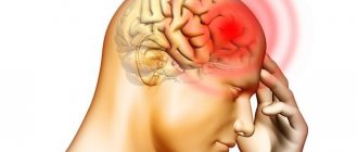
Tick-borne encephalitis is an acute natural focal transmissible viral infection with predominant damage to the central nervous system.
Brief historical information
In the 30s of the 20th century, domestic scientists L.A. Zilber, E.N. Pavlovsky, A.A. Smorodintsev, A.G. Panov, I.I. Rogozin et al. conducted in-depth studies of the etiology, epidemiology, pathomorphology and clinic of the disease, which occurred in the taiga regions of the Russian Far East, and developed methods for specific prevention and treatment of the disease.
Etiology
The causative agent is an RNA-genomic arbovirus of the genus Flavivirus
family
Flaviviridae.
The virus has antigenic variants that cause various clinical and epidemiological forms of the disease. The specificity of the antigenic properties of the pathogen allows it to be distinguished in serological reactions from other arboviruses. Within one antigenic variant, the clonal nature of variability of the tick-borne encephalitis virus has been demonstrated. The virus is cultivated in chicken embryos and cell cultures. The tick-borne encephalitis virus persists for a long time at low temperatures (optimal mode -60 ° C and below), tolerates lyophilization well, persists in a dried state for many years, but is quickly inactivated at room temperature. Boiling destroys it after 2 minutes, and in hot milk at 60 °C the virus dies after 20 minutes. Ultraviolet irradiation, chlorine-containing preparations, Lysol and other disinfectants also exhibit an inactivating effect.
Epidemiology
Reservoir and source of infection
- a large range of warm-blooded animals and birds.
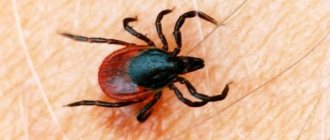
However, the main species that supports the existence of the virus in nature are ixodid ticks. The possibility of spontaneous infection with the tick-borne encephalitis virus has been established for 16 species of ixodid ticks, as well as for a number of other species of arthropods and vertebrates. However, the direct source of infection for humans most often are ixodes ticks Ixodes persulcatus
and I.
ricinus,
which are also its main carriers and long-term keepers in natural foci. These ticks are characterized by a complex development cycle (adult-egg-larva-nymph-imago), lasting at least 3 years. Under certain conditions, larvae and nymphs enter a biopause, each of which lengthens the development cycle by 1 year. During metamorphosis, there is a mandatory change of hosts for mites. The main hosts of the larvae are small mammals and, in some places, birds. The adult feeds on medium and large sized domestic animals. The nymphal phase has the widest range of hosts. The encephalitis virus is transmitted from one phase of tick development to another, and also transovarially. However, the long-term existence of the virus population due to transovarial and transphase transmission alone is impossible. Replenishment occurs when ticks are regularly infected during their feeding on vertebrate animals with viremia. Humans are a dead-end host because they cannot be a donor for ixodid ticks.
Transmission mechanism
— transmissible,
carriers
are ticks
Ixodes persulcatus
and I.
ricinus.
Ticks become infected by attacking sick animals. Humans are usually infected through vector-borne or nutritional routes through the raw milk of infected goats, sheep and cows. Goats are capable of contracting tick-borne encephalitis several times throughout their lives, excreting the virus in their milk, and, therefore, the same animal can be a source of infection during different epidemic periods.
People are attacked mainly by adult ticks and only in rare cases by nymphs. The tick begins to inoculate the virus with the very first portions of saliva, so sometimes even a very short stay of the infected carrier on the body after a bite can lead to infection of a person. There are also cases of human infection due to the penetration of the virus through damaged skin and eyes when crushing an infected tick or non-compliance with laboratory work.
Natural sensitivity of people.
There is no reliable information about the immunity of one or another part of the population to the tick-borne encephalitis virus. The disease left behind strains the immune system.
Basic epidemiological signs.
Tick-borne encephalitis is classified as a natural focal human disease.
Natural foci coincide with the range of ticks that carry the pathogen in forest and forest-steppe regions of Russia. In addition to natural ones, secondary, anthropurgic foci are known, located near populated areas and maintained by farm animals and synanthropic rodents. The main reason for this situation is the almost complete cessation of ground-based treatments of forests against ticks with acaricidal preparations due to the ban on the use of DDT, as well as the active visits by the urban population to natural biotopes for the purpose of collecting wild plants and the intensive development of garden plots. The incidence has a spring-summer seasonality associated with the period of greatest activity of ticks. The seasonal peak in the abundance of I. persulcatus
almost everywhere occurs in the last ten days of May - the first ten days of June.
The I. ricinus
usually has two seasonal peaks of adult activity: in spring and in late summer - early autumn. Among the cases, adults working in the forest (foresters, loggers, etc.) or visiting forest plantations predominate. In recent years, tick-borne encephalitis has ceased to be an occupational disease. Up to 20% of cases are children under 14 years of age. The increase in the number of diseases among urban residents, including school-age children, is mainly due to the population of large cities. City dwellers become infected in suburban forests, forest parks, in individual garden plots, as well as at a distance of tens and hundreds of kilometers from cities. The number of people at immediate risk of tick-borne encephalitis is extremely large. The nutritional route of infection, often accompanied by family-group diseases, is often associated with the consumption of raw goat milk. The number of nutritional cases and their share in the morbidity structure are increasing, which is determined by the goat population, which has increased significantly in recent years.
Pathogenesis
In the case of a transmissible route of infection, the pathogen enters the human body through the skin (Ixodid tick bites); in the case of alimentary infection, it enters through the mucous membranes of the gastrointestinal tract. Primary replication of the virus occurs in the area of entry during the incubation period of the disease. The development of clinical manifestations of the disease coincides with the onset of lymphohematogenous dissemination of viruses into the lymph nodes, internal organs and central nervous system. The mechanism of the cytopathic effect of the virus on nerve cells remains poorly understood. It is believed that after entering the central nervous system, the virus fixes on cells, causing a mesenchymal-inflammatory reaction with the development of edema, dilation and congestion of blood vessels, hemorrhages, disturbances of blood microcirculation and liquor dynamics.
The process involves gray matter in various parts of the brain and spinal cord, primarily motor neurons, peripheral nerve roots, and soft membranes of the brain. Necrobiosis of nerve cells is most active in the reticular formation, nuclei of cranial nerves, and in the anterior horns of the cervical spinal cord. Clinically, these injuries are manifested by the development of flaccid paresis and paralysis.
During alimentary infection, the virus quickly penetrates the internal organs through the hematogenous route, where it replicates. Subsequently, due to secondary viremia, the central nervous system is damaged in a similar way.
Clinical picture
Incubation period.
Varies from several days to 3 weeks, with an average of 2 weeks. During the course of the disease, three periods are distinguished: initial, period of neurological disorders and outcome (recovery, transition to a chronic form or death of patients).
Initial period.
Lasts on average about 1 week and is characterized mainly by the development of general toxic symptoms. Prodromal phenomena rarely manifest themselves in the form of headaches, sleep disturbances, radicular pain, numbness of the skin of the face or torso, and mental disorders. Much more often, the disease begins and develops acutely with an increase in body temperature to 39-40 °C, chills, painful headaches, pain in the lumbar region and limbs, in the eyeballs, hyperesthesia, photophobia, and severe general weakness. Nausea and repeated vomiting are possible. When examining patients, they are noted to be lethargic, sometimes drowsy, and stunned, but consciousness is preserved. Diffuse hyperemia affects the skin of the face, neck, chest; the conjunctiva is full-blooded, the sclera is injected. The mucous membranes of the upper respiratory tract are also hyperemic. With the development of shortness of breath in the lungs, physical signs of pneumonia are often detected. Heart sounds are muffled, pulse is slow (relative bradycardia), blood pressure is reduced. An ECG can reveal signs of conduction disturbances and myocardial dystrophy, which, if severe, may cause patients to develop a picture of acute heart failure. The tongue is coated, often trembles when protruded, the abdomen is swollen, intestinal motility is weakened, stool is prone to constipation. The liver and spleen may be enlarged.
Period of neurological disorders.
It begins with the occurrence of neurological disorders in the form of paresthesia, paresis of the limbs, epileptiform seizures, which can develop already in the first days of the disease.
In accordance with the severity of neurological symptoms, febrile, meningeal, meningoencephalitic, meningoencephalopoliomyelitis and polyradiculoneuritic clinical forms
of the disease are distinguished.
Feverish form.
It manifests itself as a general toxic syndrome, has a benign course and ends with recovery.
Clinical manifestations of the chronic form of tick-borne encephalitis
Researchers of the problem of chronic tick-borne encephalitis note a variety of clinical manifestations of the chronic course of TBE, the most common are poliomyelitis, polyencephalomyelitis, amyotrophic lateral sclerosis syndrome (ALS), hyperkinetic, psychotic, as well as various combinations thereof. Various authors have proposed options for clinical classifications of chronic TBE, but to date a unified classification has not been accepted.
Zhukova N.G. identifies the following clinical forms of chronic CE: poliomyelitis, Kozhevnikov epilepsy, amyostatic syndrome (Parkinsonism syndrome), amyotrophic lateral syndrome (ALS); primary or sequentially progressive forms; 3 degrees of severity - mild, moderate, severe; the course is recurrent, progressive.
In clinical practice, the classification of K.G. is often used. Umansky, A.V. Subbotina et al (1984), thanks to the grouping of syndromes and the identification of the main parameters characterizing the course of the disease:
1. Clinical forms
1.1. Hyperkinetic (Kozhevnikovsky epilepsy syndromes, hyperkinetic, myoclonus-epilepsy, epileptic);
1.2. Amyotrophic (poliomyelitis, encephalopoliomyelitis, multiple encephalomyelitis, amyotrophic lateral sclerosis syndromes);
1.3. Rare forms (vegetative, parkinsonian, syringomyelitic);
2. Severity: mild, moderate, severe
3. According to the time of occurrence of the chronic process
3.1. Initial progressive (direct continuation of the acute period)
3.2. Early progressive (within 1 year after the acute period)
3.3. Late progressive (a year or more after the acute period)
3.4. Spontaneous progressive (without an acute period) (the concept more often used is primary progressive)
4. By nature: recurrent (with periods of deterioration - intensification of existing symptoms and (or) the appearance of new ones), continuously progressive (slow increase in symptoms); abortive or regressive (after progression, a permanent stop of the process may occur, without subsequent resumption of progression).
According to A.P. Ierusalimsky (2001), a number of terms were not legally included in the group of amyotrophic forms: in particular, encephalopoliomyelitis syndrome, which has a clinical picture of severe damage to the stem, often bulbar, structures of the brain stem, which determines the patient’s disability; Along with this syndrome, the clinical features of disseminated encephalomyelitis syndrome, also included in the group of amyotrophic forms, are not clear.
It is believed that systematization of knowledge on the clinical forms of the chronic course of TBE and correction of the clinical classification are currently required.
Hyperkietic syndrome more often developed after the meningeal form and the focal form with encephalitic syndrome, and the formation of amyotrophic syndrome after the focal form with poliomyelitis and encephalopoliomyelitis syndromes.
Tick-borne encephalitis (TBE) as an acute neuroinfectious disease has been studied and described in detail in many articles and monographs, which detail its etiopathogenetic aspects, describe the clinical picture and diagnostic features. As is known, there is a chronic form of TE (CLE), which is of particular interest, and in relation to which there are many unresolved issues.
The chronic form of the disease develops in a person who has undergone the acute phase of CE, when, after a period of stabilization of the condition and even complete regression of symptoms, i.e. against the background of complete or relative well-being, after a period of time from several months to several years, clinical manifestations of damage to the nervous system develop or increase, and subsequently a progressive or remitting course is observed, which can lead to profound disability or even death [10].
A distinctive feature of CCE, which at the same time complicates its diagnosis, is the polymorphism of clinical manifestations [1, 3, 4, 6, 7, 10]. Pathological lesions in CCE are found in various parts of the nervous system - in the subcortical structures, spinal cord, various parts of the hemispheres, thalamus, pons, medulla oblongata, cerebellum [2, 5, 8-10, 12].
In the typical course of CLE, when we are presented with a patient with Kozhevnikov epilepsy or amyotrophic syndrome, with a predominant lesion of the cervical-brachial region and a history of an acute phase of CBE, provided that a high titer of antiviral antibodies is detected, making a diagnosis is not difficult. In this case, even a clinical and epidemiological diagnosis is valid, without laboratory confirmation [10].
However, in the absence of an acute phase of the disease or in the case of an “atypical” clinical picture of focal damage to the central nervous system (CNS), difficulties in diagnosis may arise.
We present our own observation of a patient who was admitted with suspicion of multiple sclerosis (MS) and had a clinical picture and results of magnetic resonance imaging (MRI) of the brain characteristic of this pathology, however, a clarification of the medical history, a routine neurological examination and additional laboratory tests raised doubts and reconsider this diagnosis in favor of CCE.
Patient A
., born in 1989, complains of numbness in the right limbs, the right half of the abdomen, unsteadiness of gait, which intensifies in the dark, dizziness with a feeling of sinking.
In 1996, at the age of 5, according to the patient, he suffered a tick bite. It was while staying in the forest that I was bitten by a tick twice, with an interval of 2 days. Geographically, at the time of the bite, he was in the Nizhnekamsk region of the Republic of Tatarstan. This area is endemic for the TBE virus. The boy was administered anti-tick gamma globulin, however, despite preventive measures, the patient soon developed a prolonged fever with intense headaches. He was hospitalized and treated for about 1 month in the infectious diseases department of one of the local hospitals. He was discharged with improved condition. Subsequently, he was registered with a neurologist in a children's clinic with a diagnosis of “consequences of tick-borne encephalitis.” It was not possible to find out whether specific laboratory tests were done during the hospitalization period and subsequently to establish the diagnosis of CE.
Subsequently, the boy developed normally, went to school on time, and studied well. The only complaint during school was a headache, which at times grew so bad that the patient was forced to take analgesics and even left classes. During these years, he continued to live in Nizhnekamsk with his parents, in the spring and summer he went to the dacha and went to the forest. In 2006, when he was 17 years old, he began to experience episodes of numbness in the extremities, which developed over the course of a week and “lasted” for about a month; he also began to experience unsteadiness in his gait and dizziness, and headaches persisted. The general state of health did not change. The described symptoms usually regressed after treatment, but recurred every six months. In May 2009, against the background of relative well-being, numbness of the left extremities appeared, then dizziness, unsteadiness when walking, which intensified in the dark, and it became difficult to walk. The patient went to a medical institution at his place of residence, where an MRI of the brain was prescribed (with a device characteristic of 1.0 Tesla) and multiple lesions were discovered in the white matter of both hemispheres, the midbrain and cerebellum (Fig. 1)
.
Figure 1. MRI of the brain of patient A. in 2009. Multiple sub- and supratentorial lesions in the white matter of the brain, periventricular and subependymal on both sides, as well as in the middle cerebellar peduncles on the left, measuring 0.3-1.3 cm, highly intense in T2 and isointense in T1, having an irregular oblong and round shape, with symptoms of moderate perifocal edema of the brain substance.
The length of the lesions located in the periventricular sections is directed perpendicular to the corpus callosum. Expansion of the subarachnoid space along the convexital surface of the brain. With a suspected diagnosis of MS based on MRI results, the patient was referred for consultation to a center for the study of MS. During the first examination in the indicated center, slight smoothing of the left nasolabial fold, conduction-type hypoesthesia from the level of Th7 to Th11 on the right, a decrease in vibration sensitivity in the legs to 6 points on both sides, an increase in tendon reflexes on both sides (slightly higher on the left) and on Against this background, reflexes from the triceps muscles were reduced. Muscle strength was 5 points. Coordination tests were performed with mild ataxia and inconclusive dysmetria. Moderate dysadiadochokinesis was detected in the hands. There were no pelvic disorders. In the neurological status, we also noticed the presence of a “no-no” type tremor of the head and a slight plastic tone in the extremities.
The fact of a tick bite and subsequent long-term fever in the anamnesis, as well as the identification during an objective examination of symptoms atypical for MS, such as head tremor in combination with extrapyramidal tone in the extremities, gave grounds to exclude the diagnosis of CCE.
To clarify the diagnosis and detect antibodies to the TBE virus and the causative agent of borreliosis, polymerase chain reaction (PCR) and serological blood tests (enzyme-linked immunosorbent assay - ELISA) were prescribed. Antibodies to the causative agent of borreliosis, antibodies to the TBE virus IgM and viral RNA were not detected during PCR diagnostics, but antibodies (IgG) to the TBE virus were recorded - in a titer of 1/1600.
Thus, the patient had a chronic disease of the nervous system, which debuted at a young age (17 years), had a remitting course with multifocal damage to the central nervous system, which was confirmed by MRI results. However, the neurological status revealed symptoms atypical for MS. The pathological condition developed 12 years after the acute phase of TBE, judging by the anamnesis, most likely of the meningeal form, and at the time of examination a positive IgG titer to the TBE virus was detected. After acute TBE, a weak seroconversion develops - antibodies can remain in the blood for a short time - no more than 3 years, but when the process becomes chronic, antibodies are found in high titers and remain at the same level for a long time or tend to increase [10]. All this allowed us to settle on the diagnosis of CE, a chronic form, and recommend further observation to the patient at the center.
For the purpose of dynamic observation, the patient underwent a follow-up examination in September 2011. At the time of examination, the leading complaints were severe general weakness and increased fatigue, which is pathognomonic for previous neuroinfections [9]. Over the past 2 years, episodes of deterioration have not bothered him. The status revealed hyperreflexia, there were no sensory disorders, and the patient performed coordination tests satisfactorily.
A control MRI study (with a 1.5 Tesla device) of the brain was performed, which recorded a decrease in the size of the lesions, the absence of perifocal edema around the lesions, and regression of the lesion in the cerebellum (Fig. 2)
.
Figure 2. MRI of the brain of patient A. in 2011. The ventricles and other cerebrospinal fluid cavities and slits are slightly expanded, symmetrical, and not deformed.
Paraventricularly in the white matter of the cerebral hemispheres and the corpus callosum, multiple foci of softening of various shapes are detected. There are no lesions in the brainstem and cerebellum. The MRI picture of the brain of the described patient was very similar to that of MS and served as a reason to suspect this diagnosis. And if you rely only on its results, you could come to an erroneous diagnosis. The development of periventricular lesions in the cerebellum during CCE is possible, as evidenced by previously conducted pathomorphological studies of the brain in this pathology [8-12]. An interesting fact is the complete regression of the lesion in the cerebellum, despite the fact that the repeated examination was performed on a higher-field apparatus (1.5 T, compared to the first - 1.0 T). There is a description in the literature [1, 12-14] of the complete disappearance of lesions in CCE according to MRI results.
Repeated serological tests did not detect IgM antibodies to the TBE virus; the IgG titer was 1/1600, which speaks in favor of the persistence of chronic tick-borne infection in the body. Although these results should be considered with caution, since the patient continues to live in an endemic area and constant contact with the pathogen, which can support seroconversion, cannot be ruled out.
In the described case, which was difficult to diagnose, epidemiological data (tick bite and prolonged fever), symptoms atypical for MS (head tremor, extrapyramidal tone in the limbs) and positive laboratory results (high titer of IgG to the TBE virus, which persists) helped in differentiating MS and CTE. to this day at the same level). This made it possible, with a high degree of probability, to settle on the diagnosis of chronic tick-borne encephalitis, a remitting course, in the form of disseminated encephalomyelitis syndrome.
Diagnosis of the chronic form of tick-borne encephalitis
Clinical manifestations of the progressive course of TBE have regional characteristics, mainly due to the dominant subtype of the virus in a particular endemic region. Currently, most of the works devoted to the clinical description of forms of chronic tick-borne encephalitis have been published based on data from the Primorsky Territory, the region of circulation of the Far Eastern subtype of the virus. To date, the regional clinical features of the chronic course of the disease in the area of circulation of the Ural-Siberian subtype of the virus remain insufficiently studied, the age-related aspects of the clinical picture of progressive forms have not been studied, and there is no complete understanding of the transformation of the disease into a chronic course of the disease. Modern diagnostic technologies - polysomnographic studies, neuroimaging, ELISA diagnostics allow us to take a fresh look at the pathogenesis, clinical course of chronic forms, propose algorithms for diagnosis and differential diagnosis and management of patients in the acute, subacute periods of the disease at the stage of recovery and stabilization.
Treatment of chronic tick-borne encephalitis
Specialized medical care for patients with tick-borne infections requires systematization of measures aimed at timely provision of emergency medical care, highly qualified diagnosis and treatment during the acute period of the disease, further dynamic monitoring of patients with residual effects due to the frequent formation of neurological deficits leading to disability and high risk of developing chronic forms of infections.
Most works present certain aspects of the treatment of tick-borne encephalitis: regimens for the use of specific immunoglobulin, nucleases, immunomodulatory agents; drugs that improve microcirculation; approaches to intensive therapy of severe focal forms of TBE, the use of antibacterial therapy. Many authors have discussed the need for clinical observation of patients with tick-borne neuroinfections in order to select therapeutic and health measures for patients with the consequences of diseases and improve their labor prognosis. A number of works provide recommendations on the organization of dispensary observation and therapeutic measures for persons who have suffered tick-borne encephalitis, which establish the terms and main groups of observation. But most dispensary observation schemes do not fully cover all groups of patients requiring dynamic monitoring of their general somatic and neurological status; clear recommendations for laboratory and instrumental monitoring are not provided; the scope of therapeutic and rehabilitation measures is incomplete.