Stereotactic biopsy of brain tumors
- a diagnostic neurosurgical operation performed to take a sample of tumor tissue for the purpose of its detailed study. The obtained data on the type of neoplasia and the degree of its malignancy make it possible to resolve the issue of the advisability of surgical treatment, the extent of the operation and methods of conservative therapy (chemotherapy, radiation exposure). A stereotactic biopsy is performed by creating a small hole in the bones of the skull and removing material with a special needle. Neuronavigation or a stereotactic frame is used to accurately determine the location of the collection of altered brain tissue.
- Indications
- Contraindications
- Methodology
- Complications
- Prices in Moscow
Indications
In modern neurology, surgery is performed in complex diagnostic cases when other research methods (or MRI) do not provide accurate information about the pathological formation of the brain and are ineffective in differentiating the tumor process from a brain abscess, hematoma, granuloma or focus of demyelination. In addition, a biopsy is performed to identify concomitant infectious and inflammatory processes in the brain tissue. Stereotactic biopsy is especially important in cases where the tumor is located in functionally significant and difficult to reach areas. The technique makes it possible to preserve vital brain structures that may be injured during more extensive surgery.
Often, only a biopsy and careful histological examination of the obtained material can determine the benign or malignant nature of the neoplasia. However, when diagnosing brain tumors, this is not as significant as in other areas of oncology. Due to the limited space inside the skull, malignant and benign neoplasms compress nearby brain structures and therefore must be removed, regardless of the presence or absence of malignancy. Biopsy data is necessary for the neurosurgeon to choose the optimal method for excision of neoplasia.
Stages of the procedure
To understand how a biopsy is done in the brain area, you need to get an idea of the procedure. The preparatory stage includes a comprehensive examination, including laboratory tests, consultations with a cardiologist, phlebologist, and anesthesiologist. Several hours before the intervention, the patient does not take food or water.
It is important to promptly inform your doctor about any allergies to medications and regular use of medications that prevent blood clots. If the patient is taking thrombolytics before an invasive diagnostic test, therapy is temporarily suspended. Stages of the open procedure:
- Making an incision on the scalp.
- Formation of a hole in the skull.
- Removal of a fragment of skull bone tissue to form a trepanation window.
- Peeling of the meninges.
- Resection (partial or complete removal) of the tumor.
- Closing the trepanation window with bone tissue or a special plate.
After completion of neurosurgical procedures, a sample of neoplasm tissue is sent to the laboratory to study the cellular structure. The stages of performing a stereotactic biopsy in the brain region depend on the type of equipment used. The sequence of manipulations when using a coordinating stereotactic frame:
- The exact localization of the pathological focus is determined using neuroimaging (mainly MRI).
- Local anesthetics or general anesthesia are used.
- A stereotaxic frame is installed in the head area.
- An MRI localizer with contrast marks is attached (a special device for limiting the area of intervention).
- A CT scan is performed.
- The results of MRI and CT studies are combined to achieve maximum accuracy when creating injection points.
- An incision is made into the skin of the head.
- A hole is drilled in the skull bone.
- A needle is inserted and moves under the control of an MRI or CT diagnostic complex.
- Material is being collected.
A study using a frame allows you to precisely approach the site of a tumor or other pathological process with an accuracy of 1 mm. The duration of the intervention is about 6 hours. One day after completion of the procedure, the patient is discharged from the hospital. Sequence of manipulations when using the navigation system:
- A study is carried out in MRI or CT format to obtain information such as the structural features of the patient’s brain, the location and size of the pathological focus.
- Neuroimaging results are transferred to the navigation system.
- A model of the brain is created in 3D projection.
- The image is studied to determine the optimal insertion point and needle trajectory.
- Anesthetics are administered to the patient.
- An incision is made on the skin of the head.
- A diagnostic hole is formed.
- Under the control of a navigation system, a puncture needle equipped with a camera and LED illumination is inserted into the cavity of the skull.
- Material is being collected.
A brain biopsy using a navigation system is carried out with minimal negative impact on healthy brain tissue, because calculations such as the shortest and safest path to the tumor site are previously performed. The doctor has the opportunity to choose a trajectory that goes around the areas responsible for vital functions, thereby significantly reducing the risk of complications.
The procedure time for a biopsy in the brain area using a navigation system is significantly reduced compared to intervention using a stereotactic frame. Reducing the duration of the procedure is associated with preliminary planning of actions.
During a puncture biopsy, an incision is made on the skin in the projection where the tumor is localized. Through a hole drilled in the skull bone, a puncture needle is inserted into the pathological focus to obtain a sample of the material. The stereotactic and puncture-type procedure is associated with minimal damage to the medulla, meninges, and bone tissue.
Methodology
Stereotactic brain biopsy is usually performed under general anesthesia. After making an incision in the skin and soft tissues of the head, a hole with a diameter of several millimeters is created in the skull bone, through which a special needle is inserted into the brain tissue and passed to a predetermined location for tissue collection. After taking the material, the needle is removed and the wound is sutured. A stereotactic frame is used to determine reference points that allow the needle to be aimed precisely at the tumor. The device is placed on the patient's head in the operating room, then the patient is transported to the diagnostic department and a tomography is performed. After this, the patient is transported back to the operating room and a needle is inserted, based on the tomogram data. The technique provides guidance accuracy of up to 1 mm, but takes a lot of time (several hours) and is tedious for both the patient and the surgeon.
Neuronavigation involves the use of special neuronavigation systems that make it possible to create a three-dimensional image of the brain based on tomograms obtained before surgery. During the intervention, the system determines the position of the biopsy needle, correlates it with a virtual model of the brain, and continuously displays on the screen a picture of the brain with the needle located in it. The use of neuronavigation (frameless stereotaxis) significantly speeds up the procedure and makes it more comfortable for the patient, however, the accuracy of needle guidance is reduced to 2-3 mm. This is due to the fact that during neuronavigation the scalp is not fixed to the skull and can move, introducing an error in determining the localization of intracerebral structures.
After the operation, the patient remains in the hospital for 2-3 years under the supervision of a neurologist. A sample of the taken tissue is sent for histological examination. A histologist determines the type of cells that make up the sample, identifies pathological cell growth, conducts a series of special tests, and prepares a conclusion on the type of structure and degree of malignancy of the tumor. If an infection is detected during the study, its type and sensitivity to medications are clarified.
Types of diagnostics
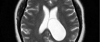
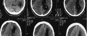
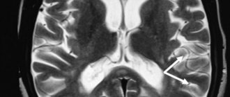
Before performing a biopsy of a tumor located in the brain, diagnostic studies are performed, including neuroimaging (CT, MRI), electroencephalography, if necessary, angiography (a study of the circulatory system of the brain) and ventriculography (a study of the ventricular system of the brain). There are different types of biopsy:
- Open. It is performed during a neurosurgical operation through a craniotomy to remove the tumor.
- Stereotactic. Minimally invasive intervention is performed using a stereotactic frame or navigation system.
- Puncture. A needle is inserted through a hole in the skull bone into the brain tissue, and a small piece of brain matter is removed.
Among all types, open biopsy in the brain area is performed more often than others and is associated with the greatest risk of injury and complications. The high frequency of use is associated with the mandatory removal of a fragment of material during surgery to remove an intracranial tumor. An open diagnostic procedure is performed under general anesthesia in parallel with the main operation.
Stereotactic biopsy is the second most frequently performed biopsy if there is a suspicion of the development of a tumor in the brain. This method is chosen when, due to the inaccessible location of the lesion or the serious condition of the patient, it is impossible to perform a full-fledged operation to remove the tumor. It is considered an informative and safest research method.
Complications
There is a small chance that important brain structures will be damaged during the biopsy. A small scar is formed in the area where the material is taken, which can later become a source of epileptic impulses and the cause of epileptic seizures. When performing a stereotactic biopsy, common surgical complications such as infection and bleeding, as well as negative consequences associated with the use of anesthesia, are possible. Preparing the patient for surgery, contraindications and complications are described in more detail here.
Indications for biopsy
A biopsy involves excising a tissue sample that is then examined microscopically. The procedure allows you to differentiate a neoplasm that has appeared in the brain. This is a mandatory event if there is a suspicion of the development of cancer. The main indication for the procedure is the need to identify the morphological structure and nature of the intracerebral formation, which can be:
- Malignant or benign tumor.
- Cyst, granuloma.
- The source of hemorrhage.
- The source of the abscess.
A brain biopsy is a study that is carried out to identify the nature of damage and the cellular structure of brain tissue in hemorrhagic stroke, multiple sclerosis (analysis for oligoclonal antibodies), Alzheimer's disease, which indicates the versatility of the method in the differential diagnosis of many pathologies. Other possible indications:
- Neurodegenerative diseases, including spongiform encephalopathy.
- Cerebral toxoplasmosis.
- Inflammatory diseases of the central nervous system.
The degree of need for such diagnostics is determined by the attending physician individually in each case. If a tumor process is suspected, the results of histological examination allow the correct choice of treatment tactics. Contraindications to invasive intervention include:
- Terminal condition of the patient.
- A site of complex localization. If the mass formation is located in the part of the brain that is responsible for vital functions.
- Blood clotting disorder. Due to the risk of bleeding during the procedure.
- History of systemic diseases (lupus erythematosus, rheumatism, ankylosing spondylitis).
The intervention is not carried out if it is impossible to perform local anesthesia or general anesthesia, if the doctor can obtain the necessary information about the pathological process using other diagnostic methods. Any manipulation affecting the brain is associated with a high risk of complications. Negative consequences after a biopsy in the brain area are often associated with damage to brain structures and secondary infection.
Brain biopsy
- Indications for the purpose of manipulation
- Types of biopsy
- Stages of minimally invasive biopsy
- Possible complications
- Video on the topic
Neurosurgery is a field of medicine that examines and treats pathologies of the brain and spinal cord in adults and children. One of the diagnostic methods in this area is a brain biopsy.
But as for neoplasms, in neurosurgery a biopsy is not as informative as in the case of other organ systems. This is due to the fact that it does not matter what kind of tumor it turns out to be (benign or malignant), because it must be removed in any case.
Indications for the purpose of manipulation
A brain biopsy is resorted to in cases where other diagnostic methods have not provided enough information. It is prescribed to confirm the diagnosis or clarify the nuances of the course of pathological processes in the following conditions:
- multiple sclerosis;
- Alzheimer's disease;
- Creutzfeldt-Jakob disease;
- hemorrhagic stroke;
- inflammatory diseases (meningitis, encephalitis);
- clarification of the histological structure of the tumor.
Types of biopsy
There are 3 main types of biopsy:
- Puncture manipulation involves drilling a small hole in the cranial cavity, into which a hollow needle is inserted and a small sample of brain tissue or tumor is taken.
- Stereotactic biopsy is a minimally invasive method. This manipulation guarantees high safety, since the needle advances under MRI and CT control. In the process, you can not only take a sample of the biomaterial, but also carefully examine the tumor through imaging.
- An open biopsy occurs during surgery. The procedure is performed under general anesthesia. To remove the tumor, the neurosurgeon is forced to first remove part of the skull bones. An open biopsy is a rather risky procedure that requires a long recovery period for the patient.
The most common method is open biopsy. And the stereotactic method is considered the safest and most patient-friendly.
Stages of minimally invasive biopsy
To perform stereotactic biopsy, two main methods are used:
- Using a frame (frame-based) - this method can be called classic and the most accurate.
- The use of a neuronavigation system (frameless) is a more convenient method for the patient and the doctor, requiring less time.
Method using a frame
The procedure does not require special preparation. This type of biopsy is done as follows:
- The day before, the patient undergoes an MRI using special contrast agents and the implantation coordinates are determined.
- Immediately before the operation, the patient is subjected to local anesthesia at the intended sites for screwing in the screws.
- The main ring of the stereotactic frame is attached to the patient's head.
- Then an MRI localizer with contrast marks is attached.
- This is followed by a CT scan.
- The results of MRI and CT are combined to clarify the injection points.
- The neurosurgeon makes a small incision in the skin and creates a diagnostic hole using a cutter.
- A needle is inserted along a pre-modeled path and biomaterial is collected.
- The final stage is suturing the incision.
As a rule, after this procedure, the patient can return home within 24 hours after a control CT scan to exclude complications. But for a week he must remain in bed.
Methodology using a neuronavigation system
This technique differs significantly from that described above at the stage of determining implementation points. This procedure is carried out as follows:
- The patient undergoes a CT or MRI.
- The results obtained are recorded on digital media and transferred to a navigation system to obtain a three-dimensional image.
- The surgeon can determine the insertion point, direction and path of the biopsy needle, and the specific area to be biopsied on the screen.
- The patient is fixed on the operating table to avoid any movement during the procedure, and anesthesia is administered.
- When you touch the patient with a special instrument, the required point is displayed on the monitor.
- Next, as in the case of the frame, an incision is made in the skin and a diagnostic hole is formed.
- Then, under the control of neuronavigation, a biopsy needle is inserted and a biopsy sample is taken.
- At the end of the manipulations, sutures are applied at the incision site.
The procedure is performed under local anesthesia with maximum comfort for the patient. The latest neurosurgical navigation systems make it possible to calculate the least traumatic path to a tumor located in the deepest corners of the brain or spinal cord, and to reach it with minimal collateral damage
Possible complications
Brain biopsy requires special precision, since damage to important brain structures can have unpredictable consequences. And although the capabilities of modern medicine are so high that possible complications after a biopsy are minimized, they still occur.
The most common consequences are:
- acute cerebrovascular accident;
- sudden paroxysmal, involuntary muscle contractions;
- addition of secondary infections;
- bleeding;
- swelling at the site of biomaterial collection;
- coma.
The wait for biopsy results usually takes several days. Modern biopsy methods are so accurate and minimally invasive that they allow patients to agree to this diagnosis with less fear and more hope for a favorable prognosis.
Indications for use
A biopsy is prescribed when there is a suspicion of a violation of the organic structure of the brain matter:
- Brain neoplasms: meningioma, pituitary adenoma, glioma or astrocytoma.
- Neurodegenerative pathologies: Alzheimer's disease, Pick's disease, diffuse multiple sclerosis, Parkinson's disease, multiple system atrophy, dementia with Lewy bodies, amyotrophic lateral sclerosis.
- Inflammatory diseases: encephalitis, meningitis.
Also, a biopsy may be prescribed after acute and emergency brain conditions: hemorrhagic and ischemic stroke.
Types of biopsy
There are 3 main types of biopsy:
- Puncture manipulation involves drilling a small hole in the cranial cavity, into which a hollow needle is inserted and a small sample of brain tissue or tumor is taken.
- Stereotactic biopsy is a minimally invasive method. This manipulation guarantees high safety, since the needle advances under MRI and CT control. In the process, you can not only take a sample of the biomaterial, but also carefully examine the tumor through imaging.
- An open biopsy occurs during surgery. The procedure is performed under general anesthesia. To remove the tumor, the neurosurgeon is forced to first remove part of the skull bones. An open biopsy is a rather risky procedure that requires a long recovery period for the patient.
The most common method is open biopsy. And the stereotactic method is considered the safest and most patient-friendly.
Open brain biopsy
The peculiarity of this type of biopsy is that the collection of material (biopsy) of brain tissue occurs directly during the operation. Among other types of diagnostics, it is the most dangerous, since neurosurgeons have to perform many manipulations. But at the same time, this is a fairly common method. So, tissue collection occurs as follows:
- A hole is made in the skull.
- Part of the bone is removed, leaving a window for trephination.
- Through it, the formation is removed.
- The open area is covered with bone or a special surgical plate.
- A biopsy is taken from the removed tumor.
Nootropics for meningioma. Can it be used or not?
If a person has pronounced cerebral edema, the defect remains open and is covered with a skin flap. This decision is made by doctors depending on the severity of the disease and the danger of possible infection.
During these actions, the patient is under general anesthesia. After the operation, he needs a long period of rehabilitation.