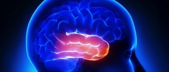
Motor speech area of the cerebral cortex, Broca's area
The motor speech area is an area of the cerebral cortex located in the posterior
section of the inferior frontal gyrus of the dominant hemisphere anterior to the cortical centers of movements of the lips, larynx, and tongue. When the motor speech area is damaged, motor alalia or motor aphasia occurs (motor alalia or motor aphasia syndrome).
Sensory speech area of the cerebral cortex, Wernicke's center
The sensory speech area of the cerebral cortex is an area of the cerebral cortex in the posterior part of the superior temporal gyrus of the dominant hemisphere. The term "sensory" comes from the Latin word "sensus", which means "feeling, perception, sensation". A synonym for the sensory speech area is Wernicke's center, Wernicke's area. When the sensory speech area is damaged, sensory alalia syndrome or sensory aphasia occurs.
When the sensory and motor areas of the cerebral cortex are damaged, sensorimotor alalia or sensorimotor aphasia occurs simultaneously.
Cerebral cortex
The cortex is a layer of gray matter
thickness on average 3 mm. Sensory fibers enter the cortex after a “switch” in the thalamus, and motor fibers exit from it and go to the spinal cord.
The two cerebral hemispheres are connected by commissures - transverse bundles of nerve fibers. The main of these commissures is the thick plate of the corpus callosum;
it extends 8 cm from front to back and consists of more than 200 million nerve fibers running from one hemisphere to the other.
The cortex of each hemisphere forms six separate lobes,
delimited by grooves
of which two are especially large - Rolandova and Sylvian.
In the anterior part of the brain there is a frontal lobe, in the upper parietal lobe, in the lateral temporal lobe, in the posterior occipital lobe; under the temporal lobe, in the depths of the Sylvian fissure, there is a lobe called the insula,
and under the corpus callosum, on the inner surface of the hemisphere, there is a lobe of the corpus callosum (Fig. A.24).
Between the grooves of the cortex, ridges called gyri are formed,
which more or less correspond to areas with certain functions.
These can be sensory, motor or association areas of the cortex (see Fig. A. 19). Sensory areas
receive information from various receptors, and
motor areas
send commands that control movements. Thus, the sensory areas of the cerebral cortex represent the final points on the path of fibers associated with the peripheral nervous system, and their destruction leads to loss of sensitivity in the area of the body where the corresponding receptors are located. Motor areas give rise to fibers, the destruction of which causes paralysis of the limb, controlled by neurons in the corresponding area of the cortex.
' The basal ganglia also include other, smaller nuclei located in the brain stem (such as the substantia nigra, red nucleus, locus coeruleus, etc.).
254 Appendix A
| Arc fold |
Figure A.24. Cerebral cortex.
The most significant part of the cortex, however, is occupied by association zones,
the organization of which is most characteristic of this brain structure. In fact, it is these zones, devoid of any obvious specialization, that are responsible for combining and processing information and programming actions. Because of this, they form the basis of higher processes such as memory, learning, thinking and speech (see document 8.4).
A. Sensory areas. Such zones are found in different lobes of the cortex. The general sensitivity zone is located in the parietal lobe, the visual zone is in the occipital lobe, the auditory zone is in the temporal lobe, the gustatory zone is in the lower part of the parietal lobe, and the olfactory zone is in the two olfactory bulbs located under the cerebrum.
General sensitivity zone
located in the gyrus running along the Rolandic fissure, in the parietal lobe and receives signals from skin receptors. The entire human body—head down and toes up—is presented here in the form of areas (projections), the surface of which is proportional to the sensitivity of the corresponding parts of the body; So. the projection of the hand is much larger than the projections of the back or legs (Fig. A.25).
Damage to all or any part of this area leads to a blockage of sensory signals from the corresponding areas of the body; as a result, tactile, temperature and pain sensations disappear here, although external stimuli continue to excite skin receptors and cause a flow of impulses in the nerve pathways coming from them.
The association zone, located in the upper part of the parietal region, is gnostic and is responsible for the recognition and perception of stimuli. causing sensations at the level of the parietal gyrus.
Visual sensitivity zone
located in the occipital lobe along the calcarine sulcus, and the information transmitted by each retinal ganglion cell is very precisely projected to its different points.
The occipital zone of each hemisphere of the brain receives information from the opposite half of the visual field. Before entering the cerebrum, some of the fibers of both optic nerves cross
Biological basis of behavior
255
Xia, forming the so-called visual chiasm
(Fig. A.26). As a result of this crossing, the left visual lobe receives fibers from both eyes, carrying information about the right half of the visual field, and the right lobe - about the left half. Thus, as a result of the integration of nerve signals from both retinas, the brain recreates a three-dimensional image of an object, the images of which are slightly different on the right and left retinas.
Visual perception of objects, words and numbers is carried out in the associative zone located around the sensory zone.
Hearing sensitivity zone
located in the temporal region of the cortex. Each of the two temporal lobes receives information received by both ears. Therefore, even significant damage to the auditory zone cannot lead to deafness, unless, of course, it affects both cerebral hemispheres.
The perception of sounds, including the interpretation of words and melodies, occurs in the association area located below the sensory area (see document 8.4).
Taste and olfactory sensitivity
localized in areas located relatively close to each other.
The taste
area is located at the base of the ascending gyrus and is responsible for deciphering nerve signals coming from the tongue. The dominant zone of olfactory sensitivity in most animals is reduced in humans to two olfactory bulbs, which are a continuation of the olfactory stripes at the base of the cerebrum.
B. Motor (motor) zones. The area responsible for voluntary movements is located in the gyrus of the frontal lobe, stretching along the Rolandic fissure. The motor fibers emerging from it are sent to the spinal cord either directly, passing in the form of two bundles through the pons and medulla oblongata (where they intersect), or indirectly through the cerebellum and various nuclei responsible for the coordination of movements.
As in the zone of general sensitivity, in the motor zone the entire human body is represented in the form of projections (head down, toes up); the area of these projections is proportional to the complexity of controlling the corresponding muscle groups (see Fig. A.25, B}.
The association area, adjacent to the motor area and closely interacting with the underlying striatum (see above), is responsible for motor automatisms, as well as for the programming and coordination of more complex and subtle movements. Damage to this area is accompanied by a disorder called motor apraxia.
(see document 8.4).
B. Zones of thinking and action planning. Strictly speaking, there are no zones where thoughts are “born”. The whole brain is involved in making even the smallest decisions. Various processes come into play, occurring both in various zones of the cortex and in the lower nerve centers.
The forms of the thinking process itself are also diverse. He can be
Appendix A
| Sensory homunculus |
| Motor homunculus |
Fig A 25 The size of the projections of sensory fibers in the somesthetic zone of the cortex is disproportionate to the size of those parts of the body from which these fibers extend (A)
The same applies to the distribution of the centers of the motor zone, which are in charge of voluntary movements
(B)
By depicting the projections of various parts of the body in the cortex, this disproportion can be illustrated in the form of a sensory or motor
gothic nose
aimed at solving a variety of problems, from simple assessment of spatial or temporal relationships before anticipating the results of actions - and, among other things, may be associated with memory and speech functions or even with the mastery of complex psychomotor skills (see Appendix A 3)
At any given time, our brain is aware of the position of the body in space thanks to the information that enters it through various sensory channels. This information apparently flows into an area located at the junction of the three lobes of the brain, including the main sensory areas. We are talking about this called the "arcuate fold", located in the upper part of the Sylvian fissure (see
Biological basis of behavior
257
| •Centriculate body (in the thalamus) |
| Etc, |
| second half of the field of view |
Left half of the visual field
bark
Fig A 26 Visual chiasma and visual pathways Information about events in the right half of the visual field enters the left occipital lobe from the left part of each retina, while information about the right half of the visual field is sent to the left occipital lobe from the right parts of both retinas. Such redistribution of information from each eye occurs as a result of the crossing of part of the fibers of the optic nerve at the level of the optic chiasm
Fig. A 24), which also receives nerve signals transmitted by the thalamus and various nuclei. Damage to this area leads to disorders of gestures and orientation in space
The brain's ability to determine the time of an event is largely dependent on memory. Recent research appears to indicate that the ability to navigate time is particularly characteristic of higher animals and that it is to some extent independent of circadian rhythms (Richelle and Lejeune, 1986).
Memory is obviously not associated with any one specific area of the brain, it depends on numerous areas that play important roles. This is especially true for some areas of the temporal cortex and, more generally, the hippocampus (see document 8-1)
Speech and language are simultaneously associated with such sensory functions as
Appendix A
as hearing and vision, and with the motor functions necessary for speaking and writing (see document 8.4). The centers responsible for these functions are located in different areas of the brain, especially in the frontal, occipital and temporal lobes. For the vast majority of people, linguistic activity is controlled by the left hemisphere of the brain.
Action planning, which, in fact, is the essence of thinking, occurs in the prefrontal cortex (i.e., in the anterior parts of the frontal lobes) as a result of its integration and processing of information received and deciphered in other areas of the cortex. It is in the prefrontal cortex that there are structures that determine the ability to count, predict and foresight1.
Finally, complex psychomotor functions are controlled at the level of the upper brain stem. This area of the brain is a veritable “telephone exchange” (Lazorthes, 1973), combining information from receptors and motor signals from the cerebral cortex. Thanks to this, it can control the execution of movements planned by the frontal cortex.
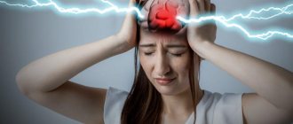
Treatment of damage to speech zones in Saratov, how to improve the functioning of speech zones
Sarklinik provides comprehensive conservative treatment of the speech areas of the cerebral cortex in children (boys and girls), adolescents (boys and girls), adults (men and women) in Saratov, Russia. At the first appointment, the doctor will tell you what reflex massage of speech zones is, what new advanced methods exist for restoring speech disorders; how to treat pathology of the motor and sensory speech zones, how to restore and improve speech. What happens when a pulsed magnetic field is applied to the speech centers in the cerebral cortex in Russia, does this technique always help? What new methods exist for stimulating speech centers that give 95% results? How to really improve the work of the Wernicke center, how to activate the Wernicke area? What is Broca's center? How to increase a child’s vocabulary in Russia? How to improve the impressive (sensory) and expressive (motor) speech of infants, preschoolers, and schoolchildren? Where to treat mental retardation, and what to do if speech zones are disrupted, how to treat speech disorders in Saratov . On the sarclinics website you can ask the doctor a question online and read reviews about the treatment of children.
What parts does the brain consist of?
The human brain consists of several sections. Each of them performs its function, ensuring the vital functions of the body.
Table 1. Main departments.
| Name | Description |
| Oblong | This part is a continuation of the spinal cord. It consists of gray matter nuclei and white matter tracts. It is this part that determines the connection between the brain and the body. |
| Average | It consists of 4 tubercles, two of which are responsible for vision and two for hearing. |
| Rear | The hindbrain includes the pons and cerebellum. This is a small section in the back of the head, which weighs around 140 grams. Consists of two hemispheres connected to each other. |
| Intermediate | Consists of the thalamus, hypothalamus. |
| Finite | This section forms both hemispheres of the brain, connected by the corpus callosum. The surface is full of convolutions and grooves covered with bark. The hemispheres are divided into lobes: frontal, parietal, temporal and occipital. |
The last section occupies more than 80% of the total mass of the organ. The organ can also be divided into 3 parts: the cerebellum, the brainstem and the cerebral hemispheres.
In this case, the entire brain is covered in the form of a shell, divided into three components:
- Arachnoid (cerebrospinal fluid circulates through it)
- Soft (adjacent to the brain and full of blood vessels)
- Hard (in contact with the skull and protects the brain from damage)
All components are important in the regulation of life and have a specific function. But the activity regulation centers are located in the cortex.
The human brain consists of many sections, each of which has a complex structure and performs a specific role. The largest of them is the final one, which consists of hemispheres. All this is covered with three shells that provide protective and nourishing functions.
Learn about the structure and functions of the brain from the video provided.
Treatment of speechopathy in Saratov, how to treat speechopathy in Russia
Sarklinik provides speech therapy treatment in Saratov , treatment of speech pathologies, speech disorders in children, boys, girls, teenagers in Russia. Sarklinik knows how to treat logopathies, how to cure logopathies, how to get rid of logopathies.
Treatment of alalia, treatment of aphasia, treatment of stuttering, treatment of speech delay, treatment of speech development disorder.
Sign up for a consultation. There are contraindications. Specialist consultation is required.
Photo: (©) Bioraven | Dreamstime.com\Dreamstock.ru
Related posts:
Puberty boys, girls, teenagers, adolescence, puberty
Mental retardation in children: moderate, mild, severe, profound, mental retardation, dementia, treatment
Enuresis in adolescents, teenage enuresis, treatment of enuresis
Why does a child pee in bed at night? How to stop a child from peeing at night?
Enuresis treatment center in Russia, Saratov, which doctor to contact