content .. 100 101 102 103 104 105 ..
Hindbrain (human anatomy)
The hindbrain includes the pons and cerebellum. It develops from the fourth cerebral vesicle (metencephalon).
The pons borders the medulla oblongata from below, passes into the cerebral peduncles from above, and its lateral sections form the middle cerebellar peduncles. In the anterior (basilar) part of the pons there are accumulations of gray matter - the nuclei of the pons; in the posterior part of the tegmentum of the pons there are nuclei of the superior olive, reticular formation and V - VIII pairs of cranial nerves. These nerves emerge from the base of the brain lateral to and behind the pons, at the border with the cerebellum and medulla oblongata. The white matter of the pons in its anterior part is represented by transversely running fibers going to the middle cerebellar peduncles. They are penetrated by powerful longitudinal bundles of fibers of the pyramidal tracts, which then form the pyramids of the medulla oblongata and go to the spinal cord. In the rear part of the bridge (pontineum) there are ascending and descending fiber systems (Fig. 113).
Rice. 113. Brain stem (front view). 1 - anterior median fissure; 2 - pyramids of the medulla oblongata; 3 - olive; 4 - cerebellum; 5 - decussation of the pyramids (the place of transition of the medulla oblongata into the spinal cord); 6 - middle cerebellar peduncle; 7 - bridge; 8 - interpeduncular fossa; 9 - cerebral peduncle; III - XII roots of cranial nerves; C - first spinal nerve
Physiology of the medulla oblongata and pons
The medulla oblongata and the pons perform two functions - reflex and conductive. Through the sensitive fibers of the roots of the cranial nerves, it receives impulses - information from the receptors of the scalp, mucous membranes of the eyes, nose, mouth (including taste buds), from the organ of hearing, the vestibular apparatus (organ of balance), from the receptors of the larynx, trachea, lungs, as well as from interoreceptors of the cardiovascular system and digestive apparatus.
Through the medulla oblongata, many simple and complex reflexes are carried out, covering not individual metameres of the body, but organ systems, for example, the digestive, respiratory, and circulatory systems. The reflex activity of the medulla oblongata can be observed in a bulbar cat, that is, a cat in which the brain stem above the medulla oblongata has been cut. The reflex activity of such a cat is complex and diverse.
The following reflexes occur through the medulla oblongata: 1) protective: coughing, sneezing, blinking, lacrimation, vomiting; 2) food: sucking, swallowing, secretion of juice from the digestive glands; 3) cardiovascular, regulating the activity of the heart and blood vessels; 4) in the medulla oblongata there is an automatically working respiratory center that provides ventilation to the lungs; 5) the vestibular nuclei are located in the medulla oblongata and the pons.
From the vestibular nuclei of the medulla oblongata begins the descending vestibulospinal tract, which is involved in the implementation of posture reflexes, namely in the redistribution of muscle tone. A bulbar cat can neither stand nor walk, but the medulla oblongata and cervical segments of the spinal cord provide those complex reflexes that are elements of standing and walking. All reflexes associated with the standing function are called positioning reflexes. Thanks to them, the animal holds its body, usually with the crown up.
The special importance of this part of the central nervous system is determined by the fact that the medulla oblongata contains vital centers: respiratory, cardiovascular. Therefore, not only removal, but even damage to the medulla oblongata ends in death.
In addition to the reflex function, the medulla oblongata performs a conductive function. Conducting pathways pass through it, connecting the cortex, diencephalon, midbrain, cerebellum and spinal cord with a two-way connection.
The cerebellum is located dorsal to the pons and medulla oblongata. It has two hemispheres and a middle part - the worm. The surface of the cerebellum is covered with a layer of gray matter (cerebellar cortex) and forms narrow convolutions separated by grooves. With their help, the surface of the cerebellum is divided into lobules. The central part of the cerebellum consists of white matter, which contains accumulations of gray matter - the cerebellar nuclei. The largest of them is the dentate nucleus. The cerebellum is connected to the brain stem by three pairs of peduncles: the upper ones connect it to the midbrain, the middle ones to the pons, and the lower ones to the medulla oblongata. The peduncles contain bundles of fibers connecting the cerebellum to various parts of the brain and spinal cord.
During development, the isthmus of the rhombencephalon forms the boundary between the hindbrain and midbrain. From it develop the superior cerebellar peduncles, the superior medullary velum located between them and the triangles of the loop, lying outward from the superior cerebellar peduncles.
During development, the fourth (IV) ventricle (ventriculus quartus) is a common cavity of the medulla oblongata and hindbrain. At the bottom, the IV ventricle communicates with the central canal of the spinal cord, at the top it passes into the cerebral aqueduct of the midbrain, and in the roof area it is connected by three openings to the subarachnoid space of the brain. Its anterior (ventral) wall—the bottom of the fourth ventricle—is called the rhomboid fossa. The lower part of the rhomboid fossa is formed by the medulla oblongata, and the upper part by the pons and isthmus. The posterior (dorsal) wall - the roof of the IV ventricle - is formed by the superior and inferior medullary sails and is supplemented at the back by a plate of the pia mater lined with ependyma. This area contains a large number of blood vessels that form the choroid plexus of the fourth ventricle. The convergence of the superior and inferior sails protrudes into the cerebellum and forms a tent. The rhomboid fossa is of vital importance, since most of the nuclei of the cranial nerves (V - XII pairs) are located in this area.
Physiology of the cerebellum (human anatomy)
The cerebellum is a suprasegmental part of the central nervous system that does not have a direct connection with the receptors and effectors of the body. It is connected in numerous ways to all parts of the central nervous system. Afferent pathways are sent to it, carrying impulses from proprioceptors of muscles, tendons, vestibular nuclei of the medulla oblongata, subcortical nuclei and cerebral cortex. In turn, the cerebellum sends impulses to all parts of the central nervous system.
The functions of the cerebellum are studied by irritating it, partially or completely removing it, and studying bioelectrical phenomena. The Italian physiologist Luciani characterized the consequences of removal of the cerebellum and loss of its functions with the famous triad A: astasia, atony and asthenia. Subsequent researchers added another symptom: ataxia.
A dog without a cerebellum stands on widely spaced legs and makes continuous rocking movements (astasia). She has impaired proper distribution of flexor and extensor muscle tone (atony). Movements are poorly coordinated, sweeping, disproportionate, abrupt. When walking, the paws are thrown beyond the midline (ataxia), which is not observed in normal animals. Ataxia is explained by the fact that movement control is impaired. Analysis of signals from proprioceptors of muscles and tendons is missing. The dog cannot get its muzzle into the food bowl. Tilt of the head downwards or to the side causes a strong opposite movement.
The movements are very tiring: the animal, after walking a few steps, lies down and rests. This symptom is called asthenia.
Over time, movement disorders in a cerebellar dog smooth out. She eats on her own and her gait is almost normal. Only biased observation reveals some violations (compensation phase).
As E. A. Asratyan showed, compensation of functions occurs due to the cerebral cortex. If the bark of such a dog is removed, then all the violations are revealed again and are never compensated.
The cerebellum is involved in the regulation of movements, making them smooth, precise, proportionate. According to the figurative expression of L. A. Orbeli, the cerebellum is an assistant to the cerebral cortex in controlling skeletal muscles and the activity of autonomic organs. As studies by L.A. Orbeli have shown, cerebellar dogs have impaired autonomic functions. Blood constants, vascular tone, the functioning of the digestive tract and other autonomic functions become very unstable and easily shift under the influence of certain reasons (food intake, muscle work, temperature changes, etc.).
When half of the cerebellum is removed, motor functions on the side of the operation are impaired. This is explained by the fact that the cerebellar pathways either do not cross at all or cross twice.
content .. 100 101 102 103 104 105 ..
Anatomy of the White Matter of the human brain hemispheres - information:
The entire space between the gray matter of the cerebral cortex and the basal ganglia is occupied by white matter . It consists of a large number of nerve fibers running in different directions and forming the pathways of the telencephalon.
Nerve fibers can be divided into three systems:
- associative,
- commissural and
- projection fibers.
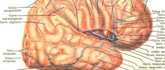
A. Associative fibers connect different parts of the cortex of the same hemisphere. They are divided into short and long. Short fibers, fibrae arcudtae cerebri, connect neighboring convolutions in the form of arcuate bundles. Long association fibers connect areas of the cortex that are more distant from each other. There are several such fiber bundles. Cingulum, belt, is a bundle of fibers passing through the gyrus fornicatus, connecting various parts of the cortex of the gyrus cinguli both with each other and with neighboring convolutions of the medial surface of the hemisphere. The frontal lobe is connected to the inferior parietal lobe, the occipital lobe and the posterior part of the temporal lobe through the fasciculus longitudinalis superior. The temporal and occipital lobes are connected to each other through the fasciculus longitudinalis inferior. Finally, the orbital surface of the frontal lobe is connected to the temporal pole by the so-called uncinate fasciculus, fasciculus uncindtus.
B. Commissural fibers , which are part of the so-called cerebral commissures, or commissures, connect the symmetrical parts of both hemispheres. The largest cerebral commissure, the corpus callosum, connects the parts of both hemispheres belonging to the neencephalon. Two brain commissures, commissura anterior and commissura fornicis, much smaller in size, belong to the rhinencephalon and connect: commissura anterior - olfactory lobes and both parahippocampal gyri, commissura fornicis - hippocampi.
B. Projection fibers connect the cerebral cortex partly with the thalamus and corpora geniculata, partly with the underlying parts of the central nervous system up to and including the spinal cord. Some of these fibers conduct excitations centripetally, towards the cortex, while others, on the contrary, centrifugally.
Projection fibers in the white matter of the hemisphere closer to the cortex form the so-called corona radiata, corona radiata, and then the main part of them converges into the internal capsule, which was mentioned above. The internal capsule, capsula interna, as stated, represents a layer of white matter between the nucleus lentiformis, on the one hand, and the caudate nucleus and thalamus, on the other. On a frontal section of the brain, the internal capsule looks like an oblique white stripe that continues into the cerebral peduncle. On a horizontal section, it appears in the form of an angle, open to the lateral side; as a result, in the capsula interna they distinguish the anterior leg, crus anterius capsulae internae, - between the caudate nucleus and the anterior half of the inner surface of the nucleus lentiformis, the posterior leg, crus posterius, - between the thalamus and the posterior half of the lentiform nucleus and the knee, genu capsulae internae, lying in place bend between both parts of the internal capsule.
Projection fibers according to their length can be divided into the following systems, starting with the longest:
- Tractus corticospinalis (pyramidalis) conducts motor volitional impulses to the muscles of the trunk and limbs. Starting from the pyramidal cells of the cortex of the middle and upper parts of the precentral gyrus and lobulus paracentralis, the fibers of the pyramidal tract go as part of the corona radiata, and then pass through the internal capsule, occupying the anterior two-thirds of its posterior limb, with the fibers for the upper limb going in front of the fibers for the lower limb . Next they pass through the peduncle of the brain, pedunculus cerebri, and from there through the bridge into the medulla oblongata.
- Tractus corticonuclearis - pathways to the motor nuclei of the cranial nerves. Starting from the pyramidal cells of the cortex of the lower part of the precentral gyrus, they pass through the knee of the internal capsule and through the cerebral peduncle, then enter the bridge and, moving to the other side, end in the motor nuclei of the opposite side, forming a decussation. A small part of the fibers ends without crossing. Since all motor fibers are collected in a small space in the internal capsule (the knee and the anterior two-thirds of its posterior leg), if they are damaged in this place, unilateral paralysis (hemiplegia) of the opposite side of the body is observed.
- Tractus corticopontini - pathways from the cerebral cortex to the pontine nuclei. They come from the cortex of the frontal lobe (tractus frontopontinus), occipital (tractus occipitopntinus), temporal (tractus temporopontinus) and parietal (tractus parietopontinus). As a continuation of these pathways, fibers from the pontine nuclei go to the cerebellum as part of its middle peduncles. Using these pathways, the cerebral cortex has an inhibitory and regulatory effect on the activity of the cerebellum.
- Fibrae thalamocorticalis et corticothalamici - fibers from the thalamus to the cortex and back from the cortex to the thalamus. Of the fibers coming from the thalamus, it is necessary to note the so-called central thalamic radiance, which is the final part of the sensory pathway heading to the center of the cutaneous sense in the postcentral gyrus. Coming from the lateral nuclei of the thalamus, the fibers of this pathway pass through the posterior limb of the internal capsule, behind the pyramidal tract. This place was called the sensitive chiasm, since other sensory pathways pass here, namely: visual radiation, radiatio optica, coming from the corpus geniculatum laterale and pulvinar of the thalamus to the visual center in the occipital lobe cortex, then auditory radiation, radiatio acustica, heading from corpus geniculatum mediale and the inferior colliculus of the roof of the midbrain to the superior temporal gyrus, where the hearing center is located. The visual and auditory tracts occupy the most posterior position in the posterior limb of the internal capsule.