The main character of the new book from the publishing house Mann, Ivanov and Ferber, Taming the Amygdala, usually weighs 1360 grams. This is the brain. The book was written by the doctor John Arden, who is aware not only of the latest advances in the field of neurophysiology, but also how to write about them in simple language to the average citizen so that he can use the acquired knowledge in practice. Everyone needs to read the book - or rather, all those who want to reprogram their brain so that it works “faster, higher and stronger.”
The vicious circle of irregular work schedule Anna Zhurba, online school EnglishDom
Management
Characteristic
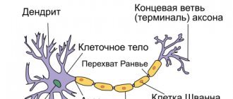
The amygdala is a cluster of nuclei, a paired structure located in the limbic system within the brain. The area of brain tissue is responsible for the formation of emotions, such as fear and pleasure. It has been experimentally proven that stimulation of the right amygdala provokes the appearance of negative emotions (anxiety, sadness, worry), the left - more often positive (joy, happiness), less often negative.
According to anatomy, the amygdala in men is larger on average by 0.3 cm3 than in women. However, given that men's brains are on average 10% larger than women's (due to the larger size of the torso and skull), the increased volume of male tonsils is a predictable and logical result. The brain structure develops faster in women.
The left amygdala matures faster than the right, which is associated with the repeated occurrence of adequate reactions to dangerous situations in childhood. The larger the volume of the amygdala, the greater the number of connections formed with other parts of the brain. Scientists associate the number of synaptic connections formed by the amygdala with an individual’s communication activity.
The more social contacts there are, the more actively the brain region interacts with other brain systems. Research shows the relationship between the volume of the amygdala and such individual abilities as recognizing people's appearance and determining the nature of the emotions they show. The amygdala is a part of the brain that is responsible for the occurrence of fear, which allowed scientists to suggest the influence of increased activity of the structure on the development of anxiety-phobic disorders.
Stimuli that are associated with negative experiences can activate the activity of the amygdala, which provokes the body’s preparation for stress and to face a dangerous situation. Some scientists suggest a similar pathogenetic mechanism for the occurrence of panic attacks. Increased activity of the amygdala complex is detected in conjunction with post-traumatic neurotic, bipolar, and panic disorders.
The studies revealed that in patients diagnosed with depression of various origins, emotionally significant verbal stimuli (words, phrases) provoked prolonged activation of the tonsils. Similar physiological reactions are observed in patients who are presented with visual stimuli associated with feelings of sadness and grief.
After a course of treatment with antidepressants (Venlafaxine), the increased activity of the amygdala regresses, and the structure begins to function normally. Aggression is not an independent neurotic disorder. Aggressive behavior can be a consequence of a normal reaction to an external stimulus or a manifestation of mental pathology. The implementation of aggressive behavior patterns occurs with the direct participation of the amygdala.
Damage to the brain matter in the temporal lobe is often accompanied by a loss of control over manifestations of aggression. Research results provide an example of this. In 20-28% of patients with temporal lobe epilepsy and atrophic changes in the substance of the amygdala complex, interictal (between attacks) aggressive behavior is observed.
The amygdala cannot be called a gland because it does not produce specific substances. This department is involved in the process of detecting threats, expressing emotions of fear, and enhancing emotional memory. The amygdala complex is a structure that monitors danger and provides a response to facial expressions of threat and rage.
The vicious circle of irregular work hours
457
Want to know what happens in the brain during meditation or prayer? Managing anxiety? Stimulate positive mood? Manage your concentration? Increase stress resistance? And in general, to improve the quality of life? Of course you do. Only 293 pages - and the knowledge of how to do it is yours. First, of course, you will have to thoroughly understand which areas of the brain are responsible for which emotions, how neural connections are formed (which control our entire body) and what, for example, you need to eat for greater neuroplasticity.
This will take some time. But an introduction to basic biology is essential: spindle cells, the orbitofrontal cortex, neurotransmitters and mirror neurons are already waiting for you. However, there is no need to be afraid: neurophysiologist Arden has 11 books under his belt (some of them are deeply scientific literature about the brain, awarded with specialized awards), and he has already learned to explain all these things briefly and not boringly.
So, the central idea of the book is this: the human brain is capable of changing under the influence of external experience. This means that the genetic makeup determines only the potential strengths and weaknesses of a person, but does not affect a person’s behavior, thoughts and emotional state. This means that the brain can be reconfigured - for this, Arden rightly clarifies, you need to know how it works.
On the one hand, this is difficult, because the brain consists of billions of neurons and even more supporting cells - this is comparable to the number of stars in our galaxy. Therefore, in the book there are sometimes complex words: corticotropin, neurotransmitter glutamate, hypocanic alkalosis and all that.
But complex terms are always followed by simple examples: if you walk into a room and don’t remember why you did it, it’s the dorsolateral prefrontal cortex acting up. And that’s why she’s being naughty - and then there’s such an elegant explanation that you want to smile and figure it out. In some places, the book, however, still resembles a textbook, but the dry and very logical narrative with an abundance of terms is often interrupted by practical advice: Marley, Jane and other patients of the author of the book are at your service. They also wanted to manage stress and improve their quality of life.
On the other hand, to control your brain, it is enough to grasp a simple idea: all the billions of neurons in our heads strive to be activated. And those that are activated together form connections. By connecting in the same pattern several times, they work wonders. Hence the phenomenon of neuroplasticity, which can be consciously controlled. In fact, the reader is convincingly shown this: the brain is truly a muscle that needs to be exercised. And then it says how.
An interesting example: not only practical actions, but also simply the thought of these actions can change the structure of the brain through neuroplasticity. Scientists have shown that simply visualizing playing the piano stimulates changes in the part of the brain that controls finger movements when actually playing the piano. Useful knowledge both for those who do not want to do homework at music school, and for those who have hopelessly grown out of it.
Your own elevator. Review of the book “The Next Level” Contemporary literature
Functions
The amygdala of the brain specializes in the perception of emotions and is activated when a person experiences strong emotional experiences. The main function is to prepare the body for a “fight or flight” response. The functions of the amygdala are distributed based on its structure. The amygdala structure consists of nuclei of different types, divided mainly taking into account functional differences:
- Basolateral. Associated with emotional manifestations. An input structure that receives diverse sensory-type information from the cortical regions and thalamus.
- Central. Consists of lateral and medial sections. The medial section projects incoming information to the brain stem and hypothalamus, which causes the occurrence of autonomic and motor reactions appropriate to the situation.
- Cortical (cortical). Reacts to taste sensations.
- Medial. Associated with the olfactory system.
Activation of the basolateral, medial and central nuclei gives an anxiogenic (state of horror, fear) effect. Inhibition of the activity of these structures is accompanied by an anxiolytic (suppression of anxiety) effect. Dysfunction of the tonsils underlies the development of anxiety-phobic disorders.
The coordinated interaction of the amygdala nuclei allows you to promptly recognize danger and respond adequately to it. For example, the unpleasant smell and taste of spoiled foods make a person experience a feeling of disgust and hostility, which protects against food poisoning.
The reaction to the unpleasant odor of waste products is the desire to withdraw and avoid contact, which prevents infection by pathogenic microorganisms. Close interaction with the hippocampus involves fixing in memory memories of experienced events that provoked a feeling of horror, strong fear, which subsequently affects the occurrence of an appropriate reaction to such stimuli.
If an individual repeatedly encounters similar situations, the corresponding images stored in long-term memory activate the amygdala complex. As a result of the perception of external stimuli that are associated with the experienced traumatic situation, the body mobilizes efforts to avoid troubles.
The amygdala complex is involved in the formation of the reaction of aggression, affective (sensual, emotional) perception, experience and manifestation of emotions. The tasks of the amygdala complex include emotional assessment of stimuli that are primarily processed in cortical structures. Stimuli containing an emotional component are better recorded in memory.
The emotional center of the brain regulates aggression and sexual behavior
The amygdala, known as the “fear center,” can make a male peaceful and indifferent to the opposite sex, or turn him into an exclusive aggressor - in every sense.
The amygdala, or amygdala, was once called the center of fear - because if this area of the brain did not work, then the individual (whether a person or a mouse) ceased to be afraid of anything. However, over time, it became clear that the amygdala controls not only fear, but also other emotions, and that it is generally involved in a wide variety of functional processes in the brain. It is known to contain groups of neurons necessary for learning and memory; it is known that it has its own center of pleasure - which means that the amygdala is associated with the functions of the famous reinforcement system, which through a feeling of pleasure controls our behavior and motivations. Finally, the amygdala, this “fear center,” helps predators hunt.
(Photo: CreativeNature/Depositphotos)
Amygdala in the human brain. (Photo: Life Science Databases/)
‹
›
Another example of how broad the functions of the amygdala can be is described in Nature Neuroscience
researchers from New York University. They studied aggression and sexual behavior in male mice. Both aggressiveness and sexual behavior are highly dependent on the hypothalamus. Being connected to almost all parts of the central nervous system, from the cerebral cortex to the spinal cord, the hypothalamus influences both purely physiological reactions, such as hunger and thirst, and complex cognitive reactions, emotions and memory. It has groups of neurons called nuclei that perform specific functions. In particular, sexual behavior depends on neurons in the medial preoptic nucleus, or MPN neurons. Aggression depends on VMHvl neurons - some cells in the ventromedial nucleus.
The researchers noticed that both MPN neurons and VMHvl neurons are connected to the amygdala, or more precisely, to the posterior part of it. And if the activity of amygdala neurons, which are connected to the “sex” neurons of the hypothalamus, was suppressed in male mice, then the males almost lost interest in sex. If these neurons were stimulated, and the “sex” neurons of the hypothalamus received many signals from them, then the sexual interest of males increased enormously - they began to care for even those females who were not ready to reproduce at all and for whom no one normally cares.
As for aggression, male mice usually attack males that are unfamiliar to them. But they attacked even strangers half as weakly if in their amygdala they suppressed the activity of cells associated with VMHvl neurons of the hypothalamus. And vice versa - if the activity of neurons in the amygdala was stimulated, males began to fight not only with strangers, but also with familiar males, and even with females.
That is, in the “center of emotions” (although this name for the amygdala is not very correct) there are two groups of cells that regulate aggression and sexual desire. Neuroscientists already knew that the amygdala influences social behavior, but little was known about which structures are involved and which areas of the brain they communicate with. In some psychiatric disorders, patients experience increased aggression and sexuality, and the authors of the paper believe that the new data will help to better understand how such symptoms develop and how they can be acted upon. On the other hand, these results were obtained in experiments on mice, and the extent to which the amygdala influences the “sex” and “aggressive” neurons of the hypothalamus in humans remains to be seen.
Pathologies
Excessive activity of the structures of the amygdala, which is located in the temporal zone, where the area between the putamen and the temporal pole is located, leads to disruption of the functioning of many body systems.
Effect on the heart
Cardiac dysfunction is associated with dysfunction of the brain's tonsils. Increased activity of the tonsil structures provokes the development of heart failure.
The pathogenetic mechanism is based on the participation of the amygdala in the formation of affects such as fear or rage. When a person is exposed to stress, the amygdala perceives the situation as a threat and activates the activity of the bone marrow, which intensively produces white blood cells (leukocytes). As a result, an inflammatory reaction develops in the blood vessels, which, as it progresses, can provoke angina pectoris, heart attack, and stroke.
Urbach-Wiethe disease
A rare, genetically determined pathology characterized by destruction of the amygdala. Patients with Urbach-Wiethe disease experience virtually no feeling of fear while simultaneously having the instinct of self-preservation.
Research shows that in order to scare patients, it is necessary to use unusual methods, for example, inhalation of air with a high (about 35%) concentration of carbon dioxide. Inhaling such air can cause a panic attack in patients. The clinical picture of the disease is variable and is represented by neurological deficits (epileptic seizures) and dermatological symptoms (scars, papules on the skin).
Dementia and Parkinson's disease
Symptoms of dementia include loss of the ability to think, reason, or remember. Other signs include changes in a person's behavior that affect their daily life.
Various diseases can cause dementia, and a person may sometimes have a mixed form of dementia. There is a strong connection between Parkinson's disease and dementia. Up to 50% of people with Parkinson's disease have dementia. People with Parkinson's disease may experience joint stiffness, shaking or tremors, and difficulty walking. The disease occurs when a person's brain cells die, although it is not yet clear why this happens. Parkinson's disease can damage large areas of a person's brain. It is at this stage that scans can detect the disease; a decrease in brain volume often causes symptoms of dementia.
Dementia progression scans in Parkinson's disease look at the loss of areas of the brain. However, brain imaging usually reveals these changes in the later stages of the disease. As a result, doctors assess the progression of dementia by symptoms.
The authors of the new study believe that scanning will help detect dementia much earlier and more accurately.
People with Parkinson's disease often have a buildup of protein in the brain, which is also seen in Alzheimer's disease.
The study authors note that the presence of iron in the human brain is a natural part of the aging process and is associated with an increased presence of the protein.
According to the study's lead author Rimona Weil from the UK, "iron in the brain has attracted the interest of scientists studying neurodegenerative diseases such as Parkinson's disease and dementia."
As we age, iron accumulates in the brain, but it is also associated with the accumulation of harmful brain proteins, so scientists are starting to find evidence that it may help in monitoring disease progression and for diagnosis.
Instead of measuring brain volume loss, the researchers are using a new method called quantitative susceptibility mapping, which uses magnetic resonance imaging.