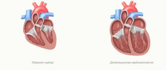
Dilation of the heart chambers
- a pathological condition accompanied by an increase in the volume of the heart chambers without changing the thickness of the heart wall.
As a rule, this condition is not an independent disease, but indicates a congenital or acquired pathology.
Dilatation of the left atrium often occurs as a result of a sustained increase in pressure in the systemic circulation due to constant heavy physical exertion, and indicates a state of cardiac overload.
Dilatation of the right atrium occurs as a compensatory reaction of the heart to increased pressure in the pulmonary circulation. Usually as a result of chronic bronchopulmonary diseases accompanied by spasms of the bronchial muscles (in this case, dilatation is part of the complex pathology of the pulmonary heart); less often - as a result of pathologies of the blood vessels of the lungs, pulmonary hypertension. Another cause of dilatation is infectious heart diseases.
Dilatation of the left ventricle occurs when myocardial pathology occurs, or when volume overload occurs, most often in this case the cause is aortic stenosis.
Dilatation of the right ventricle can also develop with myocardial pathology and with blood volume overload, which is caused by heart defects, both congenital and acquired. In particular, atrial septal defect, tricuspid valve insufficiency, pulmonary valve insufficiency, and a number of other pathologies can lead to dilatation of the right ventricle of the heart.
If the cause of dilatation is eliminated, the chamber may decrease in size, down to normal. In the case of chronic exposure to unfavorable factors, fibrous tissue changes occur and dilatational hypertrophy is formed (increase in volume and thickening of the walls). Both dilatation and hypertrophy are accompanied by increasing heart failure (decreased EF). Treatment comes down to combating the underlying disease that led to dilatation. Sometimes surgery is indicated, but if the underlying disease persists, survival does not increase. In severe cases (accompanied by progressive heart failure), a heart transplant is indicated.
Dilatation is the expansion (or stretching) of a section of the heart muscle. This occurs under the influence of a number of factors. This condition is most typical for the left ventricle. The pathology develops gradually. Dilatation of the left ventricle leads to rhythm disturbances and the appearance of signs of heart failure. The disease ends with the complete inability of the myocardium to perform its functions.
Features and classification
One of the known variants of cardiomyopathy is ventricular dilatation.
Expansion of cavities occurs in many patients for no apparent reason. As a result, the superficial function of the myocardium is disrupted, which leads to a rapid increase in its size. The appearance of dysfunction is associated with a decrease in the strength of contractions of the muscular wall of the ventricles. At the same time, there is a decrease in the release of blood into the aorta. During the examination of some patients, the thickness of the heart wall does not change when the cavities are dilated. The following options for dilatation of the left ventricle of the heart are distinguished:
With tonogenic dilatation, expansion of the heart cavity is noted due to increased blood flow to them and increased pressure. The myogenic form is characterized by an irreversible change in the volume of the chamber. It appears against the background of elongation of fibers and their stretching with a simultaneous lack of contractility.
The latter variant of dilatation is most often combined with a decrease in wall tone. It is divided into primary and secondary. The first form develops with myocarditis in acute or chronic stages, cardiosclerosis caused by atherosclerosis. During primary expansion, the cavity grows uniformly in size. The function of myocardial contraction is significantly reduced. The pulse and heart rate become weak and difficult to feel.
The secondary form occurs against the background of established myocardial hypertrophy. The size of the heart is significantly increased in comparison with the primary one.
There are many factors that have a negative effect on the myocardium, but there are certain conditions that contribute to dilatation of the left ventricular cavity:
- Pathology associated with damage to the myocardium itself.
- Excessive load.
Some patients are characterized by an asymptomatic course of the disease against the background of complete health. Over time, if it is impossible to compensate for the condition, signs of the disease appear. This is typical for dilated cardiomyopathy. Other causes are inflammation, arterial hypertension, which over time make the muscle wall weak. This condition leads to loss of elasticity and excessive extensibility, which leads to dilatation of the cavity.
Process Features
The left ventricle is enlarged only due to its hypertrophied wall, which leads to distortion of the interventricular septum of the heart. Its thickening can be uniform or localized. The interior space is not modified.
Experts distinguish 2 forms of pathology:
- Concentric hypertrophy occurs under the influence of high pressure.
- The eccentric form develops due to congestion of the ventricle with a large volume of incoming blood.
If a person has an enlarged left ventricle of the heart, then he should remember the general list of consequences of this pathology:
- cardiac ischemia;
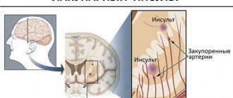
- stroke;
- asystole (cardiac arrest);
- expansion of the aorta;
- irregular heartbeat;
- loss of consciousness;
- heart failure;
- stenosis of the coronary arteries.
Complications are associated with the gradual loss of elasticity of the left ventricle due to its hypertrophy. The pressure inside the heart will increase, causing severe disruptions in hemodynamics. Overgrown tissue can also begin to compress the coronary vessels, causing disruption of myocardial nutrition.
Right ventricular dilatation
One of the reasons for the expansion of the cavity is considered to be insufficient functioning of the valve apparatus. A similar condition is typical for patients who have suffered endocarditis or rheumatism, where damage to the leaflet structures was a complication. Dilatation of the right ventricle occurs in the absence of the pericardium, which occurs in some patients.
The result of this pathology is gradually stretching of the muscle fibers. The altered septum between the atria leads to dilation of the artery in the lungs. An increase in pressure in this vessel indicates an increase in pressure in the cavity of the right ventricle.
Defects also negatively affect the chamber of the heart. They contribute to increased pressure in the pulmonary artery. This process ends with dilatation due to a deficiency of compensatory functions of the body.
Symptoms
Moderate expansion of one or two chambers in the heart may not manifest itself for a long time. Often, pathology is discovered by chance, during a routine examination or treatment of another disease. Severe dilatation of the cavity leads to a decrease in pumping function, which leads to the appearance of signs of heart failure or arrhythmia. These include the following:
- Palpable palpitations.
- Dyspnea.
- Blueness of the nasolabial triangle, lips, earlobes, fingertips.
- As the severity of the course worsens, cyanosis spreads to the skin.
- Swelling in the arms and legs.
- Memory impairment.
- Fatigue and weakness that persists after rest.
- The appearance of discomfort when lying down.
- Dizziness.
- Headache.
- Feeling of interruptions in the heart.
Shortness of breath in the compensation stage appears only with excessive physical exertion. With gradual wear and tear of the myocardium, the condition worsens. Shortness of breath begins to bother you with slight exertion, and then at rest.
With chronic exposure to unfavorable factors, a change occurs in the myocardium, which leads to a gradual expansion of the cavity and thickening of the walls. Dilatation of the left ventricle of the heart in the absence of timely therapy increases the risk of complications. Most often, thrombosis and fibrillation of the ventricles or atria are observed.
In some patients, the valve apparatus is affected, which is manifested by expansion of the ring, deformation of structures and ends in the formation of acquired heart disease.
After the transition from the stage of compensation to decompensation, fluid appears in the abdominal cavity (ascites), and the size of the liver increases (hepatomegaly). The skin of such patients becomes damp and cold to the touch. Systolic blood pressure decreases. Tachycardia is noted.
When auscultated, wheezing is heard in the lungs. Determination of the boundaries of the heart shows cardiomegaly (increase in heart size), the rhythm is disturbed.
Causes
Dilation of the chamber in the left ventricle often occurs under the influence of several provoking factors. This condition is directly related to the patient’s age, heredity, and the presence of excess body weight. The reasons that negatively affect the myocardium are:
- Congenital heart defects. Exposure to unfavorable environmental factors occurs already during pregnancy. If the lesion becomes extensive, the fetus dies. In the case of a slightly pronounced lesion, a defect is formed.
- Inflammatory diseases, which include myocarditis, pericarditis, endocarditis. The risk group includes children and adolescents, in whom cases of this pathology are often recorded.
- Chronic diseases of the cardiovascular system. These include arterial hypertension, angina pectoris, ischemia.
- Metabolic syndrome, the basis of which lies in the presence of excess body weight and diabetes mellitus in the patient.
- Chronic pathology of lung tissue.
- Diseases of the kidneys, endocrine and hematopoietic systems.
- Genetic predisposition.
- Autoimmune disorders.
One of the common factors of dilatation is chronic intoxication with alcohol and nicotine.
This group also includes side effects from medications. Pheochromocytoma is the most common endocrine pathology. It is a benign or malignant form of tumors. It is characterized by excessive production of adrenaline.
Prevention
Left ventricular hypertrophy is only a consequence of certain pathological processes, so it is important to follow the rules of prevention in order to prevent their development:
- give up alcohol and drugs;
- quit smoking;
- Minimize the consumption of caffeine and energy drinks;
- undergo a full examination annually;
- get enough sleep (7-8 hours a day);
- fully treat emerging diseases;
- adjust your diet;
- exercise;
- try not to overload yourself physically and mentally;
- avoid stress.
The growth of the wall of the left ventricle also occurs due to other factors, among which the first place is occupied by diseases of the cardiovascular system. The development of the pathological process is accelerated by dysfunction of internal organs, bad habits, excess body weight and stress. Treatment includes medications and other means aimed at eliminating the cause and reducing the load on the heart. Advanced cases require surgical intervention.
Forecast
Every patient with left ventricular dilatation, already knowing what it is, must follow all medical recommendations. With this diagnosis, early diagnosis and initiation of treatment are important. In advanced forms, there is a high probability of developing heart failure. In these same patients, the valve apparatus is deformed, which leads to mitral insufficiency. This diagnosis significantly affects the quality of life and reduces its duration. The prognosis for patients is unfavorable.
The average survival rate for left ventricular dilatation is 10 years. If the course is asymptomatic, then life expectancy is on average 5 years. Patients with chronic heart failure observed in the hospital survive up to 50% of those admitted.
It is important for each patient to remember that the first symptoms are not considered normal and require a set of diagnostic procedures. Timely treatment will reduce the risk of complications, and treatment will prolong life for many years.
The following sources of information were used to prepare the material.
Expansion or stretching of any hollow organ is called dilatation. In this case, there should be no thickening of its walls. Most often in practical medicine they talk about such changes in the chambers of the heart. As a result, it cannot pump the required volume of blood and serious complications develop.
Varieties and reasons
Anatomically, the heart is a hollow organ in which there are 4 sections - the right and left atrium and the same ventricles. When the cavities expand, the release of blood into large vessels decreases, and stagnation occurs.
There are several classifications of expansion. Depending on the reasons, dilatation occurs:
- Tonogenic - develops due to increased filling with circulating fluid. At the initial stages of the pathology, the walls of the heart chambers do not thicken.
- Myogenic - due to thickening of the myocardium, its contractile function suffers, blood ejection is disrupted and the cavities expand.
Dilatation is not an independent disease; it occurs against the background of various diseases. Based on morphological characteristics, simple, hypertrophic and atrophic forms are distinguished:
- With simple dilatation, the thickness of the walls does not change much, but the volume of the cavities increases.
- The hypertrophic form is accompanied by thickening of the walls and expansion of the chambers.
- With the atrophic variant, the walls become thinner and the volume of the chambers increases. This form is considered the most unfavorable.
The expansion can involve individual cardiac chambers or several sections at once. From this point of view, dilatation is distinguished:
- left atrium;
- right atrium;
- left ventricle;
- right ventricle;
- left half (ventricle and atrium);
- two ventricles at once;
- expansion of all chambers at the same time - “bull’s heart”.
The reasons for dilation may be common to all types of dilatation or different for isolated forms. Common reasons include:
- Past inflammatory processes of the heart muscle - bacterial, fungal or viral myocarditis.
- Parasitic infestations with myocardial damage.
- Chronic intoxication with alcohol, drugs and certain medications.
- Autoimmune diseases.
- Endocrine disorders.
- Oncological neoplasms.
The cause of isolated dilatations can be congenital or acquired heart defects (damage to the right atrium due to tricuspid valve stenosis), as well as hypertension (the left side is affected).
Diagnostics
If a clinical picture characteristic of the process of proliferation of the heart chambers is identified, it is necessary to contact a cardiologist. He will interview the patient to find out about disturbing symptoms and the presence of other diseases. Then he will conduct an examination, which includes auscultation (listening to sounds) and measurement of pressure and frequency of contractions. Next, the doctor will order for examination:
- Electrocardiography (ECG) will allow you to see the following deviations: the QRS complex is deviated (to the right and forward) from its usual position;
- the degree of excitation increases (from the endocardial layer to the epicardium);
- signs of ischemic disease appear;
- failures occur during the impulse conduction process;
- the electrical axis of the heart is deviated to the left;
- the chest lead is displaced;
- partial or complete blockade of the His bundle appears.
Left half extension
The function of the left atrium is to pump blood from the lungs into the cavity of the left ventricle. From here, oxygen-enriched fluid enters the aorta and all organs. A valve is located between these chambers.
Atrial lesion
The cause of left atrium enlargement may be narrowing of the mitral valve leaflets or its deformation. Sometimes dilatation occurs with atrial flutter.
There are several degrees of expansion. With a mild degree, clinical manifestations do not yet occur. Detection of dilatation at this stage is most often a diagnostic finding during examinations for another reason. Initial organic changes can be diagnosed using echocardiography.
Moderate dilatation is detected more often. The patient develops the first nonspecific complaints - shortness of breath, arrhythmias, chest pain. It is more difficult to achieve a complete recovery at this stage, but you can control the process and get a stable remission with good health.
Severe dilatation is accompanied by severe symptoms and instability to physical activity. The skin becomes pale or bluish, breathing is heavy. Persistent changes are observed in other organs - the liver, etc. The prognosis at this stage is unfavorable - even with high-quality treatment, life expectancy is no more than 3-4 years.
The terminal stage occurs if the patient does not receive proper treatment. Symptoms depend on the disease that was the cause. Treatment is mainly palliative, aimed at improving well-being.
Ventricular dilatation
As the left ventricle expands, the pressure in its cavity increases. At the initial stages, this increase is compensated by hypertrophy of the heart muscle. For some time, compensatory mechanisms keep circulatory functions normal, the patient does not feel discomfort. At this time, pathology can be discovered by chance.
Left atrium dilatation: what is it and how to treat it?
Determination of treatment method
If the patient does not complain, no other diseases of the cardiovascular, endocrine or other systems that can lead to dilatation have been identified, treatment is not indicated; observation by a cardiologist and control echocardiography at least once a year is sufficient. When identifying the cause leading to the expansion of the atrium, it is necessary to directly influence it.
If such causes are complications of infectious diseases leading to inflammation of the heart muscle and changes in its chambers - anti-infective treatment; if the reason is a change in the valve apparatus - consultation with a cardiac surgeon about the advisability of replacing the valve; if dilation occurs due to consistently high blood pressure numbers - adequate antihypertensive therapy, if the cause of dilatation lies in endocrine disorders - treatment and normalization of the endocrine glands.
Eliminating the cause prevents the progression of dilatation. Also, treatment should be aimed at eliminating the complications of the enlarged cavity of the left atrium, which include rhythm disturbances, heart failure, and thromboembolism. If there is a tendency to form blood clots, antiplatelet agents are prescribed, and antiarrhythmogenic therapy is carried out if rhythm disturbances are detected. To improve nutrition and myocardial oxygenation, metabolic drugs are prescribed.
In modern medicine, dilatation is an increase in the volume of any internal organs. If we are talking about changes in the size of the atria, the patient undergoes a number of examinations to clarify the diagnosis.
Often additional, more serious cardiac disorders are discovered, which were the original cause of the expansion of one of the heart chambers.
Early diagnosis is the key to a successful and quick recovery.
The heart is one of the most important organs in the human body, as it performs the function of a pump. It consists of four chambers: the left and right atria, the left and right ventricles.
Violation of the process of enriching the blood with oxygen can be the first cause of heart dilatation.
Enlargement of multiple chambers of the heart is extremely rare. However, if the pathology is not treated for a long time, the disease will affect the right side of the heart. This is fraught with more serious consequences. A threat to life appears if the pathological process is too advanced.
The situation is aggravated by the fact that for a long time such a cardiac disorder does not manifest itself in any way. Signs of underlying medical conditions may be present, such as:
- stenosis;
- atrial fibrillation;
- tachycardia;
- Mitral valve insufficiency.
Dilatation of the cavity of the left atrium becomes a consequence of obstructed outflow of blood into the cavity of the left ventricle. The reason for this is most often a malfunction of the valve.
As a result, large quantities of fluid accumulate in the atrium cavity and burst the muscle walls. In modern medicine, this process is called regurgitation, which means the reverse flow of blood.
Such pathological processes negatively affect the functioning of the body as a whole, so it is better not to delay treatment.
Causes
Not a single disorder in the human body appears just like that. The primary task of the cardiologist is to understand what prompted the expansion of one of the chambers of the heart. It is quite difficult to identify the cause of this violation.
Dilatation of the left atrium may be a consequence of other diseases, alcoholism. Medicine also knows of cases where such a diagnosis was the only deviation from the norm in a patient.
In any case, you can live with the presence of such a disorder if you monitor the dynamics of its development.
In most cases, atrial fibrillation provokes the occurrence of pathology. In this case, chaotic work of each of the atria is noted. They contract at different rhythms.
This leads to an increase in the volume of one of the cavities of the heart muscle.
At best, the consequence of this is a rapid pulse and shortness of breath, but often such a pathology threatens a person with cerebral thromboembolism.
Systolic dysfunction almost always provokes the development of dilatation. Incorrect or insufficient operation of the valve itself is a pathology. Blood does not completely flow into the aorta.
Most of the fluid returns from the ventricle to the left atrium, accumulates there and stretches the walls. Often, against the background of such disorders, venous hypertension of one or both lungs also develops.
The nature of dilatation is not fully understood. Sometimes the disease develops due to alcohol abuse.
In addition, various autoimmune diseases or disorders of the endocrine system can provoke expansion of the left atrium.
Pathology is not always viewed negatively, since it performs a compensatory function. With the help of dilatation, the heart adapts to new operating conditions. However, people with a similar diagnosis need to remember that any load is performed due to the additional reserves of the heart. The more you load the myocardial muscle, the more it stretches.
Right camera expansion
Venous blood enters the right atrium from the greater circle. Then it is pumped through the tricuspid valve into the right ventricle, and from there into the pulmonary trunk. In the lungs it is saturated with oxygen. Dilatation of the right atrium may have the following causes:
- Narrowing of the tricuspid valve, as a result of which all the blood cannot enter the ventricle and accumulates in the atrium. An increase in blood volume leads to increased pressure and expansion of the cavity.
- Valve insufficiency - as a result, part of the blood is thrown back from the ventricle.
- Respiratory diseases. In the pulmonary circulation, a spasm of the arteries occurs, so pushing blood through them becomes difficult.
- Heart pathology - coronary disease, post-infraction cardiosclerosis.
- Infectious and other myocarditis.
Dilatation of the right ventricle occurs due to pulmonary valve defects. Other causes include heart disease and hypertension in the pulmonary circulation.
The main symptoms of expansion of the right chambers are severe swelling:
- swelling of the veins of the cervical region;
- liver enlargement;
- swelling of the tissue of the extremities;
- accumulation of fluid in the abdominal cavity - ascites;
- congestion in the pleural cavity and lungs - hydrothorax;
- fluid in the cavity of the heart sac - hydropericardium;
- the most severe manifestation is general swelling of the entire body (anasarca).
Causes of dilatation
Left atrium dilatation
There are many reasons that can lead to the development of dilatation. If it is not possible to establish what pathological factor affects the heart muscle, leading to its stretching, we speak of dilated cardiomyopathy. In other cases, the reasons for the development of dilatation of the heart chambers may be as follows.
Dilatation of the left atrium can be observed with:
- for heart defects (stenosis, insufficiency of the left atrioventricular valve),
- infectious diseases of various etiologies,
- endocrine pathology,
- drinking large amounts of alcoholic beverages,
- excessive physical activity,
- tumor formations in the cavity of the left atrium,
- rhythm disturbances, autoimmune diseases,
- rheumatic heart disease,
- rupture of chordae tendineae.
Pulmonary hypertension
Causes of right atrium dilatation:
- pulmonary hypertension,
- chronic obstructive pulmonary diseases,
- valve stenosis,
- infective endocarditis with damage to the chords and cusps of the tricuspid (three-leaf) valve,
- heart defects (tetralogy of Fallot),
- portal hypertension.
Isolated dilatation of the right atrium is much less common than combined dilation of the right atrium and right ventricle. Not only the atria, but also the ventricles, and even more often the former, can undergo dilation. There can be a great many reasons for this.
Aortic valve stenosis
The expansion of the left ventricular cavity can lead to:
- narrowing (coarctation) of the aortic mouth,
- aortic valve stenosis,
- cardiac ischemia,
- myocarditis,
- arterial hypertension.
Isolated dilatation of the cavity of the left or right ventricle is rare. Most often, along with the right ventricle, the atrium also dilates.
Pulmonary embolism
The causes of such dilatation may be those indicated for the right atrium and also listed below:
- pulmonary embolism (PE),
- atrial septal defect,
- ventricular septal defect,
- patent ductus arteriosus,
- congenital absence of the pericardium,
- arrhythmogenic dysplasia of the right ventricle,
- tumors of the right side of the heart,
- right ventricular myocardial infarction.
Possible complications
Enlargement of the cavities of the heart is a symptom of serious diseases. The later treatment is started, the higher the risk of complications. The expanded walls first thicken. Then their compensatory capabilities end, they begin to thin out. As a result, the following complications arise:
- chronic failure of heart function;
- secondary mitral valve insufficiency;
- expansion of the connective tissue ring in the valve area;
- thrombosis and thromboembolism of various vessels;
- atrial fibrillation;
- addition of secondary infections.
Advanced dilatation significantly reduces the patient's life expectancy. Death may occur suddenly.