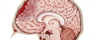
Cardiovascular diseases, such as heart disease and strokes, are the most common cause of death. Moreover, of all types of stroke, the most dangerous is hemorrhagic stroke - a condition that occurs when a blood vessel in a person’s head bursts, and this condition often leads to the most tragic consequences: disability or death.
The most important part of treatment is timely medical assistance, which is why you need to know the main signs of a ruptured vessel and the rules for providing first aid to the victim.
Definition
If a vessel in the head bursts, the pathology is called intracerebral hemorrhage, which is reflected in the IBC-10 as section 161. Hemorrhagic stroke ranks second in prevalence after stroke developing of the ischemic type. Pathology is more often detected in patients aged 45-60 years. Statistics show that most patients are representatives of the Negroid and Mongoloid races.
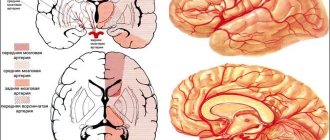
The most common localization of hemorrhage is the cerebral hemispheres, basal ganglia, cerebellum, pons, and trunk. Hemorrhages that occur against the background of arterial hypertension are usually acute and extensive. Intracranial hematomas are space-occupying formations, which often tend to expand due to the growth of the pathological focus.
If a vessel in the head bursts, the consequences arise as a result of the mechanical effect of the space-occupying formation on the adjacent brain structures. An intracerebral hematoma compresses surrounding tissues, causing neurological symptoms.
First aid
First aid instructions are an important point in the future fate of the patient. Often, it is paying attention to a person’s changing well-being that helps save his life and deliver him to a medical facility on time. How to recognize the onset of a stroke can be seen in the photo below.
How to recognize a stroke
First aid for vessel rupture and hemorrhagic stroke includes the following algorithm of actions:
- Call an ambulance immediately.
- Place the patient on the bed or floor so that the head and shoulders are in a slightly elevated position (about 30% of the surface). It is very important not to move the person too much and not to let him walk if the stroke occurred on the street.
- Loosen or remove all constrictive clothing.
- Remove dentures from the mouth, if present, and turn the victim’s head to the side.
- Provide access to fresh air.
- If vomiting, clean the mouth with gauze or a piece of cloth.
- Apply a cold compress to your head. This should be done on the opposite side to the damaged part of the body.
- Rub your limbs, thereby maintaining blood circulation.
Attention: do not engage in amateur activities under any circumstances. Any wrong actions can only worsen the victim’s condition.
Giving help
Causes of rupture of blood vessels in the head
Intracranial hemorrhage most often occurs due to the rupture of a small vessel that has undergone atherosclerotic changes, with blood entering directly into the brain tissue. The main reason that provokes the pathological process when blood vessels in the head burst is long-term arterial hypertension (80-85% of cases).
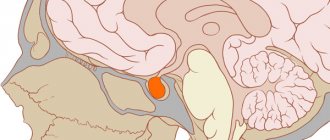
Other causes of occurrence are associated with congenital and acquired vascular pathologies - atherosclerosis, aneurysms, arteriovenous and other types of vascular malformations. A history of arterial hypertension may be combined with damage to elements of the vascular system or blood pathologies (dyscrasia). If a vessel bursts in a young patient, the reasons may be related to cocaine addiction. Other causes and provoking factors:
- Aneurysm of the mycotic type, provoked by infectious diseases of the central nervous system or injuries in the cranial area.
- Cerebral infarction of hemorrhagic type.
- Excessive therapy with anticoagulants - drugs that prevent blood clotting.
- Dissection (dissection) of intracranial arteries.
- Disturbances in the functioning of the hemostasis system (maintains the fluid state and other properties of the blood).
- Tumor processes localized in brain tissue.
- Deficiency of vitamins and nutrients.
- Intoxication is chronic and acute.
- Inflammatory processes that affect blood vessels in the brain.
- Excess body weight.
- A diet high in saturated fat.
- Taking sympathomimetic (stimulating the sympathetic nervous system) drugs.
- Smoking, alcohol abuse.
In old age, burst vessels can be a consequence of cerebral amyloid angiopathy. The disease is accompanied by the deposition of protein compounds that accumulate and form fibrils in the walls of the cerebral arteries.
The pathogenesis of hemorrhagic stroke is based on the mechanism of angiodystonic disorders, which affect the dynamics of regional blood circulation in brain tissue. When asked whether a vessel in the head can burst and for what reason, the doctor will primarily name a chronic increase in blood pressure and hypertensive crises.
Hypertensive crises are conditions requiring emergency medical care, characterized by an excessive increase in blood pressure. Crises provoke the development of paralysis and spasms of the walls of arteries and arterioles that supply the brain. Disorganization of the walls of blood vessels is more often associated with metabolic disorders and disorders of neurohumoral regulation.
Due to pathological changes, the permeability of the artery wall increases, which easily allows plasma cells and red blood cells to pass through. This condition is called diapedesis and often precedes wall rupture. In the mechanism of development of the focus of hemorrhage, disruption of blood clotting processes plays an important role.
Symptoms
If a vessel in the head bursts, signs of intracerebral bleeding appear - sudden pain in the head area, intensifying after exercise, nausea, which is often accompanied by vomiting. In elderly patients, pain in the head is sometimes absent or moderate.
In cases where the hemorrhage focus is large, neurological symptoms may be supplemented by confusion. A typical symptom is focal seizures (affecting specific areas of the body) or generalized seizures (distributed throughout the body).
Sometimes, before a vessel in the head bursts, precursor symptoms appear, which include a feeling of heat, visual dysfunction, and increasing pain in the head. More often, a stroke develops rapidly without previous signs. Typically, rupture of the vascular wall occurs during the daytime and is caused by physical or emotional stress.
If a vessel bursts in the head, the condition is accompanied by specific symptoms, which manifest themselves acutely with a tendency to increase. The clinical picture develops quickly. Confusion, stupor, and fainting are often observed. In severe cases, stunning and coma occur. Focal neurological symptoms depend on the location of the hemorrhage:
- Hemispheres. The pathology is accompanied by hemiparesis (paresis in one half of the body).
- Posterior fossa of the skull. Symptoms of damage to the structures of the brainstem and cerebellum are observed - paralysis of the eye muscles, narrowing of the pupils to a pinpoint state, stertorous breathing (noisy breathing, accompanied by inspiratory and expiratory wheezing, characteristic of obstruction, blockage of the airways).
- Ventricular system. The most life-threatening form, accompanied by meningeal (stiff neck, Kernig and Brudzinski symptoms, changes in reflexes) and brain stem (tachypnea - rapid, shallow breathing, tachycardia, stupor or stupor, increased blood pressure, increased tone of skeletal muscles) symptoms, hyperthermia (accumulation of excess heat in the body), hormetonic-type convulsions, rapid depression of consciousness.
When blood enters the ventricular system, hydrocephalus of the occlusive type may develop, occurring in an acute form. The focus of hemorrhage localized in the frontal lobe leads to a pronounced impairment of cognitive abilities (memory, mental activity, speech).
A supratentorial (located in the cerebral hemispheres) hematoma, accompanied by cerebral edema, can lead to transtentorial (the temporal lobe is wedged through the foramen of the cerebellar tentorium and under it) herniation, which in turn causes compression of the trunk and the appearance of secondary foci of hemorrhage in the area of the midbrain and pons .
Hematomas localized in the cerebellum, when enlarged, can block the 4th ventricle, which leads to the development of hydrocephalus, which occurs in an acute form. Foci of hemorrhage located in the cerebellar area with a diameter exceeding 3 cm can provoke dislocation of the median structures. The described conditions often lead to severe depression of consciousness, the development of coma and death of the patient.
Extensive intracerebral bleeding usually (70% of cases) ends in death. In surviving patients, neurological symptoms gradually regress in parallel with the process of resorption of blood poured into the cranial cavity. Hemorrhages cause less damage to brain structures than infarctions, so neurological deficits are often quickly corrected.
If a small vessel ruptures with the subsequent formation of a small focus of hemorrhage, focal neurological symptoms usually develop without depression of consciousness. In this case, the headache is moderate. Nausea may not be a clinical sign.
Diagnostics
The first choice methods for making a differential diagnosis are MRI and CT studies. Differentiation is made in relation to ischemic stroke and subarachnoid (intrathecal) hemorrhage. When making a diagnosis, it should be taken into account that acute neurological deficit can develop as a result of an epileptic seizure or hypoglycemia (a critical decrease in blood glucose levels). A blood test shows your glucose level. Computed tomography allows you to establish:
- Exact localization of the source of hemorrhage.
- Volume and prevalence of the pathological process.
- The presence and severity of cerebral edema.
- The presence and severity of dislocation of brain structures.
If MRI and CT examinations are not possible, a cerebrospinal fluid sample is taken as an alternative. Blood is detected in the cerebrospinal fluid if the focus of hemorrhage is in the area of the subarachnoid space or the posterior fossa of the skull. The attending physician will tell you what to do if a blood vessel in your head bursts, taking into account the results of the examination.
Treatment methods
Treatment of conditions that develop due to a burst blood vessel in the brain is often symptomatic. Regulated risk factors are monitored. In cases where intracranial bleeding threatens serious complications, neurosurgical intervention is performed. Typically, surgery is prescribed if the diameter of the volumetric focus of hemorrhage exceeds 3 cm.
If the patient has undergone anticoagulant therapy, the drugs are discontinued and medications are prescribed that neutralize the effect - intravenous administration of blood plasma, prothrombin complex, vitamin K. The drug Dabigatran (anticoagulant) is eliminated through the hemodialysis procedure - extrarenal, hardware blood purification. The following events are taking place in parallel:
- Monitoring blood pressure levels (antihypertensive therapy with Nicardipine).
- Anticonvulsant therapy (if seizures are present).
- Mechanical ventilation (artificial ventilation) in case of depression of consciousness below 8 points (Glasgow coma scale), tachypnea 35-40 times per minute.
- Correction of hypoglycemia and hyperglycemia.
Early neurosurgical removal of large lobar (blood does not extend beyond the white matter) hematomas improves survival. However, surgical intervention is associated with the risk of relapse - repeated hemorrhage. The operation is not performed if the focus of hemorrhage is located in the deep parts of the brain.
How are they treated?
First you need to provide first aid to the person. Naturally, urgent hospitalization will be required, but it is impossible to leave the situation unattended until the ambulance arrives. The patient should be laid so that the head is elevated - this will ensure the necessary outflow of blood and prevent the development of brain swelling. You should also worry about the flow of oxygen; accordingly, you need to relieve the neck - unbutton your shirt, loosen your tie, etc. If you lose consciousness, you should protect the person by clearing the airways - remove dentures, turn your head to the side. It is also worth applying cold objects to the head to reduce the risks of swelling and hemorrhage.
Aneurysm treatment is predominantly treated surgically. The method is selected depending on a number of parameters - the location of the formation, the person’s condition, the severity of the violations and the time that has passed since the moment of rupture.
Cleaning of blood vessels. How to get rid of cholesterol plaques Read more
The following treatment options are used for therapy::
- Clipping is a microsurgical method that involves placing a clip on the base or body of the aneurysm: this will shut it off from the bloodstream.
- Endovascular method - here an intervention is performed by inserting a catheter through the femoral artery.
- Combined method - first a blood clot is injected into the aneurysm, and then it is clipped.
There is such a nuance that it is important to perform the operation in the first 72 hours after the start of bleeding, since the risk of recurrent hemorrhage is high. If you are late, spasm begins to increase, ischemia develops, and the operation will not make sense.
Possible consequences
The prognosis is relatively unfavorable. The negative consequences of a condition where a vessel in the brain has burst are most often associated with swelling and dislocation of brain structures. Along with heart failure, which occurs in acute form, these are the main causes of death in the event of hemorrhagic stroke. Unfavorable factors that influence the outcome are long-term coma (longer than 12 hours), the patient's age over 65 years, and the location of the hemorrhage near the ventricular system.
Hemorrhagic stroke, also known as intracerebral hemorrhage or rupture of a cerebral artery, is a pathological process that threatens the health and life of the patient, which can occur for various reasons. Timely diagnosis and correct treatment will help avoid serious consequences.
428
Rehabilitation
The recovery time after a ruptured aneurysm is different for everyone. The type of aneurysm, the qualifications of the neurosurgeon, the affected brain structures, the age of the patient and the severity of complications play a big role.
First of all , it is recommended to recover in specialized sanatoriums , where a rehabilitologist will adjust an individual program of treatment procedures. Physiotherapy courses in this case include instrumental techniques for stimulating damaged tissues and tactile effects on the affected vessels and muscles.
In general, the rehabilitation complex consists of the following activities:
- classes with highly specialized specialists to restore walking, speech, reading, writing and fine motor skills;
- therapeutic massage (head, collar area, shoulder girdle, limbs) and exercise therapy with simulators;
- acupuncture;
- muscle electrical stimulation;
- hydrogen sulfide, oxygen or iodide-bromine baths;
- UHF and electrophoresis according to indications.
It is extremely important to maintain moderate activity throughout the rehabilitation period.