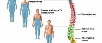
Scheme of innervation of the human body
In humans, as well as in other vertebrates, the segmental innervation of the body is preserved.
Innervation (from Latin in - in, inside and nerves) - supply of organs and tissues with nerves, which ensures their connection with the central nervous system.
This means that each segment of the spinal cord innervates a specific area of the body. For example, segments of the cervical spinal cord innervate the neck and arms, the thoracic segments - the chest and abdomen, the lumbar and sacral - the legs, perineum and pelvic organs (bladder, rectum). By determining in which area of the body sensory or motor function disorders have appeared, the doctor can guess at what level the spinal cord injury occurred.
Legend:
Sh
– cervical spine
G
– thoracic spine
P
– lumbar spine
Kr
– sacrum
- Segment (example: Ш1-2)
- Organs and parts of the body that are innervated by nerves emanating from this segment
- Symptoms and pathological conditions that arise when innervation is disrupted Unfortunately, this information could not be transferred in the form of a table (in which everything is much clearer), so I will explain how to read it: Segment (example: Ш1-2) - this means that the nerve root is located between 1st and 2nd cervical vertebrae.
The next line lists the Organs and parts of the body that are innervated by the nerves emanating from this segment. Also listed on a new line are the Symptoms and pathological conditions that arise when innervation is disrupted (in bold)
Ш0-1 Sympathetic nervous system, inner ear, internal jugular veins, vertebral arteries. Headache, fatigue, poor sleep, increased blood pressure, dizziness, hearing loss.
Ш1-2 Eyes, optic and auditory nerves, facial and trigeminal nerves. Eye diseases, hearing loss, neuritis of the facial and trigeminal nerves.
Ш2-3 Nasopharynx, cheeks, teeth. Neuritis, neuralgia, adenoids, dental problems.
Ш3-4 Nose, lips, mouth, Eustachian tube. Hearing impairment, enlarged adenoids, dermatitis in the nose, lips, nasal congestion. Ш4-5 Larynx, vocal cords, vertebral arteries, tonsils. Sore throat, tonsillitis, laryngitis, voice disturbance, increased blood pressure.
Ш5-6 Muscles of the neck, shoulder girdle, elbow joint. Pain in the neck, shoulders, elbow joints.
Ш6-7 Thyroid gland, shoulder joint. Diseases of the thyroid gland, pain in the shoulder joints and limitation of their mobility.
Ш7-8 Hands, thyroid gland. Pain and feeling of coldness in the hands, numbness of the limbs.
Ш8-Г1 Esophagus, trachea. Esophagitis, tracheitis.
G1-2 Heart, pericardium, coronary arteries. Arrhythmias, chest pain, coronary heart disease.
G2-3 Lungs, bronchi, pleura, mammary gland. Colds, bronchitis, pneumonia, asthma, pleurisy, mastopathy, heartburn.
G3-4 Liver, gall bladder, common bile duct. Gallstones, hypofunction of the liver, gallbladder.
G4-5 Liver, solar plexus. Impaired liver function, increased bilorubin levels.
G5-6 Stomach. Gastritis, gastric ulcer, acidity disorder.
G6-7 Duodenum, pancreas. Peptic ulcer of the duodenum, indigestion, stool, diabetes mellitus
G7-8 Diaphragm, spleen. Indigestion, hiccups, breathing problems.
G8-9 Adrenal glands. Allergic reactions, immune system disorders.
G9-10 Kidneys, ureters. Kidney disease, stone formation, nephroptosis.
G10-11 Kidneys, ureters. Kidney disease, urinary disorder.
G11-12 Small intestine, transverse colon. Kidney disease, urinary disorder.
G12-P1 Cecum, appendix, groin area, fallopian tubes. Colitis, appendicitis, inguinal hernia, diseases of the female genital organs.
P1-2 Appendix, cecum, abdominal cavity. Intestinal colic, appendicitis, pain in the groin area.
P2-3 Genital organs, bladder, knee joint. Impaired potency, urination, arthrosis of the knee joints.
P3-4 Lower legs, feet, bladder. Pain in the feet, legs, problems with urination.
P4-5 Prostate gland, uterus with appendages, legs, feet, toes. Prostatitis, impaired potency, adnexitis, erosion, uterine fibroids, pain in the feet, legs, toes.
P5-Kr1 Prostate gland, uterus with appendages, urethra, perineum, gluteal region, rectum, hip joint. Prostatitis, impotence, ovarian cyst, menstrual irregularities, hemorrhoids, pain in the perineum, arthrosis of the hip joint, vascular diseases of the legs.
Sacrum Pelvic organs, perineum, lower limbs, rectum, anus. Disorders of the pelvic organs, sphincteritis, anal fissures, etc.
Coccyx Perineum, rectum, anus. Hemorrhoids, dysfunction of the pelvic organs, pain in the perineum.
The spinal cord is the initial structure of the central nervous system. It is located in the spinal canal. This section has a cylindrical cord shape, flattened from front to back. Its length is 40-45 centimeters, and its weight is about 34-38 grams. Next, let's take a closer look at the structure of this department: what elements it includes, how they are formed and what tasks they perform.
Departments
The spinal cord has the following parts:
Each part has segments. Pairs of spinal nerves extend along the entire length of the cord. There are 31 in total. The number of spinal nerves, depending on the segment, is as follows:
- Coccygeal - 1-3.
- Sacral – 5.
- Lumbar – 5.
- Infants – 12.
- Neck – 8.
Below, the spinal nerves form the cauda equina. During the growth of the body, the cord does not have time to reach the length of the canal. In this regard, the spinal nerves are forced to descend, exiting the foramina.
A few words about neurons
The structural unit of nervous tissue is neurons. Completely special cells whose main function is the formation and transmission of nerve impulses. Each neuron has many short processes - dendrites, which perceive irritation, and one long one - an axon, which conducts a nerve impulse in only one direction. Depending on the task and function, neurons are classified as sensory or motor. Intermediate or intercalary neurons are peculiar “extensions” that transmit impulses between other neurons.
Internal Contents
The spinal cord contains white and gray matter. The latter consists of neurons. They form three columns in the halves of the spinal cord: lateral, posterior and anterior. In cross section, each of them looks like horns. There are narrow posterior and wide anterior horns. The lateral one corresponds to the vegetative intermediate column of the gray part. The anterior horns contain motor neurons, the lateral horns contain autonomic intercalary neurons, and the posterior horns contain sensory neurons. Renshaw cells are located in this same area. These are inhibitory neurons that slow down motor neurons from the anterior horns. The gray matter is surrounded by white matter, which forms the cords of the spinal cord. There are three of them in each half: side, rear and front. The cords consist of fibers running longitudinally. They, in turn, form bundles of nerves - pathways. Descending - extrapyramidal and pyramidal - are located in the anterior cords, in the white matter. In the laterals – ascending and descending:
- Lateral spinothalamic.
- Rear and anterior (Flexig and Gowers).
- Lateral (pyramidal) corticospinal.
- Red nuclear.
The white matter of the posterior cord includes ascending pathways:
- Burdach's bundle (wedge-shaped).
- Gaulle bundle (thin).
How it works
The branched dendrites of the sensory neurons of the spinal centers of the autonomic nervous system end with receptors, which are biological structures in which a nerve impulse is formed upon contact with a specific stimulus. Receptors provide vegetative visceral sensitivity - they perceive irritation from such parts of our body as blood vessels and heart, gastrointestinal tract, liver and pancreas, kidneys and others. The impulse is transmitted along the dendrite to the body of the neuron. Next, the nerve impulse travels along the axons of afferent (sensitive) neurons to the spinal cord, where they form synoptic connections with the dendrites of efferent (motor) neurons. It is thanks to this direct contact that we withdraw our hand from a hot pan or iron even before our main commander - the brain - analyzes the pain sensations that have arisen.
Shells
The outside of the spinal cord is covered by three structures:
- Arachnoid medium.
- Solid outer.
- Soft inner.
The epidural space is located between the periosteum of the spinal canal and the dura mater. It is filled with venous plexuses and fatty tissue. Between the arachnoid and dura mater there is the subdural space. It is permeated with thin connective tissue crossbars. The soft membrane is separated from the arachnoid by the subarachnoid subarachnoid space. It contains liquor. Cerebrospinal fluid is formed in the choroid plexuses located in the ventricles of the brain. Renshaw cells protect the central nervous system from overexcitation.
Blood supply to the cervical spine
The cervical spine is represented by a complex circulatory system. Blood from the head and neck flows through the jugular veins. The anterior jugular vein collects blood from the skin and subcutaneous tissue of the anterior neck. The external jugular vein collects blood from the occipital region of the head, skin and subcutaneous tissue of the lateral neck.
From the head, muscles and organs of the neck, blood flows mainly into the internal jugular vein. The common carotid artery runs along the upper edge of the thyroid cartilage, which divides into the external and internal carotid arteries.
At the level of the division of the common carotid artery there is a formation containing chemoreceptors that respond to changes in the chemical composition of the blood. The vagus nerve is located between the common carotid artery and the internal jugular vein.
Functions of the spinal nerves
There are two of them. The first - reflex - is performed by nerve centers. They represent segmental working zones of unconditioned reflexes. The neurons of the centers communicate with organs and receptors. Each transverse section - a metamer of the body - has sensitivity transmitted from three roots. Innervation of skeletal muscles is also carried out by 3 adjacent spinal segments. Efferent impulses are also transmitted to the respiratory muscles, glands, blood vessels and internal organs. The overlying regions of the central nervous system regulate the activity of the periphery through segmental spinal regions. The second task - conduction - is performed thanks to descending and ascending paths. With the help of the latter, information is transmitted from temperature, pain, tactile and proprioceptors of tendons and muscles through neurons to the rest of the central nervous system to the cerebral cortex and cerebellum.
Basic functions of the spinal cord
The segmental structure of the spinal cord allows it to effectively cope with assigned tasks. Their well-coordinated communication ensures a high speed of impulse transmission and perception of information coming from the environment. The main functions of the spinal cord are conductive and reflex. They ensure the ability of the organ to perform all the necessary actions for the normal functioning of the body.
The segmental apparatus is connected with almost all internal organs and ensures human activity and the functionality of each system. The first function allows you to receive complete information about the world around you: what effects are on the skin or body, whether it hurts or not. The “communication” is two-way, since the brain not only reacts to events around it, but also gives commands what needs to be done. The conduction function ensures mental and motor activity.
The reflex function is no less important. The concept itself explains what it is. Thanks to the spinal cord, if the hand feels pain or burning when exposed to fire receptors, the person quickly pulls it back reflexively. During the examination, neurologists check the knee reflex and other body reactions to stimuli; if they are absent, this helps to diagnose hidden disorders in a timely manner.
The spinal cord has a rather complex structure, since it is responsible for the most important functions in the body, namely: digestion, urination, blood circulation, breathing, sex life, motor activity and many others. It is thanks to the reflexes for which he is responsible that a person is saved in moments of danger. It consists of many segments that help transmit impulses from the periphery towards the brain. Thanks to their coordinated work, a person’s muscles contract and he feels the world around him.
Ascending Paths
- Anterior spinothalamic. This is the afferent pathway of pressure and touch.
- Lateral spinothalamic. This is the path of temperature and pain sensitivity.
- Posterior and anterior spinocerebellar. These are afferent pathways of muscle-articular sensitivity of the cerebellar direction.
- Bundles of Burdach (wedge-shaped) and Gaulle (thin). These are afferent pathways for transmitting cortical muscle-articular sensitivity from the lower half of the torso and legs and from the upper body and arms, respectively.
Diseases associated with degenerative-dystrophic processes
Headache
The human brain is nourished by the vertebral and carotid arteries. Even if the cervical spine has slight irregularities, this leads to mechanical compression of the arteries.
The vessels begin to become clogged with toxins, and as a result, oxygen starvation of the brain begins. In this case, the spine does not receive sufficient nutrition in the cervical region. As a result, intracranial pressure increases, dizziness, nausea, and headache appear.
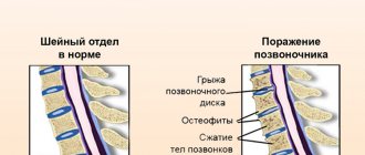
Intervertebral hernia
The spine has the most vulnerable and flexible part - the cervical region. The neck, in a normal physiological and anatomical state, can perform the greatest range of movements, since the spine in this section has an elastic ligamentous apparatus. There is no intervertebral disc between the first and second vertebrae.
They are connected to each other by a ligamentous apparatus. Between the remaining vertebrae are intervertebral discs. Their feature is a delicate nucleus pulposus and a thin fibrous ring. If the spine is in normal physiological condition, the vertebrae provide the depreciation process.
Due to prolonged overstrain on the spine, cracks form in the fibrous ring and it is no longer able to form the pulpous ring in the correct central position. As a result, it begins to protrude, pinching or irritating the nerve roots. In this case, the person begins to experience pain.
If the spine has a healthy ligamentous apparatus, the load is evenly distributed on the intervertebral disc. The openings for the spinal nerve always remain free during natural movements. When the fibrous ring and nucleus pulposus exit, the opening in which the spinal nerve is located narrows and the nerve root is pinched.
When prolapsed, the fibrous ring always remains intact. The pressure of the disc on the spinal nerve causes swelling, inflammation and pain along the nerve root. A herniation is the complete loss of an intervertebral disc.
Osteochondrosis
The spine suffers from degenerative processes that lead to the development of osteochondrosis.
According to statistics, osteochondrosis is a common cause of the development of protrusions and herniations of the intervertebral disc.
Osteochondrosis leads to the development of arthrosis of the intervertebral joints. The course of osteochondrosis is worsened by the formation of Schmohl's hernias, in which the intervertebral disc penetrates the vertebral body.
Osteoporosis
The spine may suffer due to endocrine pathologies. With hormonal disorders, calcium is washed out of bone tissue .
Osteoporosis itself does not lead to significant clinical manifestations affecting the spine. But it is precisely with this disorder that the risk of developing vertebral fractures and spinal injuries increases.
Radiculopathy
The spine in the cervical region may suffer from compression of the spinal roots. Significant compression of the spinal root leads to radiculopathy. The cause of this pathological condition can be any disease that affects the bone structures of the cervical region.
Spinal nerve formation
How does this happen? The spinal nerve is formed by connecting the posterior sensory and anterior motor areas. At its exit from the intervertebral foramen, fiber separation occurs. As a result of this, branches of the spinal nerves are formed: posterior and anterior. They perform mixed tasks. The meningeal and white communicating branches also depart from the spinal nerves. The first return to the spinal canal and innervate the dura mater. The white branch approaches the nodes of the sympathetic trunk. Against the background of various curvatures of the spine (scoliosis, kyphosis, pathological lordosis), deformation of the intervertebral foramina occurs. As a result, the spinal nerves become pinched. This leads to various kinds of violations.
Innervation of the cervical spine
The spine in the cervical region has a nerve plexus, which is formed by the anterior branches of the four upper cervical nerves (C1 - C4). The branches extending from the plexus are divided into cutaneous (sensitive), muscular (motor) and mixed.
The cutaneous branches are the greater auricular nerve, the transverse nerve, the lesser occipital nerve and the supraclavicular nerves. Sensitive branches innervate the skin of the anterolateral neck.
The muscle nerves insert into the deep muscles of the neck and chest and innervate the prevertebral muscles, the middle scalene muscle, and the levator scapulae muscle. The motor branches innervate the deep muscles of the neck.
The inferior root of the cervical plexus C1 - C2 connects to the superior root of the hypoglossal nerve and innervates the muscles that lie below the hyoid bone. The phrenic nerve is connected to the middle cervical sympathetic node, which provides innervation to the diaphragm, pleura and pericardium.
Features of the segmental structure
Structure of the spinal cord
The spinal cord has its own characteristics. It is located along the length of the spinal column, which makes it possible to effectively protect the sensitive part from any mechanical damage from the outside. But its length is less than that of the entire ridge. The ratio of the sections does not correspond to the ratio of the components of the spine. Complete agreement is maintained in preschool children.
When studying the structure of segments of the human spinal cord, scientists often draw parallels with the anatomy of a frog. The general criteria for the segmental structure are indicated: the presence of a segment and a pair of roots. The posterior roots are sensitive, the anterior ones are responsible for the function of movement. If one type of root is affected in a segment, the functionality of others is also impaired. A pair of nearby parts are connected and influence each other.
The origin of the spinal cord is in the occipital region. It ends in the lumbar area at the L1-L2 level. In general, the length is 43-45 cm. The end of the spinal cord is a cone, from which emanates a terminal filament, equipped with a nerve bundle.
The entire strand is not the same in thickness. It has two seals in the neck and lumbosacral area. This is explained by the need to house a large number of neurons that control the functionality of the arms and legs.
