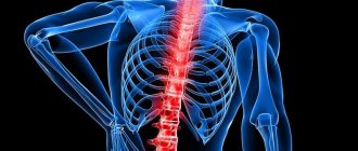
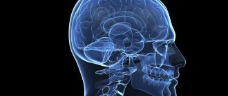
Segments of the spinal cord are of great importance for humans, because they make it possible to feel the world. The spinal cord is covered by a spinal frame of vertebrae. Human health largely depends on the proper functioning of the segments of his brain. The brain allows a person to control his body and perceive the world around him, including through reflexes.
Features of anatomy
The spinal cord is a unique organ of the human body, so it is not surprising that it has its own characteristics. It is located along the spine, and this anatomy allows you to effectively protect this extremely important and sensitive organ from any mechanical influences from the outside. But it is shorter in length than the entire ridge, so the ratio of the spinal cord segments does not correspond to the numbering of the sections of the column itself. Complete agreement is observed only in preschool children.
The spinal cord begins at the back of the head and ends at the lumbar level, approximately at the level of L1-L2. In women the cord is slightly shorter than in men. In general, its length reaches 43–45 cm. The spinal cord ends in a cone, from which a terminal filament extends, surrounded by a nerve outlet.
The entire cord along its entire length is unequal in thickness, as it has two seals in the cervical and lumbosacral regions. This is due to the need to place a large number of neurons responsible for the functioning of the lower and upper extremities.
Anatomy of the spine and spinal cord
The posterior portions of the vertebrae are formed by bony structures that extend from the vertebral body as pedicles and expand posteriorly to form the vertebral arch. Together with the posterior longitudinal and yellow ligaments, the vertebral arch forms the spinal canal, in which the spinal cord and its roots are located. The posterior parts of the vertebra are articulated with adjacent vertebrae by two small intervertebral (facet) joints. Intervertebral (facet) joints have a small articular surface, most pronounced in the lumbar spine. These articular platforms provide a certain mobility of the spine.
The vertebrae have strong transverse and spinous bone processes posteriorly. The transverse processes extend from the vertebral pedicles in a lateral direction. The spinous processes extend in the posterior direction. These processes are attached through ligaments and tendons of the back muscles, which move, support and protect the spinal column.
Vertebral anatomy: vertebral body, vertebral arch, articular processes of the vertebra, transverse processes of the vertebra, spinous process of the vertebra, intervertebral disc (nucleus pulposus and annulus fibrosus).
The center of the intervertebral disc is occupied by a gelatinous nucleus pulposus, which is considered a remnant of embryonic tissue (notochord). It is surrounded and supported by a fibrous ring consisting of fibrocartilaginous and connective tissue.
After the development of the spine is complete (between the ages of 24 and 26), the intervertebral discs lose their blood supply. Subsequently, the intervertebral discs become less elastic and cope less effectively with the role of shock absorbers of mechanical shocks when walking. These changes can lead to disturbances in the most mobile parts of the spine: in the cervical and lumbar segments.
Anatomy of the spine: spinal cord, vertebrae, intervertebral disc.
The following structures give stability to the human spine:
- articulations of vertebral bodies through intervertebral discs and intervertebral joints
- ligaments and muscles of the spine
The ligamentous apparatus of the spine has sufficient strength to withstand displacement. Vertebral bodies with an intervertebral disc do not have the overall strength of the ligaments to withstand displacement due to the loads placed on the spinal column during movement. This weakness of the spine is compensated by voluntary and reflex contractions of the sacrovertebral, abdominal, gluteal and quadratus lumborum muscles, as well as the hamstrings, which provide such stability.
The spinal cord is located in the lumen of the spinal canal. For a child, it fills it almost completely. As a person grows, already in adulthood, the cone of the spinal cord reaches only the level of the 1st or 2nd lumbar vertebra, because the nervous tissue does not keep up with the rapid growth of skeletal bones.
Anatomy of the spine: lumbar vertebrae, spinal cord and cauda equina, sciatic nerve.
The structures of the spine are innervated by the recurrent branches of the spinal nerves. Pain endings and fibers of this nerve were found in ligaments, muscles, periosteum of the processes, outer layers of the fibrous ring of the intervertebral disc and in the synovium of the intervertebral (facet) joints. Sensitive fibers from these structures of the spine and the sacroiliac and lumbosacral joints, connecting, form the sinuvertebral nerves of Luschke, which enter the recurrent branches of the spinal nerves at the level of S1 and L1-L5 into the gray matter of the corresponding segments of the spinal cord. Motor fibers emerge from these segments and reach the muscles as part of the same nerves.
Location of the spinal cord in the spinal canal.
The lumbar and cervical spine have the greatest freedom of movement. Compared to them, the thoracic spine is securely and firmly fixed by the bones of the chest. Greater mobility of the neck and lower back is the reason for its frequent lesions. In addition to voluntary movements that provide flexion and rotation of the torso, many movements of the spine are of a reflex nature and ensure that the body maintains its position in space, as happens when walking or running.
The structure of the spine and spinal cord, cross-sectional view.
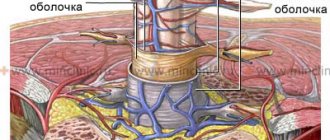
Epidural fat surrounds the membranes of the spinal cord. Infection of the epidural tissue leads to purulent spinal epiduritis.
We invite you to familiarize yourself with some useful articles and links related to the work of our clinic:
- dictionary of terms - a list of the most common medical terms is provided
- Frequently asked questions - the most frequently asked questions by patients are listed
- anatomy of the spine - basic data on the structure of the spine and spinal cord are presented and illustrated
- anatomy of the nervous system - basic data on the structure of the brain and spinal cord, nerves are presented and illustrated
- rules for preparing a person under study for certain diagnostic procedures
- rules of patient care - presents ways to safely care for bedridden or debilitated patients at home or in a hospital
What are the segments?
The sections of the spinal cord differ from those used to conventionally divide the areas of the spine. Their length varies; the fewest elements are found in the coccygeal part. The segments are connected to certain areas in the body by nerve conductors. In general, the brain is divided into the following segments:
Prognosis for spinal cord myelitis
- Neck – 8.
- Chest – 12.
- Sacral – 5.
- Lumbar – 5.
The main length falls on the thoracic segments of the spinal cord, after which 23.2% is allocated to the neck and only 7.3% to the lower back. They are posterior and anterior nerve roots that alternate regularly. They are located above the vertebrae with the same number. Their main task is to report movements and be responsible for muscle contractions. Therefore, the anterior nerve roots are also called motor, and the posterior - sensitive.
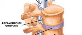
The intervertebral foramen contains the roots of each individual segment. Their direction is not the same, because the spinal column is filled with the brain. In the cervical region they lie horizontally, in the thoracic region they are directed diagonally, and in the lower part they are almost vertical.
The shortest segments are the neck segments, and the longest ones are in the lower back and sacrum. In the lower part they form the so-called “horse tail” - this is a bunch of roots located below the 2nd vertebra.
A visual representation of what a spinal cord segment looks like in cross section
CT segments - diagram
Lung segments on CT images.
Lung segments on CT images (diagram).
Right lung.
Segment S1 (apical or apical) of the right lung. Refers to the upper lobe of the right lung. Topographically projected onto the chest along the anterior surface of the 2nd rib, through the apex of the lung to the spine of the scapula.
Segment S2 (posterior) of the right lung. Refers to the upper lobe of the right lung. Topographically projected onto the chest along the posterior surface paravertebrally from the upper edge of the scapula to its middle.
Segment S3 (anterior) of the right lung. Refers to the upper lobe of the right lung. Topographically, 2 to 4 ribs are projected onto the chest in front.
Segment S4 (lateral) of the right lung. Refers to the middle lobe of the right lung. Topographically projected onto the chest in the anterior axillary region between the 4th and 6th ribs.
Segment S5 (medial) of the right lung. Refers to the middle lobe of the right lung. Topographically, it is projected onto the chest with the 4th and 6th ribs closer to the sternum.
Segment S6 (superior basal) of the right lung. Refers to the lower lobe of the right lung. Topographically projected onto the chest in the paravertebral region from the middle of the scapula to its lower angle.
S7 segment of the right lung. Topographically localized on the inner surface of the right lung, located below the root of the right lung. It is projected onto the chest from the 6th rib to the diaphragm between the sternum and midclavicular lines.
Segment S8 (anterior basal) of the right lung. Refers to the lower lobe of the right lung. Topographically delimited anteriorly by the main interlobar groove, inferiorly by the diaphragm, and posteriorly by the posterior axillary line.
Segment S9 (lateral basal) of the right lung. Refers to the lower lobe of the right lung. Topographically projected onto the chest between the scapular and posterior axillary lines from the middle of the scapula to the diaphragm.
Segment S10 (posterior basal) of the right lung. Refers to the lower lobe of the right lung. Topographically projected onto the chest from the lower angle of the scapula to the diaphragm, delimited on the sides by the paravertebral and scapular lines.
Left lung segments
Segment S1+2 (apical-posterior) of the left lung. It is a combination of C1 and C2 segments, which is due to the presence of a common bronchus. Refers to the upper lobe of the left lung. Topographically projected onto the chest along the anterior surface from the 2nd rib and upward, through the apex to the middle of the scapula. Segment S3 (anterior) of the left lung. Refers to the upper lobe of the left lung. Topographically projected onto the chest from the front from 2 to 4 ribs. S4 segment (upper lingular) of the left lung. Refers to the upper lobe of the left lung. Topographically projected onto the chest along the anterior surface of the 4th to 5th ribs. S5 segment (lower lingular) of the left lung. Refers to the upper lobe of the left lung. Topographically projected onto the chest along the anterior surface from the 5th rib to the diaphragm. Segment S6 (superior basal) of the left lung. Refers to the lower lobe of the left lung. Topographically projected onto the chest in the paravertebral region from the middle of the scapula to its lower angle.
Segment S8 (anterior basal) of the left lung. Refers to the lower lobe of the left lung. Topographically delimited anteriorly by the main interlobar groove, inferiorly by the diaphragm, and posteriorly by the posterior axillary line.
Segment S9 (lateral basal) of the left lung. Refers to the lower lobe of the left lung. Topographically projected onto the chest between the scapular and posterior axillary lines from the middle of the scapula to the diaphragm.
Segment S10 (posterior basal) of the left lung. Refers to the lower lobe of the left lung. Topographically projected onto the chest from the lower angle of the scapula to the diaphragm, delimited on the sides by the paravertebral and scapular lines.
Location Features
The skeletotopy of the areas is individually variable, since the lower part of the lumbar region can be located in adults from the lower third of the body of the 9th thoracic vertebra to the disc between L1-L2. Because of this, a certain feature appears. When the upper processes are directed in the transverse direction, the further down the canal, the higher the exit site is located relative to the intervertebral foramen.
Each segment of the spinal cord is responsible for its part of the periphery. This could be muscle tissue, internal organs or skin. The division into such sections is almost identical in all people, so it is not difficult for doctors to identify the location of the lesion based on the specific sensitivity of a particular area. The relationship can be seen in more detail in the table:
| Department | Number of spines | Related department |
| Cervical | 8 | Skin and muscles in the neck, upper limbs, diaphragm |
| Chest | 12 | Skin on the chest, back and abdomen |
| Lumbar Sacral Coccygeal | 5 5 1 | Lower body, legs, feet, pelvic organs |
For example, when a patient complains of discomfort in the navel area, there is a high probability that the pathology is hidden below the 10th thoracic vertebra. The doctor will check for the presence of a lesion in this particular part of the body.
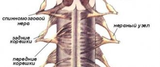
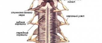
Segment structures
The structure of the segments in the spinal cord is paired, and they are connected to the organs by nerves. The anterior and posterior roots provide the transmission of information, and their main task is to stimulate muscle contraction. Only the dorsal roots are sensitive; they are the ones that are activated by receptor stimuli. The spinal cord is not homogeneous in cross section, but is a substance consisting of white and gray matter.
Segmental structure of the spinal cord with the designation of roots in different sections
White matter
The white matter of the spinal cord is represented by processes of nerve cells that make up the pathways. It is located on the periphery of the gray matter and is formed from nerve bundles. The white matter contains descending ascending fibers that pass into the brain. They transmit information coming from receptors. Their location is natural: on the side of the back there are ascending ones, on the abdominal side there are outgoing ones. The white matter is delimited by the grooves of the spinal cord into:
- anterior - make up the descending pathways.
- lateral - there are both ascending and descending paths.
- posterior cord - make up the ascending pathways.
White matter contains projection, association and commissural nerve tracts. The first provide communication with the brain.
Gray matter
Gray matter consists of interneurons and motor neurons. They provide motor reflexes and communication between neurons. In total, it occupies approximately 18% of the total volume of the spinal cord, and includes approximately 13.5 million neurons. This is a special device that contains some functions of the central nervous system. Thanks to two-way communication, irritation can pass through both ascending and ascending pathways. The response is a contraction of muscle tissue and a motor response.
Do you know that:
- The spinal cord stops growing at the age of 5 years.
- In the process of evolution, it was the spinal cord that appeared first, and only then the brain.
- People with spinal cord injury cannot sweat below the injury site. Therefore, in hot weather they need to be in the shade or otherwise cool their body to avoid overheating.
- The spinal cord relaxes all the muscles of the body during sleep. This function allows a person not to repeat the movements that he performs in his sleep.
How many segments make up the spinal cord
The spinal column consists of 31 pairs of segments
It is customary to distinguish 31 pairs of segments in an axis. In some cases, the number of spinal cord segments is 32-33 pairs. The roots are connected into a single system, resulting in the formation of the spinal nerve. The spinal cord consists of five sections.
Cervical
Includes 8 pairs. It has a high level of mobility, as a person often turns and tilts his head. The transverse processes contain vessels responsible for the flow of blood to the brain and cerebellum. In the presence of morphological abnormalities in the neck, blood flow is disrupted. Brain cells undergo oxygen starvation, as a result of which they begin to atrophy. This provokes cognitive impairment, intense migraines, and deterioration in the quality of vision.
The morphology of the upper cervical segments is different from the rest. The first fragment does not include the vertebral body. It consists of anterior and posterior arches, which are connected by thickenings based on bone tissue. The second includes the odontoid process. Thanks to it, the neck gains flexibility.
Chest
Includes 12 pairs, to which the ribs are attached to form the rib cage. It is in this zone that most of the internal organs are located. This explains the immobility of the thoracic region. Despite this characteristic, it is susceptible to damage. Pathological changes in its morphology are dangerous: the functionality of other systems may be impaired. The vertebral bodies have the property of enlarging as they undergo stress. This is due to the location of internal organs and respiratory function. The vertebrae of this segment have special lateral fossae in which the bases of the ribs are located.
Lumbar
Includes 5 pairs of vertebrae. The department is small in size, but carries out important functions. It bears the maximum share of the load. The vertebrae are large. There is a pathology in which there are 6 pairs of vertebrae. An increased amount is not beneficial, but does not interfere with the quality of life. The lumbar area has a physiological lordosis, representing a slight forward bend. If the bending rate is too high, then this indicates that the person has some kind of pathology.
The lumbar region is responsible for the motor ability of the legs. The upper half of the body experiences the load. Caution should be exercised when charging or lifting heavy objects. With incorrect movements, the functionality of the lumbar region is disrupted. The wear of the vertebral discs is provoked, which causes the formation of a hernia.
Sacral
Injuries to the spinal cord and related areas of the body
Includes 5 fused pairs that form a triangle. The bone connects the upper part of the spine to the pelvic region. The segments are connected by age 25. In infants and adolescents, the region is mobile and therefore vulnerable to injury. The sacrum contains several openings through which nerves pass. Thanks to this, the bladder, rectum and legs have sensitivity.
Coccygeal
Includes 1 or 3 pairs. The amount depends on the structural features of the skeleton. The segment performs important functions. For example, in women it is mobile, which ensures normal gestation and the passage of the child through the birth canal. It acts as a link between the muscles and ligaments involved in the functionality of the genitourinary organs and intestines. The tailbone regulates hip extension and promotes proper load distribution. It protects the spinal column from destruction in a sitting position. If this department did not take on part of the load, the spinal column would be injured.
The elements are marked in Latin letters according to the name of the department, as well as in Roman numerals, which indicate the segment number (C (I-VIII) cervical, Th (I-XII) thoracic, L (IX) lumbar, S (IV) sacral, Co (I -III) coccygeal).
Blood supply
The supply of nutrients is carried out through the blood, which flows through numerous vessels. They extend from the upper part, along the thyroid and vertebral arteries. They start from the aorta and vertebral arteries, of which there are normally about 6–8 in humans. The largest of them provide nutrition to the cervical and lumbar thickening.
The largest radicular-spinal artery is the inferior artery of Adamkiewicz; there are 15–20 posterior arteries in total, but all of them are significantly narrower. Their main task is to provide nutrition to the posterior third of the spinal cord.
All vessels are connected to each other; such joints are called anastomosis. The entire system provides nutrition to different parts of the brain, while being protected from the formation of blood clots. Even if a separate vessel is closed by a plug, the area for which it is responsible will not be left without power; this is what the anastomosis provides. It saves neurons from hypoxia and death.
In addition to the arteries, the spinal cord is also supplied by veins that are connected to the cranial plexuses. This is a whole system of vessels through which blood flows from this organ to the vena cava. Thanks to the presence of special valves, blood is not able to flow back.
Skeletotopy of the spinal cord
Basic functions of the spinal cord
The segmental structure of the spinal cord allows it to effectively cope with assigned tasks. Their well-coordinated communication ensures a high speed of impulse transmission and perception of information coming from the environment. The main functions of the spinal cord are conductive and reflex. They ensure the ability of the organ to perform all the necessary actions for the normal functioning of the body.
The segmental apparatus is connected with almost all internal organs and ensures human activity and the functionality of each system. The first function allows you to receive complete information about the world around you: what effects are on the skin or body, whether it hurts or not. The “communication” is two-way, since the brain not only reacts to events around it, but also gives commands what needs to be done. The conduction function ensures mental and motor activity.
The reflex function is no less important. The concept itself explains what it is. Thanks to the spinal cord, if the hand feels pain or burning when exposed to fire receptors, the person quickly pulls it back reflexively. During the examination, neurologists check the knee reflex and other body reactions to stimuli; if they are absent, this helps to diagnose hidden disorders in a timely manner.
The spinal cord has a rather complex structure, since it is responsible for the most important functions in the body, namely: digestion, urination, blood circulation, breathing, sex life, motor activity and many others. It is thanks to the reflexes for which he is responsible that a person is saved in moments of danger. It consists of many segments that help transmit impulses from the periphery towards the brain. Thanks to their coordinated work, a person’s muscles contract and he feels the world around him.