What it is
Intramedullary tumors of the spinal cord are diagnosed quite rarely, accounting for approximately 8% of all tumor diseases of the central nervous system.
Since tumors predominantly grow from the spinal cord, the pathology is diagnosed within the patient’s spinal cord.
Often the formation occurs in the cervical regions of the brain. The majority of spinal tumors are gliomas - formations that are formed from glial types of human brain cells.
Intracerebral formations are diagnosed in all patients, regardless of their gender or age. But according to statistics, children most often encounter astrocytomas, and adults most often encounter ependymomas.
Read reviews about treatment of spinal cord glioma abroad
Konovalov I., Russia
“Several years ago I had back pain, which I did not treat with anything. I went to both doctors and healers, neither one nor the other helped, and the pain kept increasing. Finally they did an MRI and it turned out that I had a spinal cord tumor, probably an ependymoma. I decided to get treatment in Israel; I no longer trusted local doctors. I understood the risks I faced, both from the tumor itself and from unsuccessful treatment, and decided not to risk it. I was operated on in Israel, my fears were unfounded, the operation was successful. Yes, it's expensive, but compared to the alternative of dying or being paralyzed, the price is not that high. I recommend, there are good doctors in Israel.”
Classification
Intramedullary spinal formations are classified into the following types:
- neurilemmoma;
- lymphoma;
- cholesteatoma (pathological formation, which is an epidermal cyst);
- teratoma (formation arising from gonocytes);
- dermoid cyst;
- lipomas (fat or fatty tumor);
- vascular formations (cavernomas and hemangioblastomas);
- glioma (a common neoplasm arising from glial cells).
On this topic
- central nervous system
How does a headache with a brain tumor hurt?
- Natalya Gennadievna Butsyk
- December 3, 2021
Among all types of spinal tumors, glioma is considered the most common, followed by vascular tumors. The rest of the above types of neoplasms are diagnosed much less frequently.
Pathology is also classified by localization:
- caudomedullary tumors;
- neoplasms in the lumbar region;
- breast neoplasms;
- tumors of the cervicothoracic and cervical region;
- medullocervical.
Intramedullary neoplasms can also be metastatic, which most often occurs with the development of melanoma, lung or breast cancer.
Neurosurgical practice
In terms of neurosurgery, tumors are classified into focal and diffuse.
Focally growing tumors can affect several spinal segments, but such tumors have a clear border with the spinal tissues, due to which their surgical removal occurs without any difficulties. Focal development is characteristic of neuroma, lymphoma and ependymoma.
Diffuse tumors do not have a clear boundary with the spinal tissues, and when they develop, the pathological process can spread from one segment to complete damage to the entire spinal structure. This development is typical for some types of tumors.
Surgical practice
From a surgical point of view, itramedullary tumors are classified into endophytic and exophytic. With the development of exvophytic formations, the pathological process spreads beyond the area of the pial membrane, affecting the surface of the patient's spinal cord.
Doctors include such tumors as teratomas, dermoids and hemangioblastomas. The development of endophytic neoplasms is accompanied by the spread of the pathological process in the inner part of the spinal cord.
In this case, the formations, as a rule, do not extend beyond the pial membrane. These include metastatic neoplasms and ependymomas.
Find out the cost of treatment for spinal cord glioma abroad
Two main parameters influence patients' choice of place for treatment abroad - the level of medical care and cost. As for the level, it is clear that there is no point in going for mediocre treatment, it can be obtained on the spot, so it is worth choosing the best, but the second factor, price, significantly limits the choice. The advantage of Israeli clinics, which has made them so popular among medical tourists, is the optimal price-quality ratio of medical services. Prices here correspond to the world average, and the quality of care provided is excellent, especially in such serious areas as neuro-oncology. One of the reasons for the moderate cost is that prices for treatment of spinal cord glioma in Israel, as well as prices for any medical procedures, are controlled by the state.
Find out the cost of cancer treatment abroad:
- Contact us, for this you can use the feedback form posted on the website.
- Wait for the medical consultant to respond and describe your problem to him.
- Send photocopies of medical documents to the email address provided by the consultant.
- Conduct an online consultation with an Israeli specialist for the treatment of spinal cord tumors (free of charge).
Based on the information received, we will draw up a further examination and treatment plan for you with a list of necessary procedures and an indication of their cost. Of course, after further examination in Israel and during the course of treatment, the plan may be adjusted, but still the preliminary estimate allows you to get an idea of the cost of treatment for spinal cord glioma in Israel, and, as practice shows, is not too different from the final one.
Drawing up an individual treatment plan is necessary to understand the cost of treatment for spinal cord glioma abroad, since it consists of the cost of individual procedures included in the course of treatment, which is determined for each patient separately. Gliomas themselves are heterogeneous tumors - and while for some, surgery is enough, others require complex therapy, and sometimes several courses.
To give an idea of the prices for treatment of spinal cord glioma abroad, here are the prices for some diagnostic and therapeutic procedures:
Stereotactic radiosurgery – from $7050 to $21,600;
MRI – from $114 to $1870;
CT – from $110 to $1580;
Surgical removal of spinal cord glioma – from $12,000 to $40,000.
Causes
Despite the rapid development of modern medicine, doctors cannot yet determine the exact cause of the development of intramedullary spinal tumor. But there are a number of factors that contribute to tumor formation.
The main ones include:
- genetic predisposition (if one of the parents had to deal with a neoplasm, then most likely the child will also be diagnosed with this pathology);
- negative impact of carcinogenic substances. This factor includes contact with oncogenic substances, such as acetaldehyde, environmental exposure or work in conditions of high levels of radiation;
- consequences of previous diseases;
- severe stress ;
- decreased immune system;
- development of Hippel-Lindau ;
- neurofibromatosis (a genetic disease accompanied by the appearance of tumors).
When exposed to all of these factors, the chances of developing an intramedullary tumor increase significantly.
Symptoms
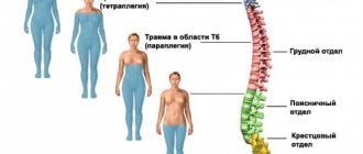
The clinical picture of a tumor disease may vary depending on the location of the pathology. For example, with medullocervical tumors, visual impairment, ataxia and symptoms of intracranial hypertension (increased pressure inside the skull) occur.
Damage to segments of the cervical spine is accompanied by pain in the occipital region, hypoesthesia and paresis.
After a certain amount of time (from 2 months to several years), the patient is faced with lower paraparesis, and in the later stages of the disease – with pelvic dysfunction.
On this topic
- central nervous system
What are the dangers of a porencephalic cyst of the brain?
- Olga Vladimirovna Khazova
- May 27, 2021
Often, with intramedullary tumors, patients are diagnosed with a mild form of scoliosis, and then pain appears in the area of paravertebral muscle tissue (these are the muscles that extend the spine). Over time, stiffness of movement is added to the pain syndrome.
One of the latest manifestations of a tumor disease includes pelvic disorders and dysesthesia. In rare cases, the disease is accompanied by a decrease in sensitivity in the anogenital area.
Diagnostic features
At the first manifestation of symptoms, you should immediately consult a doctor for a diagnostic examination. First of all, the specialist will conduct a medical history and a thorough visual examination.
If necessary, the patient will be prescribed additional procedures:
- Computed tomography (CT). With its help, you can study in detail the spinal column, its size and shape, as well as additional structures that are located next to it.
- Magnetic resonance imaging (MRI). Allows you to obtain a three-dimensional image during diagnosis, assess the condition of the patient’s spine and identify all kinds of pathological changes, including tumors.
- X-ray examination . One of the cheapest diagnostic procedures performed for suspected spinal tumors. X-rays can reveal abnormalities in bone structure, the presence of tumors or vertebral fractures.
- Biopsy . It is carried out to clarify the type of neoplasm. During a biopsy, the doctor removes a tissue sample from the tumor for analysis.
In addition, the patient undergoes diagnostics of other organs to determine the primary neoplasms. Based on the data obtained, the doctor makes an accurate diagnosis and prescribes appropriate therapy.
Methods for diagnosing spinal cord glioma
Diagnosis of spinal cord glioma consists of neuroimaging data and clinical studies. It begins with a neurological examination, during which the doctor conducts a series of coordination (functional) tests. The main imaging diagnostic method in this case is MRI (magnetic resonance imaging). Spinal cord gliomas are usually clearly visible on MRI because the soft tissue of the spinal cord provides high-quality MRI resolution to identify areas of compression.
In addition, the following methods are used:
- CT (computed tomography) with contrast;
- PET-CT (the method allows you to monitor the condition of the tumor over time during treatment);
- Angiography;
- Scintigraphy;
- Lumbar puncture followed by histological analysis.
A list of all Israeli cancer centers with state-of-the-art diagnostic capabilities for spinal cord glioma can be obtained upon request by contacting us in any convenient way.
Features of therapy
The main and, perhaps, the only method of eliminating the formation is surgery. This is the only way to completely eliminate an itramedullary tumor.
If the patient seeks medical help in a timely manner, when the disease has not had time to intensify (progress), then the surgical intervention takes place without any complications.
Otherwise, when a spinal tumor has led to paralysis, it is very difficult to completely restore the body’s motor abilities after therapy.
On this topic
- central nervous system
Everything you need to know about pituitary adenoma
- Olga Vladimirovna Khazova
- February 28, 2021
When an intramedullary tumor develops, a well-performed operation does not always give the desired result, since it is not always possible to remove the tumor without affecting the patient’s spinal cord. Often such operations end with decompression (elimination of increased pressure on the spinal cord).
To achieve better results, doctors often resort to radiation therapy (a method of exposing the body to ionizing radiation). There are great hopes for such therapies.
But the main disadvantage of radiation therapy is the fact that during the procedure the patient's spinal cord is damaged, which is associated with the negative effects of radiation (spinal cord cells do not tolerate radiation well).
Therefore, for poorly differentiated astrocytoma or ependymoma, the radiation method is practically not used. Some modern clinics perform microneurosurgical operations, allowing all necessary surgical actions to be performed with minimal traumatic effect.
Depending on the type of tumor, the stage of the pathological process or the location of the tumor, the outcome of surgical treatment may vary. For example, when treating meningioma or neuroma, the chances of a complete recovery are several times higher than with an intramedullary tumor of the spinal cord, since the prognosis here is not always comforting.
In addition, the patient’s ability to work after a rehabilitation course may depend on the quality of the operation performed. Treatment of severe forms of intramedullary tumor often results in disability for the rest of life. Therefore, serious diseases such as a spinal tumor cannot be neglected.
New methods of treating spinal cord glioma in Israel
Israeli neuro-oncology is at a very high level of development, and is rightfully considered one of the best in the world, so patients from many countries come here for treatment. One of the advantages of Israeli neuro-oncology is the use of innovative treatment methods, many of which are developed with the direct participation of Israeli specialists, and approaches to the treatment of spinal cord tumors in Israel correspond to best international practice.
Treatment of spinal cord gliomas in Israeli clinics is also carried out using surgery, radiation and chemotherapy, all of them are used in the latest modifications, modern equipment is used, which is regularly updated.
Indications for surgery here can be somewhat expanded, firstly, due to high-precision diagnostics, which makes it possible to accurately localize the tumor and determine its nature and relationship with the underlying tissues, and secondly, due to the use of innovative surgical robotic technology, with which it is possible to perform the finest manipulations without the risk of damaging the spinal cord.
Some spinal cord gliomas can be treated with stereotactic radiosurgery. This is a non-invasive method that replaces surgical intervention. Its essence lies in high-dose irradiation of a precisely specified area, after which the tumor is destroyed, and its decay products are eliminated from the body naturally. For some tumors, this method is used as the main one, and in some cases, stereotactic radiosurgery is used to remove the residual part of the tumor, the one that could not be removed during surgery.
In a certain percentage of patients treated, the tumors recur. This occurs when microscopic tumor particles remain in the body. In this case, you have to undergo a second course of treatment, which may include stereotactic radiosurgery, chemotherapy, and radiation.
Possible complications
Since an intramedullary tumor can be not only benign, but also malignant, depending on the location of the pathology, the functioning of internal organs (for example, the bladder or intestines) may be disrupted in the patient, the sensitivity of the limbs may decrease, or numbness may occur, which will lead to more serious consequences .
If treated incorrectly or completely absent, an intramedullary spinal tumor can lead to complications, which, in turn, can be unpredictable and completely different.
In the group of patients who, before tumor resection, suffered from paraplegia (paralysis of the upper or lower ends) for a long period, the likelihood of completely restoring motor functions is, unfortunately, low. The use of the median myelotomy method during surgery often leads to burning pain.
Repeated surgical interventions to remove tumors can provoke arachnoiditis (inflammation of the membrane of the spinal cord or brain) or meningitis (inflammation of the brain).
Forecast
Growth parameters and tumor characteristics determine the prognosis of spinal neoplasms. Even successful surgery to remove a tumor does not guarantee that there will be no relapse or continued development of the tumor.
Ependymomas have a favorable prognosis because many patients do not experience recurrence of the disease for many years after surgery.
The prognosis of astrocytoma is not so favorable, since almost 50% of all patients experience relapses within 4-5 years after surgery.
On this topic
- central nervous system
What threatens a cyst in the head?
- Olga Vladimirovna Khazova
- February 28, 2021
If we talk about teratomas, they have an unfavorable prognosis: after a short period of time, the patient may develop systemic metastases. The same applies to metastatic intramedullary neoplasms, the prognosis of which in most cases does not inspire much hope.
Depending on the start of surgical treatment and the severity of the clinical picture, the level of recovery of the neurological deficit may vary. This process is also influenced by the quality of rehabilitation treatment.
There are many cases in medicine where timely and high-quality surgery to remove a tumor allows patients to return to a full life and work activity after a long rehabilitation period.