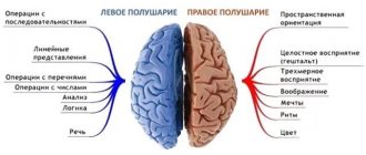
Motor neurons: characteristics
Efferent neurons (motor) have a structure identical to other nerve cells. Their network of dendrites is the most branched, and axons extend to the muscle fibers. They cause the muscle to contract and straighten. The longest axon in the human body is the motor neuron axon, which runs to the big toe from the lumbar region. On average, its length is about one meter.
Almost all efferent neurons are located in the spinal cord, because it is responsible for most of our unconscious movements. This applies not only to unconditioned reflexes (for example, blinking), but also to any actions that we do not think about. When we peer at an object, the brain sends impulses to the optic nerve. But the movement of the eyeball to the left and right is carried out through commands from the spinal cord; these are unconscious movements. Therefore, as we age and the accumulation of unconscious habitual actions increases, the importance of motor neurons appears in a new light.
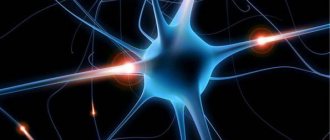
Nerve cell (neuron or neuron)- structurally functional unit of the nervous system. There are several types of nerve cells: sensory, effector and intercalary. Recently, nerve cells have been called
neurons
, from the Greek neuron - a nerve cell consisting of a body and processes extending from it - relatively short dendrites and a long axon.
Sensory nerve cells
, have branching endings in the form of receptors that perceive and convert irritation into nerve impulses. Receptors located on the integumentary tissues and perceiving external irritations are called
exteroceptors.
Receptors located on internal organs and perceiving irritation when the chemical composition changes (chemoreceptors) of the internal environment of the body are called
interoreceptors.
Another type of receptor that senses tension in muscle tissue is called
proprioceptors.
Sensitive nerve cell receptors are located in the integumentary tissues and tissues of internal organs. Signals from stimuli (nerve impulses) received by receptors are transmitted to the body of the nerve cell, where they are processed and transmitted through axons to other nerve cells, for example, effector cells.
Effector nerve cells
perform the opposite function of sensitive nerve cells; they transmit nerve impulses from the nervous system to the tissues and organs of the body, for example, the impulse of muscle contraction.
Interneurons
, nerve cells that perform an intermediate role, organizing, as it were, a bridge between different types of nerve cells.
For example, with a thermal burn of the hand, the signal received by the skin receptors of the sensitive nerve cell is transmitted to the interneuron. The body of the interneuron processes the signal and then the signal propagates towards effectors that excite muscle tissue, which, in turn, withdraws the hand from the source of fire (the so-called reflex arc). Also, the burn nerve impulse from the intercalary neuron is transmitted to the central nervous system. In the central nervous system, this impulse is analyzed for the degree of burn, the position of the hand in space, and the like. The response impulse generated in the central nervous system regulates the position of the hand in space, and also turns on the mechanisms of internal organs that neutralize the consequences of the burn. An interneuron can be considered as a switchboard of nerve signals. The largest number of interneurons are located in the spinal cord and brain. Nerve impulses propagate in the form of an electrochemical current in the form of movement of potassium K or sodium Na ions. The speed of propagation of nerve impulses is low and is approximately 0.5 - 120 m/sec. To speed up the transmission of impulses along the pathways of nerve cells, the pathways are covered with Schwann cells in the myelin sheath - lemmocytes.
Communication between individual nerve cells is carried out using synapses.
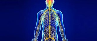
Synapses will be discussed in detail in the section describing the structure of the central nervous system. Nerve cells, whether receptor or effector, serve a very small area of tissue. To cover all tissues of the body, nerve cells are combined into fibers (similar to an electrical multi-strand cable) called nerves.
Nerves
(Latin nervus), gray fibers of nervous tissue. Nerves connect the brain and ganglia with other organs and tissues of the body.
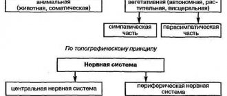
The collection of nerves forms the peripheral nervous system.
Types and characteristics of neurons
Nerve cells called neurons receive, send, and conduct bioelectrical signals. There are efferent (motor) neurons - these are components of the central nervous system that redirect signals to executive organs, for example, skeletal muscles. Afferent (sensitive) neurons are cells that perceive external and internal stimuli, which ensures the body’s connection with the external environment and reactions to changes in the functional activity of internal organs.
Intercalary cells provide interconnections within the overall neural network. Neurons of all types (sensitive, efferent, associative) are functional units that support the activity of the nervous system; they are found in all tissues of the body, where they play the role of connecting links between receptor (perceiving irritating stimuli) and effector organs that respond to irritating stimuli.
Effector organs include muscles and glands, and receptor organs include sensory organs. The significance of the signals transmitted varies significantly depending on the type of cell and its role in the functioning of the central nervous system. For example, sensory neurons that perceive impulses from the external environment transmit signals from skin receptors and sensory organs in the direction of the brain; motor neurons redirect commands generated in the brain, causing contraction of skeletal muscles and initiating movement.
Despite the different meanings of bioelectric impulses, their nature is the same and consists in changing the electrical potential in the area of the plasma membrane of the nerve cell. The mechanism of propagation of nerve impulses is based on the ability of an electrical disturbance that appears in one place of the cell to be transmitted to other areas. In the absence of factors that enhance the signal, the pulses fade away as they move away from the source of excitation.
Sensory, also known as sensitive, is an afferent neuron that conducts impulses from distal parts of the body to the central parts of the central nervous system. For example, sensory fibers form fibers that extend from the light-sensitive cells of the organs of vision. Signals leave the retina, traveling along millions of axons belonging to the structures of the basal ganglia, towards a region of the visual cortex.
The sensory neuron, together with the executive (motor) neurons, forms a simple reflex arc.
For example, the knee reflex is an unconditioned stretch reflex reaction that occurs as a result of the activity of a similar reflex arc. A reaction in the form of uncontrolled extension of the lower leg occurs when there is a mechanical impact on the tendon of the femoral muscle, which lies under the patella. Reaction mechanism:
- Mechanical effects on the neuromuscular spindles located in the hip extensor muscle.
- An increase in the intensity of nerve signals in the endings entwining the neuromuscular spindles due to their stretching.
- Transmission of impulses to sensory neurons located in the spinal ganglia through dendrites extending from the femoral nerve.
- Transmission of impulses from sensory cells to alpha motor neurons located in the anterior horns within the boundaries of the spinal cord.
- Signal transmission from alpha motor neurons to contractile muscle fibers of the femoral muscle.
The knee reflex mechanism involves interneurons that transmit inhibitory impulses to motor neurons of the flexor muscles, and other interneurons, for example, Renshaw cells. The knee reflex mechanism also involves gamma motor neurons, which regulate the intensity of spindle stretch.
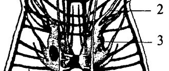
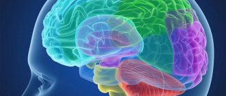
In the spinal cord, formed by gray matter, there are three types of neurons - motor, intercalary, and autonomic. Moreover, the vegetative ones are located in the visceral (related to the internal organs) nuclei. These cells interact with afferent (ascending pathways that transmit impulses from peripheral receptors to the central zones of the central nervous system) fibers responsible for general visceral sensitivity.
Visceral afferents conduct nerve signals (usually painful or reflex sensations) from internal organs, elements of the circulatory system, glands to the corresponding zones of the central nervous system. Visceral afferents are part of the autonomic nervous system. Reflex arcs within the autonomic department of the central nervous system differ in structure from the arcs of the somatic department.
Efferent components (descending pathways that transmit impulses from the cortical and subcortical zones of the brain to peripheral areas) are formed by two types of neurons - intercalary and effector (motor). The intercalary ones are located in the nuclei belonging to the vegetative part of the central nervous system. The name “intercalary” is due to its location between the sensory and motor neurons.
Sensitive
A sensory neuron is a component of the nervous system that transmits information to the brain about stimuli affecting a specific area of the body. Examples of irritants include factors: sunlight, mechanical impact (impact, touch), the action of a chemical substance. Sensory neurons are located in the ganglia of the brain - the spinal cord and brain.
The connection formed with a sensitive neuron can provoke excitation or inhibition, which is sent along nerve fibers to the cortical parts of the brain. As the level of sensory pathways increases, the transmitted information is processed to identify important features. Sensitive neurons belong to pseudounipolar neurons - their axon and dendrites extend from the body together, subsequently separate and are located in the spinal cord, brain (axon) and in the peripheral parts of the body (dendrites).
Insert
Interneurons transmit converted nerve impulses obtained as a result of processing sensory information received from various sources, for example, from the organs of vision and skin receptors. As a result, the processed information becomes the initial data for the formation of adequate motor commands.
Motor
Motor nerve cells are of two types - large and small. In the first case we are talking about α-motoneurons, in the second – about γ-motoneurons. Alpha motor neurons are present in the basal ganglia of lateral (closer to the lateral plane) and medial (closer to the median plane) localization. These are the largest cells present in nervous tissue.
Their axons interact with striated fibers contained in skeletal muscles. As a result, synapses (sites where nerve signals are transmitted) are formed. The axons of alpha motor neurons interconnect with intercalary analogues, also known as Renshaw cells, leading to the formation of collateral pathways and inhibitory synapses in the spinal cord.
Gamma motor neurons are part of the neuromuscular spindle, which is a complex receptor consisting of nerve endings (afferent, efferent). The main function of neuromuscular spindles is to regulate the force and rate of contraction or stretching of skeletal muscles.
What muscles do motor neurons connect to?
Neuron axons are connected to several types of muscles (they are working muscles), which are classified as:
- animal;
- vegetative.
The first group of muscles is represented by skeletal muscles, and the second belongs to the category of smooth muscles. The methods of attachment to the muscle fiber are also different. Skeletal muscles form peculiar plaques at the point of contact with neurons. Autonomic neurons communicate with smooth muscle through small swellings or vesicles.
Types of motor neurons
In turn, efferent cells have a certain classification. They are divided into the following two types:
- a-motoneurons;
- y-motoneurons.
The first type of neurons has a denser fiber structure and attaches to various muscle fibers. One such neuron can involve a different number of muscles.
Y-motoneurons are slightly weaker than their “brothers”; they cannot engage several muscle fibers at the same time and are responsible for muscle tension. We can say that both types of neurons are the controlling organ of motor activity.