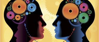
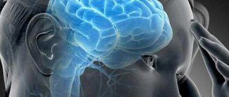
Broca's area is the area of the brain responsible for the formation and reproduction of oral speech. Numerous parts of the brain are involved in the processes of understanding and pronouncing words. Damage to the designated brain structures leads to impaired perception of other people's phrases, the inability to pronounce words and logical, complete verbal expressions. The prognosis for speech disorders depends on the location and size of the pathological focus.
Background
In medical practice, the first experiments to identify the areas of the brain responsible for speech were made at the beginning of the 19th century. In the mid-1860s, French anthropologist and surgeon Paul Brocq published the results of his research, which stated that the area of the cortex responsible for speech is located in the posterior lower region of the left hemisphere. This applies to lefties.
The result was obtained thanks to the doctor's observation. He noticed that people who had suffered a right hemisphere stroke did not have speech defects or disorders. In those whose left hemisphere was damaged due to a stroke, such anomalies were detected, and quite pronounced.
This area of the brain is called Broca's area. Let's look at this area in detail: its location and purpose according to a French specialist, and then we will resort to the results of scientists from New York Medical University, whose experiments confirm the lack of knowledge of the organ and actually put an end to more than a century and a half of medicine and science’s understanding of the speech center in the head brain.
Features of the speech system
The centers of speech function located in the brain have undergone colossal changes in the process of human evolutionary development. A prerequisite for the development of speech in ancient people was the mobility of the lips and tongue (in comparison with animals). The speech system of a modern person conventionally consists of sections - central and peripheral. In the first case, we mean the Wernicke and Broca centers, which are located in the left hemisphere.
The peripheral department includes the hearing organs, articulatory and vocal apparatus. Many vowel sounds are reflexive in nature, which makes them dependent on physiological processes. For example, painful sensations are accompanied by involuntary groans “o” or “oh”, which are formed as a result of interruption of the respiratory movement during exhalation.
The anatomical and physiological mechanisms of speech and hearing formation reflect the activity of the corresponding centers that are located in the brain, mainly where the convolutions lie - the frontal and temporal. The brain centers, in particular Broca's and Wernicke's, play a decisive role in the formation of speech skills, and the location of both areas in the left hemisphere indicates the great importance of this hemisphere in the implementation of speech activity.
The functions of the individual areas of the brain responsible for speech vary. For example, the temporal lobe of the left hemisphere is responsible for auditory and speech memory. Damage to this part of the brain is associated with impaired understanding of the meaning of verbal stimuli. A person ceases to understand the words addressed to him, especially if their meaning depends on the context (within a sentence).
When parts of the brain such as Wernicke's area and posterior associative zones, which provide phonemic perception of speech, are damaged, the integration of speech elements into a single semantic scheme is disrupted. While maintaining the integrity of the designated brain structures and simultaneous damage to the frontal areas, understanding complex phrases and profound statements becomes difficult. Such symptoms are due to the loss of functions that are regulated by the frontal areas:
- The ability to plan and program actions.
- Cognitive activity, searching for the necessary information.
- Analysis of essential speech elements within a detailed, complex utterance.
Broca's center is a department that is responsible for a person's ability to pronounce words, which involves the ability to name objects and form phrases. This area of the brain is responsible for the implementation of speech, as it is presented orally; damage to the brain matter in this section is accompanied by stuttering on any syllable, repeated repetition of previous articulation, and rearrangement of letters in a word.
Other areas of the brain take part in the implementation of the speech function of the oral form. For example, to name a word, you need to recode a visual stimulus into an audio (vocal) equivalent. The operation involves the parietal and occipital structures. Maintaining a certain acoustic (related to auditory perception) structure of a word is impossible without the participation of the temporal region, which lies on the left side of the head.
Impaired naming of objects often occurs due to deterioration of complex processes occurring in the brain. For example, choosing the only correct option for the name of an object requires inhibition of other alternatives. The structures of the frontal cortex are involved in these processes.
Other important tasks of the frontal cortex are related to translating the intent of a phrase into verbal form. One of the leading symptoms of damage to the substance of the frontal lobes is the lack of speech initiative. Patients do not pronounce words independently without external stimuli. During the dialogue, patients behave passively and limit themselves to monosyllabic repetitions.
Anterior to the central sulcus of the frontal lobe is the supplementary motor area for speech. When this area is damaged, speech rhythm and intonation change. In this case, the patient loses the ability to correctly construct sentences - he misses prepositions and conjunctions, cannot choose the appropriate pronoun and put the verb at the right time.
Moreover, grammar suffers in one’s own oral and perceived speech of others. Broca's and Wernicke's areas, which are connected into a single system by a bundle of nerve fibers, are located in the left hemisphere. However, the structures of the right hemisphere are also involved in the formation of verbal signs. Damage to the brain matter in the right hemisphere leads to disorders:
- Changes in intonation components.
- Changing the pitch and strength (loudness) of the voice.
- Changing the emotional component of verbal sayings.
Vocal reactions are controlled by the limbic system, which provides spoken phrases with a certain emotional coloring. The right hemisphere contains structures that analyze verbal material at the visual-spatial level. During the instrumental examination, an increase in the activity of thalamic structures during the perception of speech stimuli is revealed.
Physiology
Broca's and Wernicke's areas are parts of the brain that have been associated with speech since the mid-19th century. At the beginning of the 20th century, a third zone was identified - optical. The first center, according to science, is motor, associated with speech motor skills. It controls the muscles of the throat, tongue and both jaws during conversation. Most likely, it extends along the lateral surface of both hemispheres and affects the lower part of the anterior gyrus in the center and extends to the anterior part of the insula.
It is responsible for the functions of speech reproduction, coordination of many muscles involved in the formation of letters, syllables and their combinations. Damage or insufficient development of this part of the brain entails:
- stopping or significant deterioration in coordination of movements when speaking;
- cessation of movements involved in word formation;
- arbitrary pronunciation of some syllables and words.
Even with damage to Broca's area, patients not only cannot speak normally or at all (reproduce speech), but also have difficulty understanding speech or do not understand it. It all depends on the degree of damage.
Secondly, Wernicke’s area is already associated with such functions as interpretation, perception, assimilation and understanding of what other people say. The center is located at the very top of the temporal gyrus in the hemisphere, which is dominant. It follows that a person who does not yet know how to speak has only the rudiments of this area or does not have it at all. Who and when determines the dominant lobe, where Wernicke's area will be formed, remains a mystery.
If this lobe is damaged, the patient cannot construct logical sentences filled with meaning. If he manages to say something, his speech will often be meaningless - he will utter several unrelated phrases.
There is also a third center. He is responsible for the development of figurative and symbolic speech. With the destruction or damage of the area, the ability to associate images with any concepts, recognize symbols (recognize letters) and compose words from them is lost.
We learned what Broca's and Wernicke's center is, but only in the interpretation of official science, which knows very little. There is another idea about the speech centers of the brain, Broca's and Wernicke's areas.
Functions
Brain centers within the speech system are functionally connected and interact with such departments as areas of the associative cortex, areas of motor and articulatory activity, and subcortical structures. Participation of numerous brain regions in the speech system is associated with a large number of variants of the course of aphasia (for example, cortical, subcortical, dynamic, motor, acoustic-mnestic, optical-mnestic forms).
On the other hand, the presence of a large number of structural components of the speech system contributes to the restoration of speech function according to a compensatory type in cases where a certain area of the brain is damaged. Expanding the ability to restore functions is associated with the theory of neuroplasticity - the ability of normal nerve cells to take on the tasks of damaged ones.
Broca's area is responsible for the reproduction of verbal stimuli, Wernicke's area is responsible for the perception of verbal stimuli, both areas are located in the left hemisphere. The structures of the auditory cortex located in the left hemisphere take part in the perception of audible verbal signs. Neurons in this region perceive and identify phonemes—the simple sounds that make up words.
Phonemic hearing (the ability to distinguish phonemes) is impaired when designated areas of the cortical layer are damaged. The speech apparatus contains departments - regulating and performing. The regulatory department lies in the left hemisphere of the brain, which, to a greater extent than the right, is responsible for speech production and hearing. The regulatory apparatus includes pathways and nuclei of the brain stem (mainly located in the medulla oblongata).
Nerve endings belonging to the regulatory part of the brain extend in the direction of the muscles responsible for articulatory skills (tongue, lips, cheeks). Nerve endings as part of the regulatory department extend towards the muscles of the respiratory system and the vocal apparatus. Some areas of the cortical layer play a decisive role in the formation of verbal stimuli:
- Frontal gyri. The motor speech center is located in this area of the brain. Thanks to the activity of the designated structures, a person pronounces words and phrases.
- Temporal gyri. They perceive auditory stimuli - someone else's speech.
- Parietal lobe of the cortex. Participates in the process of understanding other people's words.
- Occipital lobe. Participates in the perception and identification of visual stimuli and, accordingly, written text.
- Subcortical nuclei. They regulate the rhythm, tempo, and expressiveness of reproduced words and phrases.
The pathways connect the cortical areas of the brain with the organs of the articulatory and vocal apparatus, which allows you to control muscle contractions and create the desired sounds using the tongue, lips, and cheeks. The pathways of the speech system originate in Broca's area.
Different opinion
The development of neuroimaging allows us to make the assumption that the functions that scientists previously thought were performed by Wernicke’s lobes are carried out by the temporal lobes. There are theories that claim that the inferior and middle temporal gyri process speech information. Also, a small portion of the temporal gyrus functions in speech recognition and processing. But these are just assumptions. Let's move on to the research results.
Several years ago, New York University and its medical center questioned the achievements of Brock and Wernicke, who observed people who had suffered a stroke. Scientists studied the activity of organs in completely healthy patients using modern instruments - a magnetic resonance imaging scanner, the work of which is based on observations of blood circulation during various tasks. The results led to the conclusion that the anatomy of the speech centers is not at all the same as it was believed for a century and a half.
The electrodes were applied directly to the surface of the cerebral cortex, which makes it possible to obtain a highly accurate picture with good resolution.
No one drilled into anyone’s skull; everything was done with the consent of the patient, who had to open the skull to treat any neurological abnormalities.
Some of those who agreed to the study had electrodes attached to one of the hemispheres, and some - to both. During observation, they listened to speech, repeated it to themselves and out loud. Moreover, among the words there were not only ones familiar to them, but also invented ones that did not carry meaning. Unknown syllables made it possible to separate the functions of pronouncing speech and understanding the meaning of phrases.
As a result, the centers that were the most active were identified. They are located in both hemispheres almost equally. It was also concluded that the speech department is less complex than the language department. The latter is responsible for understanding, processing information and composing logical phrases, and not just pronouncing words or syllables, like speech. That is why children utter the first inexpressive syllables before they understand the speech of strangers.
Causes of speech impairment in children
In order for a child to fully develop, it is necessary to sufficiently develop his speech apparatus and avoid factors that negatively affect this. These include:
- genetic predisposition - the mechanism is not really explained anywhere, but it is almost impossible to avoid this on your own, you can only rely on nature;
- problems with the organs of hearing - if a person hears speech unintelligibly, it will also be reproduced by him;
- inhibition in mental development;
- physical deviations in the perception of the world due to pathologies of the corresponding brain centers;
- the effect of certain medications or the combined use of several medications at the same time;
- insufficient attention from parents and untimely access to specialists.
Everything in the body has its own time, and if the child does not start speaking in time, there is a high probability that he will never be able to do this even with the help of modern medicine, since time has been lost. An example of this is Mowgli children: if a child’s puberty occurred in the wild, no one and nothing can socialize such a child.
Speech problems after a stroke
After a circulatory disorder in the brain - a stroke, most patients cannot pronounce a clear sentence, and sometimes even a phrase. It is not a fact that in such a state they can even think rationally and compose these sentences in their minds. Science has identified two groups of speech disorders in people after a stroke:
- aphasia – when the cerebral cortex, which is responsible for speech and a whole list of other mental and physiological processes, suffers;
- dysarthria is a condition caused by the innervation of the muscles of the speech apparatus located in the nasopharynx.
We see that the idea expressed at the beginning of the article that a person knows practically nothing about the brain is confirmed. For a century and a half, people have recognized that Broca's center is located in the left hemisphere, its main function is speech production, but recently it was discovered that no single brain center fully takes on all these functions. Soon textbooks will have to be written anew.
Article:
In order to correctly represent the complex mechanism of normal speech activity, take a differentiated approach to the analysis of speech disorders and competently determine the paths and directions of correctional work, knowledge of the anatomical and physiological mechanisms of speech is necessary. Speech is one of the complex higher mental functions of a person, which is ensured by the activity of the brain. Research by P.K. Anokhina, A.N. Leontyeva, A.R. Luria et al. established that the basis of any higher mental function is complex functional systems, in the formation of which various parts of the brain take part, united by the reflex mechanism. The speech apparatus consists of central and peripheral sections.
Rice. 1. Structure of the speech apparatus
1 – brain; 2- nasal cavity; 3 – hard palate; 4 – oral cavity; 5 – lips; 6 – incisors; 7 – tip of the tongue; 8 – back of the tongue; 9 – root of the tongue; 10 – epiglottis; 11 – pharynx; 12 – larynx; 13 – trachea; 14 – right bronchus; 15 – right lung; 16 – diaphragm; 17 – esophagus; 18 – spine; 19 – spinal cord; 20 – soft palate.
The central section of the speech apparatus includes the brain - its cortex, subcortical nodes, pathways and nuclei of the corresponding nerves. The frontal, temporal, parietal and occipital lobes of predominantly the left hemisphere of the brain (in left-handed people, the right) are of primary importance in the formation of speech. The frontal gyrus is a speech motor area and is involved in the formation of oral speech (Broca's area). The temporal gyrus, being a speech-auditory area (Wernicke's center), is responsible for the perception of other people's speech. The parietal lobe of the cerebral cortex ensures the understanding of speech, and the occipital lobe, being a visual area, is important for the acquisition of written speech. The subcortical nuclei are responsible for the rhythm, tempo and expressiveness of speech. Pathways connect the cerebral cortex with the peripheral speech organs. Centrifugal pathways go from the center to the periphery, and centripetal nerve pathways go from the periphery to the center. The following cranial nerves take part in the innervation of the muscles of the speech apparatus:
- The trigeminal nerve innervates the muscles that move the lower jaw;
- Facial nerve - facial muscles, including muscles that move the lips and cheeks;
- The glossopharyngeal and vagus nerves are the muscles of the larynx and vocal folds, pharynx and soft palate. The glossopharyngeal nerve is also a sensory nerve of the tongue, and the vagus nerve innervates the muscles of the respiratory and cardiac organs;
- The accessory nerve innervates the muscles of the neck;
- The hypoglossal nerve allows the tongue to make various movements.
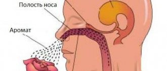
The peripheral speech apparatus consists of the respiratory, vocal and articulatory sections. The respiratory part of the peripheral speech apparatus serves to supply air, the vocal part - to form the voice, the articulatory part - forms the characteristic sounds of our speech as a result of the activity of the organs of the articulatory apparatus. The respiratory section includes the chest with the lungs, bronchi and trachea. Speech is formed in the exhalation phase, so during speech the exhalation is much longer than the inhalation (1:20 or even 1:30). A long exhalation requires a larger supply of air. Therefore, at the moment of speech, the volume of inhaled and exhaled air increases almost 3 times. In a child, speech breathing is developed gradually, in the process of speech development. At first, the child uses vital breathing skills in speech. Such breathing remains in cases of early-onset speech pathology. The vocal section consists of the larynx with the vocal folds located in it. The larynx is a cone-shaped tube consisting of several cartilages. At the top, the larynx borders on the pharynx, and at the bottom on the trachea. The vocal folds with their mass almost completely cover the lumen of the larynx, leaving a narrow glottis. During normal breathing, the glottis expands (inhalation), taking the form of an isosceles triangle, and narrows (exhalation). The mechanism of voice formation is based on the vibration of the vocal folds of the larynx, which are influenced by air entering under a certain pressure from the bronchi and lungs. The vibrations are transmitted to the environment, and we perceive them as vocal sounds. The main organs of the articulation department are: tongue, lips, upper and lower jaws, hard and soft palate, teeth, alveoli, tongue, lips, soft palate and lower jaw - these are movable organs of articulation; teeth, alveoli and hard palate are immobile, do not change their position, but also participate in the formation of sounds.
Rice. 2. Profile of organs of articulation
1 - lips, 2 - incisors; 3 – alveoli; 4 – hard palate; 5 – soft palate; 6 – vocal folds, 7 – root of the tongue; 8 – back of the tongue; 9 – tip of the tongue.
The tongue is the most active and mobile organ of articulation; the tongue muscle system makes it possible to change its shape, position and degree of tension. The tongue is involved in the formation of all vowels and almost all consonants (except labials). The front part of the tongue is movable and has a tip, anterior edges, lateral edges and a back. The back of the tongue is fixed and is called the root of the tongue. From the middle of the lower surface of the tongue to the bottom of the oral cavity, a fold of the mucous membrane (the so-called frenulum) descends, which limits the extreme movements of the tongue. Some children have this frenulum shortened from birth. In infancy, this makes sucking difficult, and later interferes with the ability to pronounce sounds correctly. At an early age, the bridle is trimmed. At a later age, the help of a speech therapist and special exercises for the tongue are needed to help stretch the frenulum. In addition to the tongue, other organs of articulation also play an important role in the formation of speech sounds: the hard and soft palate. Making various movements and taking a wide variety of positions, they modify the shape of the oral cavity, form closures, slits, etc. in it. The soft palate, rising and pressing against the back wall of the pharynx, closes the passage to the nose, falling, opens it. The movements of the active organs of articulation are called speech motor skills, i.e. the ability to make movements and hold the organ in a given position. The voice formed in the larynx is amplified and acquires an individual timbre due to resonance in the so-called supernumerary tube (pharynx, oral and nasal cavity). The extension pipe can change shape and volume, which is of great importance for the formation of speech sounds. It is these changes that create the phenomenon of resonance. With correct pronunciation, the nasal resonator is involved only in the pronunciation of the sounds m and i and their soft variants. When pronouncing other sounds, the velum palatine, formed by the soft palate and the small uvula, closes the entrance to the nasal cavity. During the formation of speech sounds, the extension pipe, in addition to the function of a resonator, performs the function of a noise vibrator (the function of a sound vibrator belongs to the vocal folds). The noise vibrator is the gaps between the lips, between the teeth and lips, teeth and tongue, tongue and hard palate, tongue and alveoli. With the help of a noise vibrator, voiceless consonants are formed, and the simultaneous vibration of the vocal folds and the noise vibrator produces voiced consonants. Important factors for the development of a child’s speech are his full hearing and vision. Good hearing is crucial for a child’s speech development. The child hears the speech of adults, imitates it and learns to speak independently. Deaf children do not master speech without special training. In children with some hearing loss (hard of hearing children), speech is grossly impaired. In the process of its development, human hearing acquired a special property: to accurately distinguish the sounds of human speech. A very young child still does not perceive words clearly, so he often confuses one phoneme with another and pronounces words incorrectly. It is necessary to constantly correct the child so that incorrect pronunciation does not become a habit that will later be difficult to overcome. Vision is also essential in the development of children's speech. A sighted child carefully observes the movements of the lips and tongue of the speakers, repeats them, trying to imitate articulatory movements. In the process of child development, a system of conditioned connections arises between analyzers that take part in speech activity, which constantly develops and is strengthened by repeated connections.