Brain tumor: symptoms and treatment
Space-occupying lesions of the brain are relatively rare.
According to various sources, the incidence rate of these tumors is 4-8 cases per 100 thousand population, which is significantly less than such widespread cancers as colon, breast and ovarian cancer.
A large proportion of volumetric formations of the cranial cavity are benign brain tumors, the symptoms of which can simulate a wide variety of diseases of the central nervous system, or may be completely absent, which presents serious diagnostic difficulties.
Classification
The classification of benign brain tumors is based on several principles. The most common and frequently used is the histological classification, according to which the following tumors are distinguished:
- Benign meningioma is the most common benign brain tumor. Develops from the membranes of the brain;
- Acoustic schwannoma is much less common than meningioma. Develops from auditory nerve sheath cells;
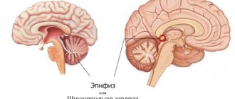
- Pituitary adenoma, accompanied by severe endocrine disorders;
- Craniopharyngioma is predominantly a childhood tumor. Develops from the remains of the canal from which the pituitary gland was formed in the prenatal period;
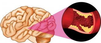
- Hemangioblastoma is usually a benign tumor of the brain vessels, which is essentially a cyst;
- Choroid plexus papilloma is an uncommon neoplasm originating from the ventricles of the brain;
- Cysts.
Knowledge of the structure of the tumor is extremely important for making the correct diagnosis and organizing treatment measures, since most benign brain tumors look the same on pictures. Only histological examination allows one to determine all the features of the structure of the neoplasm.
Microscopic examination of a tumor fragment also allows you to set the stage of the process in accordance with the international classification. There are four stages in total, but they are different for each neoplasm. An example is the stage of astrocytic tumors:
- Stage I – subependymal giant cell astrocytoma and pilocytic astrocytoma, characterized by extremely slow growth, clear boundaries and a generally benign course;
- Stage II – diffuse astrocytoma and other tumors that are characterized by slow growth. However, already at this stage it begins to grow into the tissue surrounding it;
- Stage III – anaplastic astrocytoma. It is distinguished not only by relatively rapid growth, but also by the loss of cells of their characteristic appearance;
- Stage IV – glioblastoma, the most common malignant brain tumor.
This classification convincingly shows that the boundary between benign and malignant tumors is rather arbitrary, and that the final verdict can only be made based on the results of histological examination.
How do they manifest themselves?
A benign brain tumor can have a wide variety of symptoms, since the clinical picture of the tumor directly depends on the area of the brain in which it develops.
The symptoms of some tumors appear gradually, step by step, others do not show themselves for a long time, and with others a person can live his whole life without even knowing about their existence.
- Persistent headache is often associated with a brain tumor, although according to statistics it accompanies less than 10% of all volumetric neoplasms in the cranial cavity. The mechanism of pain is based on an increase in intracranial pressure with the growth of a tumor and irritation of pain receptors;
- Nausea and vomiting that do not bring relief are caused by irritation of the vomiting center of the brain stem, as well as tumor intoxication;
- Visual impairment of various forms and intensity associated with mechanical compression of the visual pathways. Gradual unilateral deterioration of vision, accompanied by swelling of the optic disc, which is revealed by examining the fundus, is very typical for a benign brain tumor;
- Movement disorders, ranging from mild weakness to deep paralysis, arise due to the pressure of the tumor mass on the motor cortex or on the motor pathways;
- Sensitivity disturbances, mainly in the form of a decrease in sensitivity in any half of the body;
- Convulsive seizures. The tumor focus continuously irritates the neurons surrounding it, causing them to create a focus of pathological excitation. When discharged, this causes impulses to spread throughout the central nervous system, which clinically manifests as seizures;
- Mental disorders - disorders of perception (illusions, hallucinations), memory, thinking, decreased intelligence and much more.
The localization of the tumor in the brain substance has a critical impact on the clinical picture. As a rule, if the focus is located in the cerebral hemispheres, then it may not manifest itself for a long time. However, when the tumor is localized in the posterior cranial fossa, life-threatening conditions quickly arise.
This is due to the fact that the posterior cranial fossa is a very small space, the walls of which cannot move under any circumstances.
Therefore, even with the slightest increase in pressure in this area, compression of the brain substance occurs, and this is where the vital centers that control the activity of the heart, breathing and other internal organs are located.
- If the tumor is localized in the frontal lobe, there may be no symptoms at all. On the other hand, it is in this lobe that the centers responsible for higher mental functions are located, and therefore their damage leads to a wide variety of mental disorders;
- Damage to the parietal region is always accompanied by motor impairments, and sometimes by a disorder in recognizing objects by touch;
- The involvement of the temporal lobe is manifested by mental disorders and speech disorders;
- Involvement of the occipital lobe often occurs without any symptoms, although unilateral visual impairment and severe mental disorders are possible;
- When the tumor is localized in the cerebellum, dizziness, gait instability and other coordination disorders occur;
- The effect of the tumor on the brain stem leads to serious breathing problems, arrhythmias, depression of consciousness up to coma, and convulsive seizures.
A benign brain tumor may also manifest itself with some other symptoms, which depend on its histological structure. Thus, many pituitary adenomas lead to general hormonal disorders, such as Itsenko-Cushing's disease (deficiency of ACTH - adrenocorticotropic hormone), diabetes insipidus, hyperprolactinemia and others.
Tumor treatment
Therapeutic tactics for a benign brain tumor is a complex task that the doctor solves after a thorough examination of the patient:
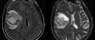
- CT and MRI of the brain with contrast allows you to detect a tumor focus, determine its size and location, and also assess the condition of the surrounding brain matter;
- EEG (electroencephalography) can show the presence or absence of pathological foci of excitation. May lead to convulsive conditions;
- A complete scan of the body to exclude other tumor processes that could metastasize to the brain;
- Histological examination of the neoplasm. The current state of medicine allows us to take a small fragment of a tumor with minimal damage to the brain substance.
Having the results of all studies in hand and knowing the histological type of the tumor, the doctor can offer the patient the following:
- Be constantly monitored, without taking medications or lying on the operating table. This tactic is justified in the case of some benign meningiomas, which grow at a rate of several millimeters per year, are located in a functionally “poor” area of the brain and do not worsen the patient’s quality of life;
- Surgical treatment consisting of complete removal of the tumor. This is the method of choice in the case of rapidly growing tumors that are located in accessible areas of the brain, as well as in situations where the expected result from the operation is significantly higher than the potential risks for the patient;
- Conservative therapy. As a rule, this treatment does not remove a benign tumor, but it significantly makes the patient feel better, relieves symptoms and improves quality of life. In particular, for swelling of the brain, the doctor may prescribe corticosteroids and diuretics; for convulsive conditions - anticonvulsants and tranquilizers; for mental disorders - antipsychotics, antidepressants and other psychotropic drugs.
The prognosis for benign brain tumors is very good: after surgery, 80-90% of patients experience significant improvement, and there is no relapse. In older people, survival rates are slightly lower due to their general condition aggravated by other diseases.
Source: https://VseoMozge.ru/dobrokachestvennaya-opuxol-golovnogo-mozga
Symptoms and treatment of a tumor of the base of the skull
Everyone knows what a skull is, but only a few understand where its base is. Let's try a quick anatomy course to clear things up. This is a very complex system, consisting of posterior and anterior sections.
The anterior one, in turn, is characterized by a boundary location between the organs of the face and the brain, and the posterior one - between the cervical region and the brain.
In more understandable language: the anterior section of the base of the skull is located in the area of the paranasal sinus, and the posterior section is located in the occipital part.
Tumors of the base of the skull are a fairly rare occurrence. Treating such a disease is very difficult and dangerous, but the neglected process brings even more trouble. It is better to get rid of the formation as soon as you notice the first symptoms.
Inner base of the skull
Types of benign tumors of the skull base:
- inverted papilloma;
- hemangioma;
- polyps;
- meningioma;
- fibroma;
- osteoma;
- schwannoma;
- neurofibroma;
- angiofibroma;
- cementoma;
- Thornwald's bag.
Most often, a benign tumor occurs on the back of the head, since it is in this part that the maximum amount of fatty tissue is concentrated, which is constantly exposed to mechanical stress. Let's look at the most popular formations.
Inverted papilloma
This is a benign tumor of the base of the skull, which is located in the nose or paranasal sinuses. It begins to develop at the age of 50, affecting mostly men. Education has local aggressiveness.
Under the influence of inverted papilloma, soft tissues suffer and the dense bone walls of the nose are partially destroyed. The main danger of inverted papillomas is the tendency to frequent relapses.
Also, about 5% of formations degenerate into cancer.
Diagnostics
Papillomas are diagnosed by assessing the symptoms of their manifestation. Let's consider the main ones:
- difficulty breathing in one of the nasal sinuses or its complete absence;
- if the tumor is enlarged, it may be opened, resulting in nosebleeds;
- the functions of smell are impaired;
- pain in the nose area, which gradually spreads to the entire face;
- constant heaviness in the nasal passage;
- excessive tearing;
- deformation of the external nasal cartilages.
After assessing the symptoms, they proceed to a hardware examination. Rhinoscopy or computed tomography is prescribed, which show the internal structure of the formation.
It has a granular structure and is located in the area of the base of the skulls in peculiar lobules. Upon closer examination, papillary growths are revealed.
The color of the inverted papilloma tissue is pink or purplish red.
We recommend reading: Brain lipoma
Treatment
Inverted papilloma can only be cured through surgery. The endoscopic method is often used. This is contact removal in a single block for less invasiveness. The main thing is not to destroy the surrounding mucous membranes. After surgery, it is very important to undergo examination every six months in order to exclude the possibility of relapse.
Meningioma
This is a benign tumor of the base of the skull that can transform into a malignant tumor over time. Grows from the hard tissues of the meninges. Characterized by slow growth and gradual expansion.
As a rule, it is very difficult to completely remove such a formation, so all patients who have undergone surgery are recommended to carry out regular diagnostics to exclude malignancy of the formation. This tumor can also spread to other parts of the brain.
Meningiomas occur most often at the age of 35 years.
Degrees of development of meningiomas:
- benign first degree;
- atypical second degree;
- malignant third degree.
The main cause of the development of meningiomas is radiation exposure. A similar tumor can also develop after radiation therapy, which is used to treat many types of cancer.
Symptoms
The disease manifests itself depending on its location and size. At first, meningioma can grow asymptomatically, but as it grows it causes considerable discomfort.
No. Useful information
| 1 | blurred vision, double objects |
| 2 | when the formation is placed closer to the auricle, hearing sensitivity may be impaired |
| 3 | frequent headaches |
| 4 | forgetfulness |
| 5 | convulsions |
| 6 | muscle weakness |
| 7 | lack of coordination |
Schwannoma (neurinoma)
A benign formation that forms from peripheral and spinal nerves. It is the result of the proliferation of the myelin sheath in the cavity of the base of the skull. Occurs at the age of 50 years.
Outwardly it resembles a dense round formation with an outer framing shell. The growth rate is quite slow: 1-2 mm annually. With more intensive growth, a suspicion arises about the malignancy of the formation.
Causes
Reasons for development:
- radiation exposure to which the body is exposed at an early age;
- long-term influence of chemical fumes;
- harmful working conditions;
- genetic neurofibromatosis heredity;
- the presence of additional benign tumors.
Posterior fossa tumors
These formations include pathological changes in brain tissue in the area of the cerebellum, IV ventricle and medulla oblongata. There are meningiomas, astrocytomas, neuromas, and gliomas.
Such formations appear at an early age, develop slowly and can turn into cancer.
If posterior fossa tumors occur in old age, this is usually the result of metastases.
Tumors of the posterior cranial fossa - Cancer treatment in Israel | Israeli Oncology Center No. 1
Tumors of the posterior cranial fossa are pathological neoplasms localized in the region of such parts of the brain as the cerebellum, medulla oblongata, lateral cistern, vermis, and fourth ventricle.
In these parts of the central nervous system, various neoplasms can develop, originating from cells of the nervous tissue. Thus, among tumors of the posterior cranial fossa one can find meningiomas, acoustic neuromas, hemangioblastomas, medulloblastomas, astrocytomas, and gliomas.
In most cases, neoplasms of this localization are detected in early childhood. Most of them are benign in nature and are characterized by slow growth. Cases of malignancy of neoplasms of this type are rare.
In elderly patients, neoplasms of the posterior cranial fossa, as a rule, are foci of secondary metastatic growth.
:
Symptoms of tumors of the posterior cranial fossa
The clinical picture of tumors of the posterior cranial fossa is determined by the degree of damage to the part of the brain that is involved in the pathological process. Most often, patients with this diagnosis develop the following pathological symptoms:
- causeless nausea and vomiting;
- painful headaches that cannot be relieved by taking painkillers;
- hearing loss;
- visual impairment;
- numbness or pain in the face and neck;
- paresis of the area of innervation of the cranial nerves;
- weakness of facial muscles;
- dizziness;
- gait disturbance;
- impaired coordination of movements;
- facial asymmetry;
- swallowing disorder.
↑ | Applying for treatment ↓
Diagnosis of tumors of the posterior cranial fossa in Israel
A qualitative examination of patients with signs of brain tumors is necessary to make a correct diagnosis, determine the location and characteristics of tumor growth, as well as determine the tactics of its treatment. To obtain the most complete information about such neoplasms as PCF tumors, advanced diagnostic technologies are used.
- X-ray – a simple X-ray examination makes it possible to determine the presence of signs of damage to the bone tissue of the skull.
- CT is a modern X-ray examination that allows highly accurate visualization of neoplasms of the central nervous system.
- MRI – performing this diagnostic procedure makes it possible to obtain the maximum amount of information about the nature of pathological changes in soft tissues.
- Spinal tap - this procedure is aimed at obtaining samples of cerebrospinal fluid. Laboratory study of cerebrospinal fluid makes it possible to assess the state of the central nervous system and determine the extent of the pathological process.
- EEG - electroencephalographic study is performed to assess the functional state of the central nervous system.
- metry – used to examine patients with signs of acoustic neuroma.
- Genetic testing is performed if the patient has signs of a hereditary disease such as neurofibromatosis.
- Biopsy - in some cases, histological examination of tumor tissue samples is required to make a diagnosis and determine the extent of the pathological process. For this purpose, a surgical procedure is performed to obtain samples of tumor tissue.
↑ | Applying for treatment ↓
Treatment of tumors of the posterior cranial fossa in Israel
Treatment of tumors of the posterior cranial fossa is best left to experienced neurosurgeons. The use of modern surgical techniques and the experience of a specialist makes it possible to completely remove the tumor without damaging the brain tissue.
- Surgical treatment - the only radical method of combating neoplasms of the posterior cranial fossa is surgery. Today, Israeli neurosurgeons use modern technologies for neuronavigation and removal of tumors of the central nervous system, which make surgical treatment as safe as possible. Thanks to microsurgical instruments and special optical systems, the surgeon performs all actions with maximum precision. This allows you to avoid damage to healthy brain tissue and the development of dangerous complications.
- Radiation therapy - ionizing radiation energy can be used to combat neoplasms of the posterior cranial fossa before and after surgery. In the preoperative period, radiation therapy helps prepare the tumor for surgical removal and reduce its size. In the postoperative period, radiation therapy sessions are used to reduce the likelihood of disease relapses.
- A modern type of radiation therapy is a technique called radiosurgery. This method of fighting tumors is based on the use of targeted energy of ionizing radiation, acting precisely on the tumor tissue. Thanks to the precise determination of the coordinates of the location of the tumor, a large number of radioactive rays are directed precisely at the tumor, without damaging healthy brain tissue. Radiosurgery is used to combat small tumors, while eliminating the need for surgical treatment.
↑ | Applying for treatment ↓
Advantages of the Israeli Oncology Center
Treatment of neoplasms of the posterior cranial fossa in Israeli clinics is highly effective for the following reasons:
- consultation with experienced specialists in the field of oncological diseases of the central nervous system;
- conducting a high-quality survey using advanced technologies;
- individual approach when leaving the therapy program;
- modern radiation therapy;
- safe and effective surgical treatment.
Tumors of the posterior cranial fossa require timely and high-quality treatment. The best solution when making such a diagnosis would be to contact highly qualified Israeli specialists.
↑ | Applying for treatment ↓
Doctors of the department
Source: https://www.cancertreatments.ru/vidy-raka/opuholi-zadnej-cherepnoj-yamki/
Discussion
When choosing the position of the patient on the operating table and when performing suboccipital approaches, a number of questions may arise regarding the risks for the patient associated only with his position, and not with his existing neurosurgical pathology. Assessing many years of experience in performing suboccipital approaches in the “lying” and “half-sitting” positions, we came to the conclusion that the safest is to perform these approaches in the “lying” position, since it avoids such a life-threatening complication as air embolism. An analysis of 200 vascular decompressions performed in various positions demonstrates that the semi-sitting position increases the likelihood of air embolism by 25 times [9]. Nevertheless, in some patients it is advisable to perform suboccipital approaches in the sitting position using intraoperative transesophageal Doppler ultrasound to monitor the occurrence of air embolism. Such patients include obese patients (in whom the “lying down” position blocks the excursion of the chest), patients with giant tumors, as well as with osteochondrosis of the cervical spine, which prevents adequate rotation and/or flexion of the head.
Using the patient's "lying" position requires precise adherence to guidelines when performing a skin incision and osteoplastic trepanation, since deviation from the standard of trepanation narrows the surgical field, making it impossible to perform the main stage of the operation. This, in turn, discredits the very idea of using the “lying down” position, which is safe when performed correctly. Thus, when performing a retrosigmoid suboccipital approach, correct marking of the surgical field ensures patient safety.
A common question is how to seal sinuses when they are damaged. If the sinus is damaged in the “half-sitting” position, in no case should the sinus defect be allowed to come into contact with air, since this will turn the sinus defect into a source of air embolism. In such a situation, an assistant or nurse should continuously irrigate the sinus defect with saline solution, and then seal its wall with a hemostatic sponge or Tachocomb.
The question also often arises: what kind of trepanation should be performed - resection or osteoplastic? With the retrosigmoid suboccipital approach, one should always strive to perform an osteoplastic craniotomy. However, if, when applying a burr hole, a pronounced adhesion of the dura mater to the bone is determined (especially in elderly patients), in order to avoid the occurrence of defects in the dura mater and sinuses, resection trepanation can be performed, subsequently covering the defect with a graft. When performing a median suboccipital approach, it is best to resort to osteoplastic trepanation, except in cases where decompression of the PCF structures is necessary, for example, during operations for Chiari malformation [10].
Tumors of the posterior cranial fossa: symptoms, causes, diagnosis and treatment
A tumor of the posterior cranial fossa is a pathological change in brain tissue in the area of the cerebellum, 4th ventricle, lateral cistern, medulla oblongata.
It occurs mainly in childhood and, in the presence of certain factors, develops into cancer. If the formation is detected in old age, then we are talking about the formation of metastases.
This tumor grows slowly. More often it is benign. Sometimes a person is unaware of it because headaches occur only occasionally. When the patient goes to the doctors, the tumor is already large and leads to the appearance of severe symptoms.
ICD-10 code: C71 – malignant, D33 – benign. According to statistics, PCF tumor accounts for 70% of intracranial neoplasms in children. With age, their frequency decreases.
The prevalence of the disease is quite high and amounts to 305 cases per 100,000 newborns. In some areas of the country this figure is significantly higher.
Causes of pathology
The prerequisites for the emergence of education may be different. More often the problem occurs under the influence of radiation and unfavorable external factors. The formation may be affected by:
- inflammatory processes of the lining of the brain,
- changes in nerve fibers
- mechanical injuries,
- absence of part of the corpus callosum.
The reasons have not been fully identified. It is assumed that teratogenic factors that affect a woman during pregnancy play a role.
Diagnosis of neoplasms
Patients with evidence of tumors should be carefully evaluated.
To obtain accurate information use:
- X-ray to determine whether there are signs of bone damage.
- CT scan – visualizes neoplasms.
- MRI provides the most complete information about the nature of pathological changes.
- spinal puncture is a manipulation aimed at studying the cerebrospinal fluid. It allows you to fully assess the spread of pathology.
- metry is used to study patients who have acoustic neuroma.
- Genetic research.
Sometimes, for a more accurate diagnosis, tumor tissue cells are taken for histological examination. For this purpose, a surgical procedure is performed, which makes it possible to take an analysis in a not very traumatic way.
Treatment of cervicocranial syndrome
For the treatment of cervicocranial syndrome, therapy for osteochondrosis and the consequences of degenerative destruction of intervertebral cartilaginous discs is of great importance. If the cause of the disease is not eliminated, then even the most effective treatment will only bring temporary relief of all symptoms. Over time, the clinic will continue to develop and all the unpleasant sensations will return with a vengeance.
Our manual therapy clinic uses a comprehensive approach to treat cervicocranial syndrome. After the initial examination, the neurologist establishes an accurate diagnosis and develops an individual course of therapy.
First of all, it is important to eliminate compression of the radicular nerves and ensure the outflow of cerebrospinal fluid from the subarachnoid space of the intracranial box. This can be done using osteopathy or traction traction of the spinal column. The patient experiences visible relief of symptoms after the first session.
Then reflexology is prescribed, which starts the process of regeneration of damaged tissues. In combination with kinesitherapy, therapeutic exercises and massage, these techniques give excellent results. The patient completely gets rid of headaches, experiences a surge of strength and a significant improvement in his condition.