A typical cow with BSE.
A feature of the later stages of the disease is the inability to stand on one’s feet. Bovine spongiform encephalopathy
,
BSE
,
mad cow disease
(Bovine spongiform encephalopathy (BSE), Mad-cow disease) is a neurodegenerative prion disease that leads to irreversible, lethal changes in the brain of infected animals, belongs to the group of transmissible spongiform encephalopathies.
Caused by the BSE prion ( BovPrPSc
,
PrPbse
). The incubation period is from 30 months to 8 years. It is transmitted by eating the meat of sick animals, causes scrapie in sheep and Creutzfeldt-Jakob disease (new variant, vCJD, nvCJD) in humans.
First recorded in the UK in 1986,[1] research began in the 1970s.[2]
Since the late 1980s, mad cow disease has been detected in more than 179,000 cattle in the UK. The disease was also found in hundreds of cows in Ireland, France, Portugal, Switzerland, Spain, and Germany. Isolated cases are registered in other countries. It is believed that the epizootic was caused by feeding livestock meat and bone meal made from the remains of “infected” animals, in particular sheep[3]. More than two hundred human deaths have been identified (as of February 2009) from the new variant of Creutzfeldt-Jakob disease.
The causative agent of Creutzfeldt-Jakob disease and routes of infection
Pathology provoked by prions, which includes Creutzfeldt-Jakob disease (CJD), manifests itself as a severe degenerative process in the nervous system of not only humans, but also animals.
The disease is based on the accumulation of altered “wrong” prion protein in the neurons of the brain. The first mention of this disease is found in the works of the German doctor Hans Gerhard Kreuztfeldt (1920). And in 1921, his compatriot Alphonse Jacob identified the disease as spastic pseudosclerosis or encephalopathy, accompanied by diffuse damage to the brain substance. He also noted a combination of mental disorders with pyramidal (paresis, paralysis) and extrapyramidal (tremor, violent movements) symptoms.
Characteristics of the causative agent of Creutzfeldt-Jakob disease
Prion protein, or PrP, is, surprisingly, a physiological component of nerve cells with a function that is still unclear. The genes responsible for its reconstruction are located on the short arm of the 20th chromosome.
The pathology is characterized by the accumulation of an altered form of this protein, which, as a result of this metamorphosis, becomes resistant to enzymes and acquires the ability to suddenly fold to form peculiar rods. In this form, it goes outside the cell, where it creates amyloid plaques.
Although prion diseases in most cases are sporadic, that is, spontaneous, they are still classified as slow infections. A similar conclusion was made possible by the fact that a healthy person can be infected with material obtained from a sick patient or animal.
The key difference between prions and viruses and bacteria is the absence of DNA or RNA, which allow microorganisms to reproduce normally. Also, this protein does not cause alertness to the immune system and is simply ignored by it, despite the cascade of pathological processes in the brain.
Prions cannot be destroyed by known antiseptic agents and methods (boiling, formaldehyde, alcohol, ultraviolet light).
In animals, the disease is most often infectious; in humans, in 90% of cases, the pathology is considered sporadic or hereditary (due to a gene mutation). The exception is kuru, a disease found only in the tribes of Papua New Guinea due to their widespread ritual of eating the brains of dead tribesmen.
Epidemiology
Creutzfeldt-Jakob disease, one of the spongiform encephalopathies, is considered the most common in the structure of this nosology and occurs in 85% of cases of prion pathologies, despite this remaining extremely rare in general - no more than one infected person per 1 million population of the planet .
The disease occurs in people of any age, men are more often affected. In the classic version, the initial symptoms manifest at 60-65 years of age, but the new form, which is “mad cow disease,” debuts much earlier - at 29-30 years of age.
Pathogenesis
It is assumed that the main role in the development of spongiform encephalopathies is played by the change in the human body's own proteins PrPc into abnormal PrPSc, which occurs under the influence of prion proteins PrP. Although the process of changing the protein structure is still poorly understood, it is known that the structure of the PrPSc protein is based on β-sheets, and normally α-helices should predominate in the membrane protein.
The disease progresses slowly.
Abnormal isoforms uniting into amyloid fibers (a protein-polysaccharide complex formed during long-term pathological processes) accumulate and form plaques on neurons. As a result, gradual death of neurons occurs (gliosis). The gray and white matter of the brain acquire a sponge-like state, and as a result of irreversible damage to the central nervous system, the patient dies.
The incubation period for Creutzfeldt-Jakob disease depends on the route of infection. It is when infected through:
- surgical instruments – 15-20 months;
- donor tissue – up to 5.5 years;
- injections (administration of gonadotropin, etc.) – 12.5 years.
Mad cow disease can appear up to 30 years after infection.
Changes occurring in the body
Pathological protein, multiplying and accumulating in the body, leads to:
- the development of spongiform degeneration of nerve cells, which consists of the formation of a large number of intracellular vesicles and expansion of neuron processes. These changes create a “spongy” fine-mesh effect during microscopic examination of a brain biopsy. In some cases, it cannot be detected using available research methods;
- replacement of microglia with connective tissue;
- a significant decrease in the number of neurons in the cerebral cortex, subcortical formations (basal ganglia, thalamus), cerebellum and brainstem structures;
- amyloid accumulation in 10-15% of cases.
Causes of rabies
There are some developments that indicate the supposed causes of the development of this disease. But to date, scientists have no confidence in these assumptions.
One version suggests that cows can become infected through feed that is intended for feeding them. They could contain infected cells and microparticles of infected cow bodies.
When prions reach the cow's brain, they cause changes characterized by the appearance of small holes in the brain tissue, giving it a spongy structure. This phenomenon can be seen under a microscope.
Further changes in the brain regions affect the body as a whole, subsequently provoking the death of the animal.
Important! Another cause of the disease lies in hereditary predisposition. Abnormal prions (incorrectly developed) accumulate in the animal or human body. As we age, they destroy the structure of the brain.
Classification, or Main forms of Creutzfeldt-Jakob disease
Until recently, only three types of Creutzfeldt-Jakob disease were distinguished, and only about 20 years ago they started talking about a new “variant” pathology, which arose as a result of the “mad cow disease” epidemic in Western Europe. All forms of pathology have one cause - the persistence of a modified prion protein in the body. But they develop differently. The clinical picture is also somewhat different.
Classical
Classic Creutzfeldt-Jakob disease, also called sporadic, has an unclear genesis. That is, its origin has not been reliably established. It is believed that pathological prion proteins arise in the brain independently, and not under the influence of any factors. Mostly patients over 50 years of age become hostages of the disease.
The clinic debuts with impaired short-term memory, mood instability (tearfulness or laughter is quickly replaced by aggression), apathy and lack of initiative. As the pathology progresses, the patient ceases to perform even his daily duties. At the end, visual impairment, hallucinatory and speech disorders occur.
Photo: https://pixabay.com/illustrations/head-think-slide-strips-film-674124/
In 40% of patients with the classic variant of Creutzfeldt-Jakob disease, a subacute type of course and a steady progression of intellectual-mnestic disorders are detected. The same number of patients suffer from cerebellar dysfunction. And in 20% of cases, a combination of the above-described symptom complexes is observed.
Along with this, the clinical picture of the pathology is characterized by pyramidal (one-sided weakness in the arms and legs) and extrapyramidal (trembling in the limbs, involuntary movements) signs, as well as myoclonic focal seizures, that is, twitching of the facial muscles, etc. Paroxysms are triggered by loud noises, flashing lights, and even touch.
In the last phase of the disease, pronounced cognitive dysfunction is revealed. Death occurs in patients 8-9 months after the development of the first symptoms of CJD.
Hereditary
The hereditary, or familial, form of Creutzfeldt-Jakob disease, accounting for about 10% of cases of prion pathology, is associated with a mutation in the PRNP gene, which is inherited. Signs of pathology do not differ from the sporadic form.
Iatrogenic
The iatrogenic type of Creutzfeldt-Jakob disease has two ways of spreading:
- during surgical interventions involving transplantation of infected dura mater or cornea, or when using contaminated neurosurgical instruments. The incubation period will then be 1.5-4.5 years, and the disease will manifest itself as mental disorders;
- upon ingestion of growth hormone, gonadotropin, which were isolated from the pituitary gland of large livestock suffering from prion pathology. It takes from 5 to 17 years before the first clinical signs develop due to this transmission route. The disease is largely characterized by cerebellar dysfunction.
Cases of infection of doctors during surgery or autopsy are rare.
Photo: https://pixabay.com/photos/surgery-hospital-sanjay-gupta-81875/
"Variant"
In 1995, 20 cases of Creutzfeldt-Jakob disease were recorded in England and France, which developed spontaneously and had an atypical course. One of the features was that the disease debuted with mental disorders in young patients - up to 40 years old. Then cerebellar disorders were added. But cognitive disorders and myoclonic attacks appeared only at the final stage of the pathology.
There are no standard EEG patterns for this type of prion pathology described by Creutztfeldt and Jacob.
Patients usually die 7-23 months after the onset of the disease. Typical brain damage characteristic of animals made it possible to diagnose a “variant” type of nosology, such as “mad cow disease” in humans. It is believed to have been caused by the consumption of contaminated cattle meat. This discovery allowed us to conclude that the prion protein can be transmitted from one species of mammal to another, because previously this was considered impossible.
About the disease
Mad cow disease is also called spongiform encephalopathy. Her homeland is England. It is infectious and has long been heard by breeders, farmers and owners of private plots. The disease received this name from the consequences it has on the cattle’s body, and primarily on the central nervous system.
Under the influence of infection, animals:
- start to behave strangely;
- are unable to control their body;
- cease to control behavior;
- lose orientation on the terrain and in the surrounding space.
Important! Often, the external manifestations of illness in cows resemble the behavior of mentally ill people.
Clinical manifestations of Creutzfeldt-Jakob disease
A typical clinical nosology begins gradually, the severity of symptoms increases slowly. Only 10% of patients have an acute onset of pathology, which leads neurologists to think about a stroke.
Nonspecific symptoms
The disease begins with the usual symptoms characteristic of many nosologies with general intoxication syndrome.
In this case it is observed:
- headache;
- dizziness;
- constant general weakness with sleep disturbance;
- eating disorders (from unwillingness to eat at all to pathological cravings for food);
- weight loss;
- diffuse, indescribable pain in the body.
Specific symptoms
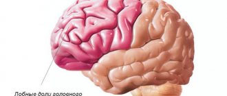
These symptoms are followed by more specific ones, depending on the predominant localization of the pathological process. If the frontal lobes of the cerebral hemispheres are mainly involved, then mental disorders are the first to develop. It all starts with a deterioration in attention, short-term memory, and thinking processes, accompanied by a general “slowness” of consciousness.
Then behavioral anomalies are added, including lack of initiative, uncriticality of one’s condition and appearance, irritability, sometimes alternating with aggression, and depression. Seizures accompanied by disorientation in space and time and visual hallucinations are also not excluded. In the end, patients experience severe dementia.
When Creutzfeldt-Jakob disease manifests itself with visual disturbances, the first complaints are double vision, blurriness of the visible picture, narrowing or loss of areas of the visual fields, and inability to recognize surrounding objects.
A third of patients at the initial stage of the disease pay attention to cerebellar symptoms, namely:
- difficulty walking due to unsteadiness;
- impaired coordination in the limbs, especially when trying to perform targeted movements, the latter is accompanied by severe tremors;
- deterioration of speech, manifested by slowing down and poor pronunciation of sounds;
- rhythmic oscillatory twitching of the eyeballs, called nystagmus.
90% of patients experience attacks of myoclonus, that is, involuntary muscle contractions that can affect not only facial muscles, but also limbs, sometimes even individual fingers. Generalized paroxysms of epilepsy usually occur only against the background of the terminal stage of the pathology.
The extrapyramidal group of symptoms that develops at the height of Creutzfeldt-Jakob disease is represented by tremor, violent movements, akinetic-rigid syndrome, accompanied by deterioration of motor activity against the background of increased muscle tone.
The late stage of the disease is characterized by akinetic mutism, when patients physically have the ability to move and speak, but simply cannot do this. The terminal stage of the pathology occurs in the form of a coma.
Photo: https://pixabay.com/photos/alone-being-alone-archetype-513525/
Kinds
The following subtypes of Creutzfeldt-Jakob disease are distinguished:
- Sporadic type or classic Creutzfeldt-Jakob disease. The most common type, affecting people in adulthood and old age (from 50 years).
- The familial type is a genetically transmitted disease.
- Iatrogenic type, in which infection occurs due to medical procedures.
- A new type that affects young people and manifests itself in personality changes and gradual disruption of basic life functions. In contrast to the sporadic type of the disease, the patient is aware of the deterioration of his condition for a long time, since the onset of dementia is remote.
Possibilities of modern diagnostics in confirming the diagnosis
Increasing dementia, combined with myoclonic seizures and characteristic changes in the EEG, gives doctors the right to suspect Creutzfeldt-Jakob disease.
Diagnosis of definite CJD
At the moment, definite, probable and possible CJD is diagnosed. In the first case, you must have:
- typical clinical picture and abnormalities in the structure of the brain matter detected during biopsy and neuroimaging studies;
- modified, enzyme-resistant prion protein in the cerebrospinal fluid, detected by protein immunoblotting;
- scrappie-associated particles.
Diagnosis of probable Creutzfeldt-Jakob disease
Issued on the basis of:
- steadily developing dementia;
- pathology-specific EEG complexes – high-amplitude sharp peaks-waves that occur despite a general decrease in the electrical activity of the brain;
- the presence of at least two of the following signs (myoclonus, visual disturbances, cerebellar disorders, pyramidal and extrapyramidal signs, akinetic mutism).
Possible CJD
It is diagnosed on the basis of a combination of dementia with a characteristic clinical picture, with a duration of pathology of less than two years and the inability to perform an EEG study or the identification of atypical patterns on film.
Instrumental diagnostic methods
To confirm the diagnosis of Creutzfeldt-Jakob disease, CT or MRI is not enough.
Although data obtained using neuroimaging technologies are also useful in establishing the true cause of the disease. Thus, on MRI, pathological changes in the area of the thalamus and caudate nucleus will be observed, resembling a “honeycomb” or a sponge, as well as diffuse cortical atrophy, the severity of the latter usually does not coincide with the severity of cognitive dysfunction. CT is less sensitive in this case and can only detect atrophy of the cerebral cortex and cerebellum.
Cerebrospinal fluid analysis and tissue biopsy
With a conventional analysis of cerebrospinal fluid, apart from a slight increase in protein levels, it is not possible to identify any pathological inclusions.
If all of the listed diagnostic methods make doctors doubt the correctness of their guesses, the only reliable method remains a biopsy of brain tissue followed by histological examination. This procedure can only be performed with the consent of the patient’s closest relatives (guardians) when the patient is declared incompetent.
Photo: https://pixabay.com/photos/mri-magnetic-resonance-imaging-2813899/
Diagnosis of mad cow disease
To diagnose and confirm the diagnosis of spongiform encephalopathy, sections of specific parts of the brain are taken from dead cows. It is not possible to study the brains of living individuals. Under a microscope, the presence or absence of holes that appear in case of disease is determined.
Prion proteins are also isolated from the resulting material. Studying their structure allows us to determine the presence of the disease. If the prion molecules are glued together, connected by the thinnest threads, this means that the animal died precisely from rabies.
Modern science has developed an innovative immunological method for detecting mad cow disease. Special antibodies are combined with prions and observed. If antibodies begin to interact with proteins in the cow's brain, this means that the infection is present in the animal's body. If the reaction to the interaction of antibodies and proteins is negative, the diagnosis of “mad cow disease” is considered unconfirmed.
Treatment of Creutzfeldt-Jakob disease
At this stage of research on CJD, no specific treatment for the pathology has been developed.
All therapy comes down to the use of symptomatic drugs, since standard antiviral drugs (Remantadine, interferons) have proven ineffective. You should also stop using medications that have a negative effect on the patient’s cognitive functions.
Some scientists claim that Quinacrine, an antimalarial drug, and the antipsychotic Chlorpromazine are able to inhibit the formation of a modified prion protein. But this information has not yet been confirmed in large experiments.
Among the drugs that actually have the ability to somewhat stop the pathology, the following can be distinguished:
- Brefeldin A, which destroys intracellular structures involved in the synthesis of prion proteins;
- NMDA receptor blockers (Ketamine, Phencyclidine) - a class of anesthetics most often used in animals;
- Tiloron is a synthetic immunomodulator.
Russian diagnostic methods
Since 1998, ultra-modern expensive equipment has been delivered to Russia (Vladimir). It was intended to detect infection in brain slices.
Currently, samples of sections from different regions of Russia are brought to the laboratory in the city of Vladimir. It takes more than 2 weeks to analyze one sample.
There is information that in recent years, about 1000 sections for research have been received by the laboratory for study, but no infected prions were found in any of them.
Prevention
Under normal conditions, the patient is not contagious and therefore does not need to be isolated in a boxed ward. After all, the only danger is the cells of the eye, brain and spinal cord, which contain the prion protein. That is, the patient’s immediate environment has nothing to fear.
There is no specific prevention of CJD, but this does not replace typical general infectious precautions: wearing gloves, washing hands after contact with the patient. Instruments used during neurosurgical interventions in such patients require special treatment. Ideally, needles and electrodes should be destroyed or subjected to long-term autoclaving.
Photo: https://pixabay.com/photos/hands-soap-bubbles-hygiene-wash-2238235/
Precautionary measures
Russia has taken a number of measures aimed at preventing cattle from becoming infected with rabies:
- Elimination of the presence of the pathogen in imported meat, livestock feed and feed additives. But it is impossible to determine the presence of infection in meat, so this measure implies a positive or negative decision on the import of meat into the country from abroad.
- Veterinary control over the quality of products at checkpoints to Russia. But since there are 291 of them, this is also quite difficult to do.
- A ban on the import of cow and sheep meat into Russia from England, Switzerland, France and some Western European countries.
- Restriction on the import of cattle into Russia from Western European countries for breeding.
In general, for a person who wants to protect himself from infection with mad cow disease, there is only one way - a complete abstinence from the meat of artiodactyl animals. This also applies to meat-containing dishes - hamburgers, various semi-finished products, etc.
Diagnosis of the disease in humans
In humans, tissue sections for analysis are taken from the tonsils or using cerebrospinal fluid. But other studies are also important for the accuracy of the diagnosis. First of all, the person’s area of residence, previous contacts with cattle, and a complete list of symptoms and complaints are clarified.
In addition to the survey, the following types of research are used on the patient:
- Electroencephalogram (EEG).
- Brain examination (computed tomography).
- Magnetic resonance imaging (MRI).
- Collection of cerebrospinal fluid for research.
- Brain biopsy.
On average, it can take about a year between the appearance of initial signs of the disease and the establishment of a correct diagnosis.
Mad cow disease infection
Despite the widespread spread of infection among cattle, infection of cows from each other is practically impossible. Scientists claim that the disease occurs spontaneously in the body of animals. But a person can become infected from a cow by eating food from infected individuals.
In the United States and Great Britain, this disease is very common and poses a great threat. Therefore, the authorities have introduced a number of measures that involve the development of outbreaks of the disease and a reduction in the number of infected people.
One of these rules obliges owners to allow sick cows to be slaughtered and to dispose of the meat (usually it is burned in a fire). Sometimes representatives of special services resort to forcibly removing infected livestock and slaughtering them. These measures prevent contaminated products from entering the market and thus preventing people from becoming infected.
Treatment of mad cow disease
With a comprehensive study of spongiform encephalopathy, a medicine that can save people and animals from suffering and death has not yet been developed. However, the exact cause of the development of the disease in living organisms has not been established.
Scientists have tried to promote the development of immunity in cattle and humans to mad cow disease, but these attempts were not successful. Drugs designed to destroy viruses also did not bring the desired results.
Currently, when signs of mad cow disease are detected, only symptomatic treatment is used. This includes taking medications to relieve seizures and against epilepsy.
The study and search for an effective cure for mad cow disease continues to this day. But the only thing that experts were able to achieve was to reduce the rate of destruction and death of infected cells.