Among neurological disorders, various types of neuropathies associated with ischemic, inflammatory or compression (tunnel) damage to the nerve fiber are often diagnosed. Median nerve neuropathy is a common pathology in modern people. This is due to a certain lifestyle and predominantly manual labor without the concomitant development of muscle groups of the upper limb. We are talking about professions related to the use of computer technology.
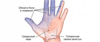
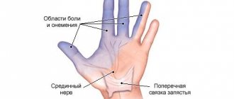
If the median nerve of the hand is damaged, a segmental disturbance of sensitivity occurs in the palm of the hand and slow fingers. Anatomically n. Medianus is responsible for ensuring motor activity and skin sensitivity in the area of the first three fingers of the hand. With neuropathy of the median nerve of the hand, an inflammatory reaction may occur in the area of the wrist joint, and motor activity of the thumb is impaired.
The anatomical features of this plexus of axons are that they are formed by two groups of bundles, extending in the form of radicular nerves from the spinal cord. The C5–Th1 segment gives rise to two pairs of radicular nerves: ventral and dorsal. The former are responsible for movement, the latter for skin sensitivity. If inflammation or damage begins at the level of the C5–Th1 intervertebral disc, then “loss” of only one function of the median nerve may be observed. When compression, ischemia or inflammation of the median nerve in the forearm, shoulder or wrist occurs, a combination of clinical symptoms of neurological and motor dysfunction occurs.
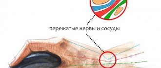
Damage to the nerve fiber can be observed along the entire length of its path to the hand. First, the median nerve descends into the axilla and passes to the beginning of the humerus. Here, injury can occur due to wearing tight and uncomfortable clothing. Along the forearm, the nerve runs deep in the thickness of the muscle layer and is reliably protected from injury. The next dangerous area is the carpal tunnel, which can become deformed. Compression of the median nerve in this anatomical node occurs in almost 80% of programmers and representatives of other professions associated with manual labor based on performing monotonous movements of the same type.
How to treat pinching?
When determining treatment, the doctor takes into account the source of the pinching and the location of the damage. Treatment of pinching caused by an infectious disease or intoxication is carried out with the help of medications. If the pinching is caused by an injury, for example, a fracture, immobilization of the arm and other measures are carried out to eliminate it. If the fracture is accompanied by a nerve rupture, the doctor will stitch it together.
Nerve damage caused by an external factor, for example, sleeping in an uncomfortable position, using crutches, or muscle activity, can be cured by eliminating the factor that provokes the disease. As a rule, treatment in this case is outpatient. A person is hospitalized only when the indications require the administration of potent drugs.
When treating pinching, you must follow all the doctor’s recommendations, especially those related to taking medications. Thus, non-steroidal drugs (to eliminate pain and inflammation), decongestants, vasodilators (to improve blood flow to the nerve), vitamin preparations and biostimulants are prescribed.
In addition to medications, physical therapy, massage, and physiotherapy (for example, laser radiation on the affected areas) are indicated. A bandage or scarf may also be useful to hang the arm on to provide relief to the patient.
Diagnostic methods
Diagnosis and determination of further treatment regimen is carried out by a neurologist. The specialist collects anamnesis and records the patient’s main complaints in the medical history. After this, the neurologist conducts a visual examination.
With ulnar nerve neuritis, the hand takes on the appearance of a bird's paw: the third and fourth fingers are bent, the little finger is moved to the side. The doctor notes cyanosis, hair loss, brittle nails, and weight loss in the affected limb.
After the examination, the neurologist conducts a series of neurological tests to determine muscle strength, degree of numbness and loss of reflexes and makes a preliminary diagnosis.
To confirm the initial conclusion, the specialist prescribes laboratory and instrumental examinations that help determine the cause of neuritis:
- General and biochemical blood test. Necessary for identifying pathogenic agents of infectious diseases, the presence of produced antibodies and leukocytes.
- X-ray using contrast.
Allows you to assess the condition of the vessels supplying the ulnar nerve. X-rays also make it possible to take into account the anatomical features of the elbow joint and cubital canal, and to identify the degree of damage to the musculoskeletal tunnel in the event of injury. - Ultrasound and MRI. Ultrasound and magnetic resonance imaging help assess the condition of blood vessels, soft tissues and bone structure in case of fractures, pinched nerves and other injuries.
- Electroneuromyography. The procedure helps determine the extent of damage to the skeletal muscles.
Special diagnostic criteria for determining the disease
Neurological tests to make a preliminary diagnosis of ulnar neuritis often include the following:
- Pitre's test. To carry it out, the patient must place his palm on the table and try to spread and close his fingers. With neuritis, the patient cannot bring the ring finger and little finger to the midline.
- Afterwards, the doctor asks the patient to try to scratch the surface of the table with his little finger. Due to inflammation of the nervous tissue, he is unable to do this.
- With neuritis, the patient cannot clench his hand into a fist, or hold a sheet of paper with two fingers. When you clench your hand into a fist, the middle, ring and little fingers do not bend.
- If the patient presses the hand firmly to the table, the little finger moves to the side. In this position, the patient cannot make horizontal movements of the third, fourth, fifth finger.
What diseases should be distinguished from?
After recording the patient’s complaints in the medical history, a differential diagnosis with radial neuritis is carried out.
In contrast to damage to the ulnar nerve, in this situation the hand hangs down and a strong muscle spasm is observed. If the radial nerve is damaged, the patient cannot straighten the hand independently; the thumb is reduced to the index finger. The sensitivity of the 1st, 2nd, and 3rd fingers of the affected limb is impaired.
It is difficult to distinguish ulnar nerve neuritis from damage to the C8 nerve root in the neck due to radiculopathy. With radicular syndrome, numbness is observed on the ulnar surface of the forearm, which is not typical for neuritis. With inflammation of the ulnar nerve, only numbness of the hand is observed.
When conducting a differential diagnosis, the neurologist pays attention to the presence of a specific symptom of ulnar nerve neuritis - numbness of the radial surface of the ring finger. When the nerve roots of the spine, spinal cord, or plexus are affected, there is a loss of sensation in the entire finger
When the nerve roots of the spine, spinal cord, or plexus are affected, there is a loss of sensation in the entire finger.
Diagnostics
- After a neurological examination, electrophysiological methods are performed to help clarify the severity of the defect, select an effective treatment approach, and obtain data on the dynamics of recovery:
- electroneuromyography to assess the speed and volume of passing impulses and detect the damaged area;
- needle electromyography (EMG) to study the state of muscle fibers, the dynamics of deterioration or renewal processes;
- radiography and computed tomography to detect the underlying cause of dysfunction.
To conduct research, you must make an appointment with a functional diagnostician.
Clinical picture: symptoms and manifestations
Median nerve neuritis is characterized by severe symptoms of irritation of neuromuscular structures:
- Severe pain syndrome. Intense burning pain spreads from the inside of the forearm to the hand, involving fingers I, II, III.
The appearance of pain syndrome is accompanied by the occurrence of vegetative-trophic disorders: the skin burns, the forearm swells, and red spots appear on it.The wrist and back of the hand become cold and pale.
- Difficulty in motor activity, paralysis.
The patient cannot make a fist, stick out or bend the thumb, or bend the index finger. The middle finger bends with difficulty. When the patient tries to bend the hand, it deviates to the left. Over time, if left untreated, the muscles of the thumb atrophy, causing it to stop moving and become inline with the others. This symptom is called monkey paw. - Sensory impairments. A disorder of the innervation of the median nerve leads to loss of sensitivity and hypoesthesia of the skin on the palms, on the back side of the last phalanges of the I-IV fingers on the back side. The development of paresthesia of the terminal phalanges is possible.
With neuritis of the median nerve in the carpal tunnel area, the pain intensifies when raising the arms up, when touching the ligaments and in a horizontal position.
If neuritis occurs above the wrist joint, then only motor activity is impaired. There is no numbness in the hand. This is due to the fact that the branch of sensory neurons of the median nerve before entering the carpal tunnel runs separately from the motor branch.
Possible patient complaints
Patients complain of numbness of the hand from the forearm and the first 3 fingers, burning pain in the area of the affected nerve.
If the doctor begins to palpate the ligaments, patients withdraw their hand due to increasing pain. The pain radiates to the fingertips, shoulder and neck area. Due to paralysis of the flexor muscles of the hand and fingers, patients cannot make a grasping movement or stick out their thumb.
Patients cannot raise their palms up, and the movements of the shoulder and forearm are impaired. With a prolonged course of the disease, complaints of muscle weakness of the hand appear.
Patients are concerned about visually distinguishable vegetative-trophic disorders:
- bluish skin;
- swelling of the forearm, hand, fingers;
- lack of sweating;
- peeling skin, increased brittleness of nails.
Peripheral nerve injuries
Peripheral nerve injuries are divided into two types: closed and open nerve injuries. According to the severity of the injury, they are distinguished: concussion, bruise, compression, anatomical break.
Anatomical break can be complete, partial or intra-trunk.
According to the nature of damage to peripheral nerves, they are distinguished: punctured, cut, chopped, bruised; by type of compression - traction, chemical, burn and radiation.
Pathogenesis and pathomorphology.
Human peripheral nerves are a continuation of the spinal roots. The nerves include axons of motor cells of the anterior horns of the spinal cord, dendrites of sensory cells of the spinal ganglia and fibers of autonomic neurons. The outside of the nerve is covered with epineurium. Fibers covered with endoneurium pass through the lumen of the nerve. These fibers can be combined into bundles. With the help of the endoneurium, the fibers and their bundles are separated from each other. The third membrane involved in the structure of the nerve is the perineurium. This is connective tissue that surrounds bundles of nerve fibers, blood vessels and performs a fixing function. The perineural sheaths along the nerve can divide, connect and divide again, forming the fascicular plexuses of the nerve. The number and relative arrangement of bundles in the nerve trunk varies every 1-2 cm, since the course of nerve fibers is not linear. Arterial branches approach large nerves every 2-10 cm. Veins are located in the epi-, endo- and perineurium. Fibers in the peripheral nerve are either pulpy or non-pulphate, depending on the presence of myelin (it is absent in non-pulp fibers). The speed of impulse transmission along the pulp fiber is 2-4 times higher (60-70 m/s) than in the non-pulp fiber.
In the pulpy nerve fiber, the axon is located in the center. On its surface there are Schwann cells, inside of which there is myelin. The junctions between Schwann cells are called nodes of Ranvier. The fiber is fed mainly in these places.
A nerve cell, in the process of its development and differentiation, eventually loses the ability to regenerate, but can restore its lost processes or peripheral endings. This restoration of the morphological structure of the nerve cell can occur if the cell body retains its viability, and there are no obstacles to the growth of the regenerating axon on the path of germination of the damaged nerve.
When a peripheral nerve is damaged, changes occur both in its proximal and distal segments. In the proximal direction, this area is approximately from a few millimeters to 2-3 cm from the site of damage, and in the distal direction, the entire peripheral segment of the damaged nerve and nerve endings (motor plates, Vater-Pacini, Meissner, Dogel corpuscles) are involved in the process.
The processes of degeneration and regeneration in the damaged nerve occur in parallel, with degenerative changes predominating in the initial period of this process, and regenerative changes begin to increase after the elimination of the acute period. Degenerative manifestations begin to be detected 3 hours after injury and are represented by fragmentation of axial cylinders, axons and myelin. Granules are formed and the continuity of the axial cylinders is lost. This period lasts 4-7 days in pulpy fibers and 1-2 days longer in non-pulp fibers. Schwann cells begin to rapidly divide, their number increases, they capture grains, clumps of disintegrating myelin, axons and resolve them. During this process, the peripheral segment of the nerve undergoes hypotrophic changes. Its cross section is reduced by 15-20%. During the same period, degenerative changes occur not only in the peripheral, but also in the central part of the nerve. By the end of three weeks, the peripheral segment of the nerve is a tunnel of Schwann cells, which is called the Büngner ribbon. Damaged axons of the proximal segment of the peripheral nerve thicken, and outgrowths of axoplasm appear, having different directions. Those of them that penetrate into the lumen of the peripheral end of the damaged nerve (into the Büngner's band) remain viable and grow further to the periphery. Those that fail to reach the peripheral end of the damaged nerve are resorbed.
After the axoplasmic outgrowths have grown to the peripheral endings, the latter are created again. At the same time, Schwann cells of the peripheral and central ends of the nerve are regenerated. Under ideal conditions, the rate of axon growth along the nerve is 1 mm per day.
If it is impossible for axoplasm to grow into the peripheral end due to existing obstacles (hematoma, scar, foreign body, displaced muscle, large divergence of the ends of the damaged nerve), a flask-shaped thickening (neuroma) forms at the central end. Tapping it is often very painful. Pain usually radiates to the area of innervation of the damaged nerve (D.G. Goldberg's tapping symptom allows one to determine the level of nerve damage and its regeneration).
It was found that after suture of the nerve into the peripheral segment after 3 months. 35-60% of fibers sprout after 6 months. - 40-85%, and after a year - about 100%. Restoration of nerve function depends on restoration of the previous axon thickness, the amount of myelin in Schwann cells and the formation of peripheral nerve endings. Regenerating axons do not have the ability to grow exactly where they were before the damage. In this regard, the regeneration of nerve fibers occurs heterotopically. Axons do not grow exactly where they were before, and do not approach the same areas of skin and muscle that they previously innervated. Heterogeneous regeneration is when sensory conductors grow in place of motor ones, and vice versa. Until the above conditions are met, conduction along the damaged nerve cannot be expected to be restored. The heterogeneous type of regeneration does not lead to restoration of nerve function. Monitoring the regeneration of a damaged nerve can be carried out using electrical conductivity studies along the nerve. Up to 3 weeks After injury, there is no electrical activity of the affected muscles, so it is not advisable to study the electrical activity of such muscles before this period. Electrical reinnervation potentials are detected within 2-4 months. until clinical signs of their recovery appear.
Clinical picture
damage to individual nerves consists of motor, sensory, vasomotor, secretory, and trophic disorders. The following syndromes of peripheral nerve damage are distinguished.
Complete nerve conduction disorder syndrome
appears immediately after damage. The patient's nerve function is impaired, motor and sensory disorders develop, reflexes disappear, and vasomotor disorders appear. There is no pain. After 2-3 weeks. atrophy and atony of the neurotome muscles and trophic disorders are detected.
Partial conduction syndrome
on the damaged nerve consists of varying degrees of severity of sensory disturbances - anesthesia, hyperpathy, hypoesthesia, paresthesia. Some time after the injury, muscle wasting and hypotonia may appear. Deep reflexes are lost or reduced. Pain syndrome may be severe or absent. Signs of trophic or autonomic disorders are moderate.
Irritation syndrome
observed at various stages of peripheral nerve damage. The leading factors in this syndrome are pain of varying intensity, autonomic and trophic disorders.
Symptoms of brachial plexus injury
.
When the primary trunks of the brachial plexus are injured, Duchenne-Erb palsy
- weakness of the proximal parts of the arm. It develops when the upper trunk of the brachial plexus or the CV and CVI roots are affected. The axillary, musculocutaneous, partially radial, scapular and median nerves are affected. In this case, shoulder abduction and rotation are impossible and flexion of the forearm is lost. The hand hangs like a whip. The superficial sensitivity on the outer surface of the shoulder and forearm is disturbed.
Damage to the lower trunk of the brachial plexus or the CVII-ThI roots leads to Dejerine-Klumpke palsy
- paresis of the distal parts of the arm. The function of the ulnar, internal cutaneous nerves of the shoulder, forearm and partially the median nerve is impaired. The syndrome is characterized by paralysis of small muscles, flexors of the hand and fingers. There are sensitivity disorders along the inner edge of the shoulder, forearm, and hand. Bernard-Horner syndrome is often detected.
Symptoms of damage to the axillary (axillary) nerve
. It is impossible to raise the shoulder in the frontal plane to a horizontal level. Atrophy and atony of the deltoid muscle are detected. Sensitivity on the skin of the outer area of the shoulder is impaired.
Symptoms of damage to the musculocutaneous nerve
. The flexion of the forearm is impaired, atrophy and atony of the biceps brachii muscle are detected, there is no reflex from the tendon of this muscle, anesthesia is detected on the outer surface of the forearm.
Ulnar nerve neuritis
Neuritis is an inflammation of a peripheral nerve, which is manifested by pain along its course, decreased or absent sensitivity, muscle weakness in the area of innervation; when several nerves are affected, such inflammation is called polyneuritis.
Ulnar nerve neuritis is one of the most common types of neuritis; in terms of the frequency of the disease, it ranks second after neuritis of the facial nerve. The ulnar nerve is one of the main nerves of the brachial plexus, responsible for mobility and sensitivity. In the area of the elbow joint it is most vulnerable, so even simple squeezing, for example, from the habit of resting your elbows on a table, can cause damage and inflammation.
Predisposing factors and causes of ulnar nerve neuritis:
- Infectious diseases (malaria, diphtheria, herpes, measles, typhoid fever, etc.);
- Hypothermia of the body;
- Bruises, fractures, gunshot wounds and damage to the upper extremities with cutting objects;
- Intoxication of the body due to alcoholism, chronic poisoning (mercury, arsenic, lead, etc.);
- Chronic diseases (thyroid dysfunction, diabetes mellitus);
- Lack of minerals and vitamins.
Often, ulnar nerve neuritis develops when the elbows are in a bent position for a long time (for example, in office workers). And if, in addition, there is poor ventilation, cold and dampness in the work area, then the risk of neuritis increases.
The main symptoms of ulnar nerve neuritis:
- Painful sensations and numbness between the little and ring fingers, as well as in the area of the ulnar edge of the hand to the wrist, due to which the patient cannot straighten and bend the fingers on the affected limb (symptom of a “clawed” paw);
- Pinkish-bluish tint of the skin, thinning and dryness, possible appearance of small abscesses or ulcers, incomplete disappearance of hair;
- Weakness of the hand causes difficulty or impossibility of clenching the fingers into a fist, holding objects with the fingers;
- Brush drooping;
- Local decrease in skin temperature in the area of the inflamed nerve.
Checking the ulnar nerve
- Press your palm against the table or knees and move your little finger.
- In the same position of the hand, spread your fingers to the side.
- Take a thin piece of paper and pinch it between two fingers.
If these simple movements cause pain or fail, then you have all the symptoms of ulnar neuritis. Since it can cause complete muscle atrophy, in which patients cannot bend their fingers into a fist, it is necessary to begin adequate treatment of ulnar neuritis as early as possible. It begins after determining the cause of the inflammation. For infectious diseases, complex antibiotic therapy is used, for viral diseases, antiviral agents are prescribed. Eliminate predisposing factors (the habit of resting your elbows on the armrests of a chair or table, hypothermia).
Regardless of the causes of inflammation, treatment for ulnar nerve neuritis includes painkillers, B vitamins, drugs to dilate blood vessels and improve blood circulation in the extremities. A splint is placed on the forearm and hand to prevent muscle overextension. In this case, the fingers should be bent, the hand should be fixed in the wrist joint in a position of extreme straightening. The hand and forearm are suspended on a scarf.
Physiotherapeutic procedures, acupuncture, massage, mud baths and special physical exercises have a good effect. If there is no improvement, surgical intervention may be required (for example, neurolysis of the ulnar nerve or suturing of the nerve trunk).
Recommended reading
A pathological condition in which a limited purulent focus of inflammation forms in the brain tissue is called a brain abscess.
Neuritis is an inflammation of a peripheral nerve, which is manifested by pain along its course, decreased or absent sensitivity, muscle weakness in the area of innervation; when several nerves are affected, this is called inflammation.
Ophthalmoplegia can occur with congenital or acquired lesions of the nervous system in the area of nerve roots or trunks, in the area of the cranial nerve nuclei. For example, congenital ophthalmoplegia occurs as a result of nuclear aplasia.
Funicular myelosis is a degenerative disease of the lateral and posterior cords of the spinal cord, the development of which is most often associated with the presence of B12 and folate deficiency anemia in the patient. This disease also has...
Pinched nerve on a finger: symptoms and treatment
Ulnar nerve entrapment is a common condition and is usually not serious. However, it can cause serious complications if left untreated, such as paralysis and loss of sensation in the affected arm or hand. Timely diagnosis and treatment in most cases lead to full recovery.
The ulnar nerve is a long nerve of the brachial plexus that provides sensation to the 4th and 5th fingers and mobility of the hand and fingers. The nerve gets its name from its location near the ulna bone. The ulnar nerve begins in the neck and runs through the entire arm to the fingers, innervating the flexor muscles of the arm.
Because the ulnar nerve runs the entire length of the arm, there are several areas along its path where it can become damaged. The pressure and irritation is called an ulnar nerve entrapment. This is the 2nd most common painful pinched nerve in the upper body, scientists say.
The ulnar nerve can become pinched anywhere along its course, but most often it occurs at or near the crease of the elbow. This disorder is known as cubital tunnel syndrome. Less commonly, the ulnar nerve becomes pinched in the wrist area.
The most common cause of a pinched ulnar nerve is compression, researchers say. It can occur when a person leans on the elbow for a long time, when the nerve slips out of place when bending the elbow, from fluid accumulation in the elbow joint, from injuries and bone spurs of the elbow, from arthrosis or swelling of the elbow or wrist, as well as from repeated prolonged flexion and extension. hands at the elbow joint.
Some symptoms of ulnar nerve entrapment may occur in the elbow joint, but most symptoms involve the palm and fingers. Many symptoms are more severe when the arm is bent at the elbow.
Symptoms of a pinched ulnar nerve include numbness and tingling in the ring and little fingers, poor hand control, difficulty controlling the fingers when performing tasks (such as typing on a keyboard or playing instruments), sensitivity to cold, pain or weakness in the elbow joint, and late stages of the disease - muscle atrophy.
Early diagnosis of ulnar nerve entrapment can usually help avoid long-term loss of function and sensation in the hand and fingers, scientists say. If symptoms of the disorder persist for several weeks, you should seek medical help.
Treatment for ulnar nerve entrapment depends on the severity of the disorder. For mild cases, treatment options include anti-inflammatory medications to reduce swelling, orthodontic braces or splints to keep the elbow in a straight position overnight, and physical therapy. In severe cases of ulnar nerve entrapment, surgery is indicated.
To prevent ulnar nerve pinching, scientists recommend avoiding any activity that involves repeated flexion and extension of the elbow, adopting a correct posture when working at the computer, keeping the elbow joint straight at night, not leaning on the elbow, and avoiding putting pressure on it. By following these rules, most people can avoid the appearance of unpleasant symptoms, experts say.
Based on materials from www.medicalnewstoday.com
Source
Treatment
The choice of treatment method for ulnar nerve neuropathies is largely determined by the reasons for their development. When the nerve is torn as a result of fractures, surgery is performed to stitch it together. After this, the patient needs rehabilitation, which can take about six months. If nerve compression is caused by other reasons, then the patient is prescribed conservative therapy, and surgical intervention is recommended only if drug and physiotherapeutic treatment is ineffective.
Conservative therapy
If the ulnar nerve is compressed, it is recommended to wear fixing devices to limit compression during movements. For this purpose, special orthoses, bandages or splints can be used. Some of them can only be used at night.
If compression of nerve fibers is provoked by habits or movements that must be performed because of their professional activities, then the patient should completely abandon them. In addition, during treatment it is necessary to avoid movements that cause increased pain or other symptoms.
To eliminate pain and signs of inflammation at the onset of the disease, non-steroidal anti-inflammatory drugs are prescribed:
- Indomethacin;
- Diclofenac;
- Nimesulide;
- Ibuprofen;
- Meloxicam et al.
For local anesthesia, a Versatis medicinal patch containing Lidocaine can be used.
In case of severe edema, diuretics (Furosemide), agents with anti-edematous and anti-inflammatory effects (L-lysine escinate) and capillary stabilizing agents (Cyclo-3-fort) are used to reduce compression.
To improve nerve nutrition, B vitamins are used:
- Combilipen;
- Neurorubin;
- Milgamma;
- Neurovitan et al.
If there are no signs of elimination of the inflammatory reaction, instead of non-steroidal anti-inflammatory drugs, a mixture of a solution of Hydrocortisone and a local anesthetic (Lidocaine or Novocaine) is prescribed by injection into the cubital canal or Guyon's canal.
In most cases, this procedure eliminates the symptoms of neuropathy and has a long-lasting therapeutic effect. Drug treatment of neuropathies is complemented by physiotherapeutic procedures:
- acupuncture;
- electrophoresis with drugs;
- ultrasound;
- massage;
- physiotherapy;
- electromyostimulation.
Surgery
If conservative therapy is ineffective and there are pronounced scar changes in the area where the nerve passes through the canals, surgical intervention is recommended. The purpose of such operations is aimed at eliminating (cutting and removing) the structures that compress the ulnar nerve.
When there is compression in the cubital canal, its plasty is performed, part of the epicondyle is removed and a new canal is created to move the nerve. In cases of Guyon's canal syndrome, a dissection of the palmar carpal ligament above the canal is performed.
Performing a surgical operation allows you to free the nerve from compression, but to completely restore all its lost functions, additional treatment is prescribed:
- medications - analgesics, drugs to improve nerve nutrition and conductivity, vitamins, diuretics;
- physiotherapeutic procedures;
- physiotherapy.
After the operation is completed, the patient’s arm is immobilized using a splint or splint for 7-10 days. After its removal, the patient is allowed to perform passive movements. After 3-4 weeks, active movements are allowed, and only after 2 months can weight-bearing exercises and throws be performed.
The duration of rehabilitation of a patient after such surgical interventions is about 3-6 months. The completeness of restoration of nerve function largely depends on the timeliness of treatment. In advanced cases, even surgical intervention does not allow complete rehabilitation, and some disturbances in sensitivity and movement will accompany the patient throughout his life.
Neuropathies of the ulnar nerve can be provoked by various reasons, which determine further tactics for treating the disease. The main manifestations of these neurological pathologies are the appearance of pain, paresthesia and sensory disturbances. And the effectiveness of their treatment is largely determined by the timeliness of contacting a doctor.
Causes of neuritis
Nerve inflammation can be caused by a variety of reasons, being the body’s reaction to an external damaging influence.
- Hypothermia leads to a decrease in tissue resistance and triggers inflammatory reactions in them.
- Infections are one of the most common causes of neuritis. They can be bacterial or viral. Typically, the pathogen penetrates the nerve from a source of infection located nearby; for example, neuritis of the facial nerve often becomes a complication of otitis or sinusitis.
- Demyelinating diseases of the nervous system.
- Vascular disorders that may be associated with atherosclerosis or other pathological changes in blood vessels, for example, diabetic angiopathy.
- The effect of toxic substances (alcohol, heavy metals and other toxins).
- Traumatic injuries, especially those that are permanent - due to an uncomfortable forced position, compression.
- Various diseases of internal organs (endocrine diseases (most often diabetes mellitus), rheumatic diseases, metabolic disorders, immune processes).
- Spinal diseases often lead to the development of neuritis as a result of compression of the roots (osteochondrosis, herniated intervertebral discs).
Structure and functions of peripheral nerves
The clinical picture of neuritis is due to dysfunction of peripheral nerves and their localization. A network of nerve fibers covers the entire body and provides skin sensitivity and muscle motor activity. It is through the peripheral nerves that signals arrive in the form of nerve impulses from the periphery to the center, providing sensitivity, and from the central nervous system to the muscle fibers, causing their contraction necessary for movement. In addition, there is an autonomic nervous system that regulates the functioning of all organs and systems autonomously, providing vital functions. All peripheral nerves are connected to the central nervous system (brain and spinal cord), which regulates all life processes.
Compressive and ischemic neuropathy of the median nerve: symptoms of neuropathy
In the practice of a neurologist, compression neuropathy of the median nerve occurs more often than manifestations of the ischemic process against the background of impaired capillary blood supply to the soft tissues of the upper limb. Among potential patients, we can note people who are in the prime of life and professional opportunities. This age category is from 25 to 45 years. It is its representatives who are most often diagnosed with compression neuropathy of the median nerve, associated with professional activities or improperly distributed physical activity during sports.
The disease is more often referred to in the specialized literature as carpal tunnel syndrome. Treatment is possible only with the help of manual therapy. In difficult cases, when precious time is lost and the pathology has reached the final stage, surgery will be required.
Ischemic neuropathy of the median nerve can also result from narrowing of the carpal tunnel. But ischemia is more often observed in persons with impaired blood circulation. This may be a consequence of cardiovascular or endocrine pathology. In most cases, ischemic neuropathy of the median nerve accompanies diabetes mellitus, hypothyroidism and gout.
Clinically, symptoms of median nerve neuropathy of the arm may include the following:
- severe pain in the wrist area, spreading to the palm, the first three fingers of the hand;
- change in the color of soft external tissues (redness or, on the contrary, an unnatural pale and bluish tint);
- limitation of motor activity (the patient cannot clench his palm into a fist or move his thumb to the side);
- over time, noticeable dystrophy of some muscle groups in the palmar zone becomes noticeable with loss of their turgor, elasticity and volume;
- sensitivity suffers (the patient cannot distinguish between hot and cold, hard and soft).
Diagnosis can be made using x-rays, MRI, CT and ultrasound. It is important for the doctor to determine the location where the median nerve is being pinched or obstructed. To exclude cervical osteochondrosis as a potential cause of this disease, it is necessary to take an x-ray of this part of the spine.
Symptoms and treatment of neuralgia
The origin of ulnar neuralgia can be different - somatic and infectious pathologies, trauma, prolonged compression.
The inflammatory process affects the fibers of the peripheral nerves and manifests itself:
- pain syndrome;
- numbness of the upper limb (impaired passage of nerve impulses to the brain);
- disruption of the functional activity of the arm muscles.
Treatment of neuralgia of the elbow joint is complex and consists of the use of medication and physiotherapeutic methods:
- using a plaster splint, the arm is fixed in a half-bent position and suspended in a special bandage - in this way, the cause that caused the neuralgia is most often eliminated;
- in case of an inflammatory reaction, antibacterial agents are prescribed, in case of an acute infectious disease - antiviral agents;
- to relieve swelling, it is necessary to take potassium-sparing diuretics;
- B vitamins are considered an effective means for improving cellular metabolism;
- Papaverine is strongly recommended to improve trophism and blood circulation in tissues;
- to maintain the physiological tension of nerve and muscle tissues, electrophoresis, ampli-pulse and UHF are prescribed;
- The patient can carry out massage sessions independently, starting with rubbing the fingertips, flexion and extension of the joints of the phalanges and hands.
Massage
Massage procedures are an important part of complex therapy in the treatment of NTN.
They help restore sensitivity and promote rapid restoration of affected tissues. But massage is prescribed only in the subacute period, when intense pain has subsided.
Massage technique:
- in the first days you can only knead and stroke the muscles and skin of the face;
- after two days they begin to use light rubbing and vibration techniques;
- on the fifth day, the procedure includes kneading the brow ridge and the lower edge of the orbit. Then the mental nerve is grasped, massaging points below the corners of the mouth on the lower jaw.
Massage should not aggravate pain. The procedures last 5-7 minutes, the course is 10-12 sessions.
Treatment
Treatment of median nerve neuropathy is primarily carried out by qualified specialists. Hematoma may be drained if the nerve ending is damaged if medications do not help. The operation is carried out by opening the affected area and washing with an antiseptic.
Tumor removal may be indicated if there is severe pressure on the nerves. Before surgery, it is imperative to consult an oncologist in order to exclude malignant neoplasms.
Various injuries are treated by restoring bones, ligaments, tendons and relieving swelling in the injured area. If a patient suffers from diabetes, then he constantly needs to monitor his blood sugar levels. This is necessary in order to avoid complications of diabetic angiopathy and polyneuropathy.
Drug therapy is also required to eliminate the inflammatory process and pain. You should not self-medicate, as this can lead to consequences. It is best to consult a doctor at the first manifestations of neuritis.
The following medications are used:
Non-steroidal anti-inflammatory drugs. Diclofenac helps relieve inflammation and relieve pain. The drug can be purchased in the form of ointments, injections, tablets. Most often, a drug is prescribed for intramuscular administration. The drug must be used carefully as there are contraindications. To ensure that NSAIDs do not cause harm to the mucous membranes, it is recommended to use the products after meals. Ibuprofen reduces inflammation and is considered a pain reliever. Most often used in the form of gels and ointments
It is worth paying attention to contraindications and side effects
Glucocorticosteroid drugs are often used together with non-steroidal anti-inflammatory drugs. Prednisolone helps cope with the inflammatory process and constricts blood vessels. Most often, the drug is injected into the affected area. There are also contraindications; injections should not be used for infections in the affected area.
The blockade is used when it is necessary to quickly relieve severe pain. This method brings positive results, but it should only be carried out by an experienced specialist. As a rule, about two blockades are done and this is enough. Novocaine has little toxicity and does not last long. Contraindication is intolerance to the component. Marcaine lasts much longer than other painkillers, but has greater toxicity.
Agents that have a restorative effect on nerve endings. Milgamma helps relieve inflammation and pain. It also contains B vitamins and lidocaine. Restores nerve nodes and is used initially in the form of injections, and then proceeds to treatment with tablets. Neuromidin improves the patency of nerve fibers
There are also contraindications and side effects, so the drug must be used with caution
Median nerve neuropathy can be treated not only with medications, but also in other ways. Massage procedures bring positive results. To do this, the massage is done first from the cervical and thoracic spine. Then you need to gently rub and knead your forearms and hands. As a rule, a course of complete therapy is carried out about twenty procedures.
Acute loss of sensation in individual limbs can be established in longitudinal zones corresponding to individual nerve roots, or in various areas innervated by individual nerves. The roots are usually damaged as a result of injury by osteophytes of the vertebrae due to spondylosis or herniated disc protrusion. The brachial plexus can be damaged by local trauma (during surgery or an accident involving the shoulder joint area, including birth injuries) and can then become inflamed. The lumbosacral plexus can be damaged during surgery when a retroperitoneal hematoma develops. Peripheral nerves are sensitive to injury or compression in certain classic areas, such as the elbow for the ulnar nerve, the wrist for the median nerve, the knee for the peroneal nerve, and the medial malleolus for the tibial nerve.
Nerve roots
In the upper extremities, reduction or loss of pain and tactile sensation in the first digit and radial surface of the hand raises suspicion of a lesion of the C6 root . A decrease in pain sensitivity on the fourth and fifth fingers, as well as on the ulnar surface of the forearm, indicates damage to the C8 root . If reduced pain sensitivity is detected on the second and third fingers, and sometimes on the radial surface of the fourth finger, it is necessary to think about damage to the C7 root .
In the lower extremities, acute loss of pain and tactile sensation due to damage to the L1 root appears as a longitudinal zone at the level of the groin, which distally reaches the innervation areas of the L2 and L3 , involving the anterior surface of the thigh, and proximally extending over the buttocks. Sensory deficits along the medial and lateral aspect of the tibia indicate involvement of the L4 and L5 . and S2 nerve roots in the pathological process is manifested by a decrease in sensitivity along the back of the thigh and lower leg.
Peripheral nerves
Axillary nerve. localized sensory deficits may be encountered , raising the suspicion of peripheral nerve damage.
The following injuries can lead to damage to the axillary nerve:
- shoulder dislocation;
- damage to the humerus;
- prolonged pressure, stretching, or traction on the arm during anesthesia or sleep.
Localized deficits in pain and tactile sensation over the lower part of the deltoid muscle allow the doctor to easily recognize such a lesion.
Median nerve. Reduction or loss of sensation on the palmar surface of the first three fingers and half of the fourth finger, as well as on the dorsum of the terminal phalanges of the second and third fingers and half of the fourth finger indicates damage to the median nerve.
Acute loss of sensation in the zone of innervation of the median nerve is caused mainly by the following injuries:
- hand lesions;
- lesions of the forearm;
- lesions of the wrist and hand, including puncture and bullet wounds.
Interventions requiring the insertion of needles , especially into the cubital fossa , can also result in median nerve damage that manifests as sensory deficits and pain, often with a burning, causalgic component.
Prolonged compression during anesthesia or sleep can also cause acute injury to the median nerve, manifesting as sensory and motor deficits.
Numbness and tingling along the median nerve, which awakens the patient during sleep and resolves with shaking of the arm and hand, are classic symptoms of carpal tunnel syndrome , usually resulting from repetitive circular movements of the wrist. Patients with diabetes, hypothyroidism, arthritis or acromegaly, and pregnant women are especially prone to developing carpal tunnel syndrome.
Ulnar nerve. Acute sensitivity disorder, indicating damage to the ulnar nerve, is manifested by paresthesia, followed by a decrease in tactile and pain sensitivity on the fifth and ulnar surface of the fourth finger, as well as the ulnar part of the hand to the wrist.
The most common causes leading to damage to the ulnar nerve are:
- fractures and dislocations in the shoulder joint affecting the elbow;
- lacerations;
- pressure on the nerve during anesthesia or while intoxicated.
Radial nerve. In patients with acute radial nerve lesions, sensory deficits may be found on the back of the arm if the nerve is damaged in the axilla. Damage to the radial nerve proximal to the spiral groove of the humerus results in decreased sensation on the distal extensor surface of the forearm. The superficial branch of the radial nerve gives rise to the dorsal digital nerve in the distal forearm, innervating the skin of the dorsal and radial surfaces of the hand and the dorsum of the first four fingers. The radial nerve appears to be the most commonly injured peripheral nerve.
The most common causes of radial nerve injury include:
- shoulder dislocations and fractures;
- prolonged pressure on the nerve (especially in the nerve groove);
- radial neck fractures.
Femoral nerve. Acute damage to the femoral nerve is manifested by a decrease in sensitivity on the anterior and medial surface of the thigh and in the zone of innervation of the hidden nerve (n. saphenus) on the medial surface of the lower leg.
Acute femoral nerve injury can occur as a result of the following injuries:
- fractures of the pelvic and femur bones;
- hip dislocation;
- pressure or traction during hysterectomy;
- delivery using forceps;
- hematoma pressure in the area of the iliopsoas muscle or in the groin.
Paresthesia and loss of sensation in the area of innervation of the hidden nerve can occur as a result of its damage on the medial surface of the knee during medial arthrotomy or during surgical interventions ( coronary artery bypass grafting ).
Obturator nerve. Sensory loss due to obturator nerve injury is detected in a small area of skin on the medial thigh.
The nerve can be damaged in the following situations:
- during surgical interventions on the hip and pelvic organs;
- in cases of obturator hernia;
- secondary to hematoma of the iliopsoas muscle.
Lateral femoral cutaneous nerve. The sudden onset of tingling, numbness or discomfort on the lateral and anterolateral thigh is typical of the lateral femoral cutaneous nerve (meralgia paresthetica). Hyperesthesia is replaced by hypoesthesia. The discomfort or pain may be bilateral.
This nerve can be damaged in the following cases:
- due to compression by the inguinal ligament;
- with hemorrhage in the iliopsoas muscle;
- when obese patients wear too tight clothes.
Sciatic nerve. Acute sensitivity disorders, involving the outer surface of the leg, as well as the dorsal, plantar and inner surfaces of the foot, appear with acute lesions of the sciatic nerve. The distribution of sensory deficits reflects the areas of cutaneous sensation provided by the two branches of the sciatic nerve: the peroneal and tibial nerves .
Acute sciatic nerve injury can occur when:
- fractures or dislocations of the hip;
- operations on the hip joint;
- other pathological conditions of the pelvic organs, including gunshot wounds or injections into areas close to the sciatic nerve.
Peroneal nerve. When the common peroneal nerve is damaged at the level of the head of the fibula, sensitivity is impaired on the lateral surface of the leg and the dorsum of the foot. Sometimes only the superficial branch of the peroneal nerve is damaged, which is manifested by a decrease in pain and tactile sensitivity in the more distal parts of the lateral surface of the leg. When the deep branches of the peroneal nerve are affected, a small area of skin between the first and second toes may be identified with decreased sensitivity to pain and touch.
Most peroneal nerve injuries are traumatic in nature and are usually caused by:
- pressure applied to the upper and outer surfaces of the leg;
- stretching in the hip and knee joints;
- surgical operations in the knee joint.
Tibial nerve. Acute injury to the tibial nerve leads to sensory disturbances on the lateral surface of the posterior part of the leg, innervated by its branch, the medial sural cutaneous nerve. Additional branches of the tibial nerve supply the skin of the lateral heel, the lateral aspect of the foot (sural nerve), and the sole, with the medial two-thirds of the plantar being innervated by the median plantar nerve and the lateral third by the lateral plantar nerve.
The tibial nerve is most often damaged in the popliteal fossa, at the level of the ankle joint or foot.
Damage to the tarsal tunnel, where the nerve crosses the medial malleolus, causes loss of sensation in the toes and dorsum of the foot.
Plexopathy.
Acute sensorimotor deficits indicating multiple nerve damage in a separate upper or lower extremity raise the suspicion of plexopathy .
Brachial plexus plexopathy. The acute appearance of a feeling of tingling, numbness and pain, followed after a few hours or days, as a rule, by muscle weakness and patch-type hypoesthesia in the area of the shoulder girdle and proximal muscles of the shoulder, is typical for damage to the brachial plexus (amyotrophic neuralgia). Acute brachial plexopathy may be caused by trauma in which the arm is excessively abducted, or may occur secondary to tractional movements of the arms, including injuries during childbirth. Damage to the brachial plexus can occur in epidemic form.
Brachial plexus plexopathy can develop after:
- infections;
- vaccinations;
- parenteral administration of serums;
- may occur as a complication of coronary artery bypass grafting.
In some patients, no apparent cause for plexopathy can be identified.
Plexopathy of the lumbosacral plexus is recognized by sensorimotor deficits and pain in the lower extremities. In acute lumbar plexus plexopathy , a common cause, in addition to trauma, is retroperitoneal hemorrhage.
Causes of defeat
Damage to the median nerve is caused by the influence of internal and external factors, namely:
- Regular long-term use of a computer mouse and keyboard. Constant, identical movements while working at a computer lead to the development of a pathology such as carpal tunnel syndrome, a disease of the peripheral nervous system. The arms are in a static position of flexion or extension, blood circulation and trophism of the nervous tissue are disrupted. The risk factors here are the female gender, since the median nerve canal is anatomically narrower than in males, the third or fourth stage of obesity - the load on the upper limb increases.
- All types of arthritis. Most problems with the body begin with inflammation. The soft tissues swell, the lumen of the canal narrows, and accordingly the nerve is exposed to external pressure. In addition, due to the chronic pathological process, many tissues become sclerotic and abraded. The articular surfaces gradually grow together as the bone surface is exposed. The hand becomes deformed over time, and due to the incorrect position of the anatomical structures, the patient’s condition worsens.
- Injuries. A common problem in orthopedics in conjunction with neurology. When a hand is sprained, dislocated, fractured or bruised, the body’s adequate reaction is the expansion of blood vessels and the accumulation of fluid in the soft tissues. As in the previous case, compression of the nerve occurs. In addition, the bones are displaced and there is a risk of improper fusion, which dramatically aggravates the situation.
- The accumulation of large amounts of fluid is associated with concomitant human diseases, for example, nephrosclerosis, acute or chronic renal failure, pregnancy, menopause, lack of thyroid hormone, dysfunction of the genital organs, and so on.
- Edema is caused by specific and nonspecific pathogens (tenosynovitis). The pathology can occur as a catarrhal form, or with the formation of pus. Microorganisms reach the affected area in several ways: from neighboring anatomical structures, through the blood, and directly through the wound.
- Diabetes. The causative factor is impaired glucose metabolism and energy starvation of cells, which gradually die. The nerve fiber is destroyed.
- Genetic predisposition. If close relatives (brothers, sisters, parents) suffered from similar diseases, then there is a high risk of developing it in the person himself.
Trigeminal nerve damage
- Content:
- Symptoms of the disease
- Causes of the disease
- Diagnostics
- Complications
- Treatment of the disease
- Unique treatment methods
- Risk group
- Prevention
- Diet and lifestyle
Find doctors
Trigeminal neuralgia (according to ICD 10, damage to the trigeminal nerve G50.0) is a pathological process that manifests itself as very intense pain in the corresponding innervation zone. This may be one branch of this nerve, or all of its branches.
Taking into account the ongoing pathological changes, it is advisable to distinguish:
- Central neuralgia, which affects the trigeminal nerve nuclei
- Peripheral, characterized by damage to the branches of the nerve.
Symptoms of the disease
The main clinical manifestation of trigeminal neuralgia is pain. It is on this basis that neuralgia can be diagnosed.
Pain syndrome is characterized by the following manifestations:
- The pain is excruciating, forcing the patient to give up his usual lifestyle
- The pain varies in nature - it can resemble the passage of an electric current, burning, shooting, stabbing, etc.
- The area of pain distribution corresponds to the places of innervation of the trigeminal nerve, and also extends to the entire face
- The pain is paroxysmal in nature - during the attack the clinical manifestations are most pronounced
- Any facial movement causes increased pain.
In addition to pain, there may be other manifestations of neuralgia, which include:
- Convulsive twitching of facial muscles or increased tone of facial muscles
- Increased sensitivity, even to touch (hyperesthesia)
- Feeling of numbness, tingling, crawling, etc.
Causes of the disease
The main causative factors for the development of trigeminal neuralgia are the following:
- Compression of the nerve by various pathological structures from the outside
- Tumor lesions
- Inflammatory processes, including the dura mater
- Pathological processes in the nose and paranasal sinuses
- Traumatic facial injury
- Malocclusions
- Increased bone formation
- Aneurysmal vasodilation
- Demyelinating diseases of the peripheral nervous system, that is, those that are accompanied by destruction of the myelin sheath, etc.
In the presence of all of the above causative factors, the presence of predisposing factors is necessary, which are accompanied by metabolic disorders in the nervous tissue. The predisposing conditions are:
- Infectious processes
- Exposure to toxic substances
- Any traumatic injuries.
Diagnostics
Diagnosis of trigeminal neuralgia is based on:
- Clinical trial data
- Electroneuromyography results.
Electroneuromyography is a functional diagnostic method that allows you to assess the condition of the nerve fiber, as well as the conductivity of electrical impulses along it. With the development of inflammatory processes in the nervous tissue, this conductivity decreases significantly.
Complications
Complications of trigeminal neuralgia usually develop when prompt diagnosis and treatment are not carried out. The most common complications are:
- Paresis or paralysis of facial muscles on the side of the trigeminal nerve lesion
- Neuroses against the background of a psychological inferiority complex
- Transition of the inflammatory process to brain tissue and meninges.
Treatment of the disease
Treatment for trigeminal neuralgia is conservative. Physiotherapeutic procedures, which are prescribed in the stage of unstable remission, have proven themselves very well. They help prevent the progression of the pathological process. The most widely used for neuralgia are:
- Ultrasound therapy
- Electromagnetic therapy
- Electrophoresis with anti-inflammatory drugs.
In the acute stage, the use of the following medications is indicated:
- Nonsteroidal anti-inflammatory drugs
- Tranquilizers (they slow down the nervous system, which provides it with functional rest)
- Nootropic (improving the course of metabolic processes in nervous tissue).
Therapeutic gymnastics and massage treatments
The doctor should prescribe therapeutic exercises, this will help to quickly cure hand neuritis. It is necessary to bend the limb at the elbow and lean on the table. In this case, you need to lower your thumb and raise your index finger with it. Such exercises must be done about ten times.
The following will need to be done in a filled bath. Apply pressure in the middle of your fingers about fifteen times until they are straight. Next, you will need to release and lift each finger with your healthy limb at least ten times. Therapeutic gymnastics makes it possible to develop hand mobility.
The attending physician must select certain exercises for each patient. There may be physical activity with a tennis ball or other objects. After a person has the opportunity to hold the elements, the exercises are transferred to the wall bars. The patient must come to the exercise therapy room almost every day. Some of the above exercises can be done at home.
Massage procedures can be performed by a specialist and special massagers. During this time, the skin, nerves, and blood vessels are captured, therefore blood flow and metabolism improves. The average massage lasts about fifteen minutes. If no methods help cure radial neuritis, then surgery is prescribed.
Causes of the disease
The most common provoking factor causing ulnar nerve neuritis is considered to be hypothermia. In second place is the development of complications after bacterial or viral infections, as well as:
- consequences after fractures;
- being in one position;
- circulatory disorders;
- endocrine diseases;
- anatomical disorders;
- osteochondrosis;
- intoxication.
Among endocrine dysfunctions, diabetes mellitus and thyroid dysfunction are dangerous. The disease can cause pinching of the nerve trunk along its course in the following situations:
- during a sudden change in body position;
- with incorrect posture;
- as a result of surgery;
- after resting your elbow on the surface for a long time.
Additional causes of neuritis are nerve nutritional disorders, vitamin deficiencies, complications after measles, diphtheria, brucellosis, and typhoid fever. Last but not least is reduced immunity and the factors that cause it.

