Home|Upper limb paralysis|Obstetric paresis
Obstetric paresis is a dysfunction of the upper limbs of a child during childbirth, caused by damage to the child’s nerve pathways. This is facilitated by difficult and protracted labor, discrepancy between the size of the fetus and the birth canal, pathological presentation of the fetus, and the use of various methods of obstetric intervention. The most common cause of the anomaly is injury to the brachial plexus , which can also lead to paralysis.
What it is
Obstetric paralysis includes only peripheral paralysis and does not include cerebral and spinal paralysis resulting from injuries and diseases of the brain and spinal cord.
Depending on which roots are affected, there are three main types of obstetric paralysis:
1) upper radicular type with damage to the V and VI cervical roots (Duchenne-Erb palsy);
2) lower radicular type with damage to the VII, VIII cervical and I thoracic roots (Klumpke's palsy);
3) damage to all of the listed roots.
In addition, there are atypical forms of paralysis with isolated paresis or paralysis of individual nerves. Duchenne-Erb's palsy is the most common.
More often there is a unilateral lesion. The right hand suffers 1.5 times more often than the left. Bilateral obstetric paralysis occurs 20-30 times less frequently than unilateral paralysis.
Duchenne-Erb paresis: what is it?
Duchenne-Erb paresis is a pathology that occurs as a result of a birth injury in which the baby’s brachial plexus, or more precisely, its upper part, is damaged.
The term "paresis" means "weakness".
And in this case, we are talking about a pronounced weakening of the muscles with the help of which the arm is raised above the horizontal level.
Most often, this type of birth injury occurs in babies who are quite large in size. It may also be characteristic of those children whose birth took place in a leg or breech position. In each of these cases, the baby's shoulder or head gets stuck at the exit.
As a result of this, the natural resolution of the birth process becomes impossible and there is a need to use auxiliary tools such as forceps
This procedure, if careless, can provoke excessive stretching of the nerve roots that emerge from the lower cervical and upper thoracic parts of the spinal cord, forming the brachial plexus
Erb's palsy most often occurs in the upper part of the left brachial plexus.
This pathology is less dangerous compared to Dejerine-Klumpke paresis, which is characteristic of the lower part of the brachial plexus. Therefore, with timely and competent treatment, the prognosis is very favorable.
Causes and mechanisms of occurrence
The main cause of obstetric paralysis is difficult labor accompanied by obstetric assistance, when due to the incorrect position of the fetus, delivery is difficult and active intervention has to be resorted to. Most often, obstetric paralysis is observed as a result of the use of forceps and various obstetric techniques (turning on the leg, releasing the arm, etc.).
Damage to the nerves during obstetric paralysis can be caused by pressing them with the collarbone to the first rib. A clavicle fracture during childbirth can also damage the brachial plexus. Pressure from the mother's pelvic ring with a narrow pelvis on the supraclavicular region can also lead to obstetric paralysis.
With birth trauma, paralysis of the muscles of the upper limb usually develops with a certain frequency and sequence, starting from the top. To explain this phenomenon, the concept of “short conductors” was put forward, according to which the muscles suffering from obstetric paralysis are innervated by the “shortest” conductors of the brachial plexus. They are made “short” by muscle branches that extend high on the shoulder from the main trunk and thus fix the nerve conductors.
While when stretching by the arm along the length of the body or abducting the shoulder to a right angle, uniform tension occurs on all branches of the brachial plexus, the addition of rotation of the shoulder in one direction or another creates uneven tension on the branches of the brachial plexus, with the nn being more sharply strained. suprascapularis, axillaris, radialis, musculocutaneus.
Their tension especially increases when the hand is raised high to the head and even more so when the hand is thrown behind the head. In these positions, the clavicle and head represent two barriers through which, as if through solid, motionless blocks, the branches of the brachial plexus are bent, thereby creating conditions for a particularly sharp tension of all “short” conductors and, first of all, the shortest of them. They become tenser the more sharply the rotation is performed with traction along the length of the shoulder thrown back behind the head. On the contrary, the “long” median and ulnar nerves, which are not connected to anything on the shoulder, turn out to be relatively unstressed. Thus, the extension of the arm sharply thrown back behind the head with its rotational movement during the birth act explains the paralysis of the muscles innervated by the “short” nerves.
Another factor that causes tension in predominantly the same conductors emanating from the upper half of the brachial plexus is a sharp deviation of the head and neck in the direction opposite to the fixed half of the shoulder girdle.
With simultaneous rotation of the head and pulling it away from the fixed shoulder, tension occurs on the upper roots of C5, C6 over the protruding transverse processes. Increased traction and increased rotation until the head is completely rotated to the opposite shoulder lead to a sharp stretching and disintegration of the upper roots and suprascapular nerve.
The same damage is caused by traction and rotation of the shoulders with a fixed head.
When you throw your arm behind your head, which is often observed with breech presentation, all the trunks are stretched, spreading out over the protruding head of the shoulder, and the upper ones, in addition, experience pressure from the collarbone. Thus, along with stretching, the nerves are subjected to pressure from bone parts: transverse processes, clavicle, head of the humerus.
The essence of the problem
This is a consequence of birth trauma in newborn children, in which damage to the brachial plexus and cervical roots is observed. Particularly common is the development of upper paralysis, which is called Duchenne-Erb palsy. The name comes from the names of the French neurologist Duchenne and the German doctor Erb, since it was they who discovered and studied this pathology in the 19th century. The problem is still relevant today, despite modern technologies and advances in medicine in the field of obstetrics.
Symptoms
Symptomatology for obstetric paralysis depends on the type of paralysis, i.e., which muscles are paralyzed. Thus, with the most common upper root type (Duchenne-Erb), the following muscles are mainly affected: always mm. deltoideus, biceps brachii, coracobrachialis, pectoralis major and in most cases mm. trapezius, levator scapulae, rhomboideus, infraspinatus, supraspinatus, serratus anterior, teres minor.
In this case, the muscles do not suffer to the same extent; accordingly, there will be functional impairments; most often in these cases, with preserved mobility of the hand and fingers, there is an inability to actively abduct the arm at the shoulder joint and move it back.
With the lower radicular type of obstetric palsy (Klumpke) with damage to the ulnar nerve, the following muscles are affected: mm. flexor carpi ulnaris, flexor digitorum profundus of fingers IV and V, muscles of the eminence of the little finger, mm. interossei and lumbricales, if n. medianus – long and short flexors of the fingers, muscles of the eminence of the thumb, mm. flexor carpi radialis, palmaris longus, pronator teres and pronator quadratus.
With the lower root type, mainly the function of the hand and fingers suffers. Movements in the shoulder and elbow joints are preserved almost completely.
With the third type of obstetric paralysis, all the muscles of the shoulder girdle and arm are affected, the arm is completely immobilized, lies motionless next to the body or, when the child is in an upright position, hangs like a whip, the forearm is extended and pronated. Skin irritation (needle prick) does not cause a response.
Various combinations of muscle paralysis are possible, and hence different clinical picture options. In general, it is quite characteristic and recognizing paralysis immediately after childbirth is usually not difficult. The arm hangs lifelessly along the body, it is extended at the elbow joint, rotated inward, the fingers are bent, there are no active movements, passive movements are free in all directions. As a result of internal rotation of the shoulder, the skin fold between the shoulder and the body is lengthened and deepened, as a result of which the arm takes on the appearance of a “doll hand.” This is more clearly visible in children with well-developed subcutaneous tissue.
Among the clinical symptoms of obstetric paralysis, uneven pupils are sometimes encountered - on the affected side the pupil is narrower than on the healthy side.
In some cases, newborns experience pallor of the skin of the lower third of the forearm and hand of the paralyzed hand. The contrast in color is sharply expressed. If you briefly squeeze the hand on the sore side, it becomes even paler (deadly pale), after which short-term hyperemia occurs, again giving way to paleness - a symptom of an “ischemic glove.”
Sometimes obstetric paralysis is combined with other injuries, for example, a fractured shoulder, a fractured collarbone, torticollis, etc.
Therapeutic massage for paresis and Duchenne-Erb palsy
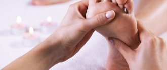
The massage during the first procedures is light and differentiated. The purpose of massage for obstetric paralysis is to improve tissue nutrition and innervation of the paretic arm. The massage is general, of the whole body, but the emphasis is on the shoulder girdle, upper limbs, and neck. The impact should be careful and painless. In the first period, up to 2 months, all manipulations with the baby’s hands should be gentle.
Gymnastics Therapeutic gymnastics for Erb-Duchenne's paresis is of utmost importance in restoring strength and movement in the affected limb. Exercises with such children should be performed by an experienced specialist who knows the characteristics of the disease. Weights For better strengthening, you can use a weight on the injured limb
It is important to choose the optimal weight so that the child can move the handle. The weight can be set at 10-20 grams at first, and gradually increase. Peculiarities of caring for a child diagnosed with Duchenne Erb's paresis. The principle is this: the younger the child, the more careful
The injured limb should not be subjected to sudden movements, stretching, pressure, hanging freely, or dangling. This must be taken into account when swaddling, bathing, feeding, transporting, etc. Styling is done according to the doctor’s recommendations. Prognosis Of course, all parents are concerned about the prognosis of treatment. With Duchenne Erb's paresis, the prognosis is favorable, i.e. children usually gradually recover, but at different speeds, and most children after a year use both hands. Regarding a complete recovery, so that nothing is noticeable at all and I can’t say for sure any restrictions in the movement of the arm, because... It was not possible to observe such children over several years to monitor their development. I think that if there are no repeated requests after 1-2 years, then this problem does not bother the child and parents. Erb Duchenne palsy has a worse prognosis than paresis, but even in such children there is a partial recovery. Much depends on how severely the plexus nerves are affected, and to a lesser extent on massage therapists, doctors and parents. Answers to frequently asked questions from parents: How long does it take to treat Duchenne Erb's palsy in newborns? The first year of a child’s life should be the most intense with treatment procedures, and most of the results from treatment should occur after the first year. The second year, if there is still a need, is usually less intensive in treatment. Therefore, the “bet” must be made on the first 2 years of a child’s life, and especially on the first. Then recovery is difficult and slow. It’s impossible to give exact dates just based on your statistics. Is full recovery possible? It is possible, but it depends on how severe the lesion is and what treatment was given. It is also very important to find good specialists. What happens if you can’t cure your hand? This happens with obstetric paralia with complete rupture of nerve plexuses and trunks. The arm will have to be fixed all the time so that the capsule of the shoulder joint does not stretch, and, accordingly, secondary problems may arise over time: scoliosis, hypotrophy, atrophy. What other treatment is possible for Erb's paresis of the arm? The principle is this: everything that causes the passage of impulses along the nerve trunks of the hand and the plexus should be used, of course, within reasonable limits. Acupressure, acupuncture, classical massage, exercise therapy, etc. It is necessary to stimulate the processes of conducting nerve impulses in the hand as much as possible.
If your child requires a qualified children's massage therapist, call us at 84993941711 or 89266057470.
If you were interested in this article, please share it on social networks, perhaps it will be useful to someone else. Mommies always have a lot of questions, let's help them find the answers!?
Diagnostics
Diagnosis of obstetric paralysis is usually not difficult. Difficulty in making the correct diagnosis can occur when the patient comes under observation after a long period of time, and patients with obstetric paralysis usually turn to orthopedists at a later stage, when secondary changes have already developed. In this case, polio may be suspected, especially if there is an epidemic outbreak.
Anamnesis is of decisive importance: with polio, the appearance of paralysis is preceded by the child’s anxiety and high temperature. Obstetric paralysis is detected immediately after childbirth. A serological blood test for polio can help make the correct diagnosis.
It is also necessary to differentiate obstetric paralysis from a fracture of the clavicle and shoulder. With a fracture of the clavicle and shoulder, local pain is sharply expressed, and movements in the hand and fingers are preserved, which usually does not happen with paralysis.
Complications
Of the complications of obstetric paralysis, the first place is taken by contractures, which develop quite early as a result of muscle imbalance. Contractures, becoming stable, fixing the limb in a vicious position, can themselves turn the child into a severe cripple, while the symptoms of paralysis have completely disappeared.
A fairly common complication of obstetric paralysis of the upper limb is posterior subluxation or dislocation of the head of the radial bone, which can cause limited mobility in the elbow joint, as well as pronation and supination of the forearm.
With age, there is a lag in the growth of the paralyzed arm, deformation of the head of the humerus, the articular cavity of the scapula and the acromion - a hook-shaped bend of it anteriorly. Sometimes there are respiratory disorders (asphyxia, shortness of breath), indicating concomitant damage to the phrenic nerve.
Treatment
Treatment for obstetric paralysis should be started as early as possible once the diagnosis is made. The results of treatment largely depend on this.
First of all, the paralyzed arm is given a position that provides rest and the most favorable conditions for restoring conduction and regenerating the damaged nerve. This position is abduction of the shoulder 90° relative to the body and its external rotation, bending the elbow at a right angle; the forearm is in supination, the fingers are extended (the “voting” pose). The arm is held in this position in the newborn with pads, by tying the arm to the crib, or with a splint. For this purpose, the best tires are made of plastic (polyethylene, vinyl plastic); they are durable, at the same time light, hygienic, easy to clean, and easy to manufacture.
As an initial aid, arm fixation can be applied by sewing the shirt sleeve in several places to the pillow on which the child lies in the “voting” position.
Among the medicines indicated: prozerin intramuscularly, dibazol.
Physiotherapeutic treatment of obstetric paralysis consists of thermal procedures, massage and gymnastics. Massage should be applied in the form of stroking and light rubbing, avoiding severe irritation. At first, massage sessions should be short (3-5 minutes). Gentle warmth: water baths 37°C, blue light (Minin lamp). At a later date (after 2-3 months), electrical stimulation of muscles with pulsed current under the supervision of a physiotherapist, without causing muscle fatigue.
To prevent contractures, passive movements of the paralyzed arm are performed.
Conservative treatment of obstetric paralysis in most patients gives quite satisfactory results, but in some cases it is necessary to resort to surgical interventions.
Of the surgical methods, we should first of all mention operations on the brachial plexus, in order to free the nerves from scars and restore the anatomical integrity of the roots. They, like other surgical interventions, should be used only after all available means of conservative therapy have been used.
This is followed by various methods of muscle transplantation to replace the function of paralyzed people; this includes: Moore's operation for Erb's palsy to improve the function of the deltoid muscle, in which part of the preserved anterior portion of this muscle is transplanted along with the outer edge of the acromial process posteriorly to the scapular spine. Transection of the subscapularis tendon according to the North. Transplant m. subscapularis on the tendon of m. teres minor.
Muscle transfer is only indicated in cases where the muscle being transplanted has sufficient strength, otherwise it is useless. As a result, muscle grafting for birth paralysis is not widely used, due to the limited possibility of using muscles suitable for transplantation due to the prevalence of paralysis in severe cases.
With complete paralysis of the deltoid muscle and good function of the shoulder girdle muscles, arthrodesis of the shoulder joint is more promising in terms of functional results than muscle plastic surgery. The purpose of the arthrodesis operation is to fuse the humerus with the scapula and ensure active abduction of the shoulder due to the movements of the scapula. After a successful operation, patients quickly begin to move their arm, raising it to the level of the shoulder girdle.
Particularly good results are obtained in cases where the muscles of the shoulder and forearm are not affected, but even in cases of paralysis of all the muscles of the arm, due to the restoration of shoulder movements, the performance of the entire previously helplessly hanging arm improves: the patient is able to hold objects by pinching them between the shoulder and body, hold the paper, notebook while writing, press the ruler when drawing.
With paresis of the muscles of the shoulder and forearm after arthrodesis of the shoulder joint, along with improvement in arm function, the strength of the paretic muscles increases, and therefore children begin to gradually master self-care skills (comb their hair, dress themselves, etc.).
For a successful operation, it is necessary to carefully remove the cartilage cover from the arthrodesed surfaces of the head of the humerus and the glenoid cavity of the scapula, ensure close contact and strong retention in their given position.
The latter is achieved by driving a nail through the head of the humerus into the glenoid cavity of the scapula or through the acromion process of the scapula, the lower surface of which is previously freshened.
After removing the plaster cast, it is advisable to wear your arm on a wedge-shaped cushion for 2-3 months, gradually reducing its thickness. At this time, treatment with massage and gymnastics is carried out.
Treatment of obstetric paralysis
Early treatment for obstetric paralysis promotes better results, so it is prescribed while the child is still with the mother in the maternity hospital. Further treatment is carried out in the neurology department.
The appointments are as follows:
- immobilization of the affected upper limb;
- massage;
- physiotherapeutic treatment;
- drug therapy.
Immobilization is carried out as follows. The child’s injured limb is carefully extended as far as it will go (it should be moved away from the body and turned with the palm outward) and fixed. The abduction is carried out in stages, a few centimeters each time, then fixed in this position. The procedure is carried out until the arm is abducted at an angle of 90 degrees. During physiotherapeutic treatment, the splint (called an abduction splint) is removed and then reinstalled. When the limb is abducted, the child should not feel pain - otherwise he will “signal” it by crying.
note
Massage for paralysis is performed to prevent congestion in the affected upper limb (in particular, during the installation of an abduction splint). It is carried out in the form of courses, which are repeated after a short pause. Massage is prescribed for a long time.
Of the physiotherapeutic methods of treatment, thermal procedures have proven themselves to be the most effective. Most often used for this disease are:
- paraffin applications;
- UHF;
- Microwave
- electrophoresis with anticholinesterase drugs and antispasmodics.
The following are prescribed as drug treatment:
- injectable antichoinesterase drugs;
- B vitamins. They are also used in the form of injections.
Forecast
The prognosis for obstetric paralysis of the upper limb depends on the nature and severity of damage to the nerves or their roots, on the time elapsed since the injury, and on the presence of secondary contractures. Fortunately, milder injuries are more common and usually respond quickly to conservative treatment.
At the same time, these mild cases initially do not differ in any way from cases that lead children to disability, so the prognosis is made with caution.
Persistent unevenness of the pupils indicates a poor prognosis.
Conservative therapy
All therapeutic measures to eliminate obstetric paralysis are traditional, but for each patient a set of necessary measures is individually selected, based on the location of the damage and the degree of its severity. From the day of birth until complete recovery, the child must be under the mandatory supervision of a neurologist.
Treatment with medications includes injections of B vitamins, in particular vitamins B1 and B12, proserine, and aloe. The dosage of drugs must be strictly observed and correspond to the patient’s age. In most cases, these medications are combined with antispasmodic drugs, namely: Eufillin and Papaverine. In some cases, doctors use nootropic drugs in combination with other therapy to stimulate nervous processes.
To relax the shoulder girdle, a plaster splint or abduction splint is often applied. In this case, the shoulder is retracted back or to the side, in accordance with the affected area.
Electrophoresis and mud applications are effective in the treatment of obstetric paralysis. If, along with this pathology, the child has torticollis, then applications should be applied to the collar area too. In physiotherapy, it is effective to use paraffin and hot wraps.
Massage for obstetric paralysis plays an important role in therapy. For children it is developed using a special method. It is important to remember that only a highly qualified and trusted specialist should perform such a massage on a newborn from 0 years old.
Prevention
Preventive measures for obstetric paralysis should be aimed primarily at familiarizing those involved in obstetrics (midwives) with complications during childbirth in the form of fractures, dislocations and paralysis that occur as a result of the use of excessive physical force and improper techniques during obstetrics.
Further preventive measures come down to early detection of obstetric paralysis and early treatment. For this purpose, it is necessary to train the staff of maternity hospitals and maternity wards of hospitals (pediatricians, obstetricians) in the early diagnosis of injuries, in particular paralysis.
Children suffering from obstetric paralysis should be registered for further clinical observation and treatment.
The information presented in this article is intended for informational purposes only and cannot replace professional advice and qualified medical care. If you have the slightest suspicion that your child has this disease, be sure to consult a doctor!
Signs of obstetric paresis
With obstetric paresis, there is a significant decrease in muscle tone (muscle hypotonia). As a rule, the affected limb hangs along the body, all joints are straightened. Muscle hypotonia in the paretic arm is pronounced. Active movements are completely absent, only slight movements in a supine position are possible. The skin of the hand affected by paresis is pale and cold to the touch. Early development of muscle atrophy, especially in the distal regions. Reduced pain threshold and temperature sensitivity throughout the entire limb. There are no tendon reflexes. The hand lacks grasping and palmo-oral reflexes.
The degree of muscle damage varies: from a slight decrease in muscle strength and tone to deep functional disorders, characterized by a complete absence of active movements. Identification of the localization and depth of motor disorders is necessary for correctly informed implementation of therapeutic measures.
What to do if your child has obstetric paresis?
If obstetric paresis is detected in a child, you must immediately contact a specialist. At the first stage, the doctor prescribes an examination of the damaged limbs to determine the degree of damage to the nerve roots. Based on the examination results, the doctor prescribes a course of treatment: conservative or surgical.
Consultations and surgical treatment are carried out by reconstructive microsurgeon Mikhail Leonidovich Novikov.
You can get preliminary organizational consultation and ask questions by calling 8-800-555-84-21 or leaving a message in the on-line consultation form in the right column of the site.
Surgical treatment is provided free of charge with the support of Rusfond. In order for treatment to be free, you need to collect documents according to the list, send them for verification and registration (can be scanned in color by email: [email protected] ), then wait for a call for treatment.