Due to the general “aging” of the planet and the increase in the number of senile and elderly people among the population, vascular diseases of the brain are becoming more important.
Decreased memory, dizziness, tinnitus, headache, decreased performance and high fatigue - all these are the first symptoms of vascular diseases of the brain of one origin or another.
However, doctors and patients do not pay much attention to vascular diseases of the nervous system, which are most often a manifestation of chronic circulatory failure.
How does the blood supply to the brain work?
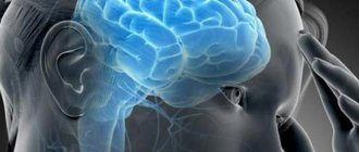
The brain is the main part of the central nervous system.
The average weight of the adult brain is 2-2.5% of his body weight (1020-2000 grams). The brain consumes about 20% of the circulating blood, glucose and oxygen received during the body's breathing. It has been known since ancient times that the functioning of brain neurons requires proper nutrition and a large amount of energy. With the help of blood, neurons receive all the necessary components, since the brain has intense blood circulation and low mass.
The blood circulation of the brain has a complex structure and is divided into venous and arterial systems. This is a complex mechanism that ensures non-stop blood circulation, optimal perfusion and blood flow volume. To increase the stability of blood circulation, protective collateral compensation of blood flow is provided through the vascular membranes and the arterial circle of the base.
In difficult situations, the brain is protected from lack of blood circulation (if it becomes difficult or stops) by the circle of Willis.
Brain vessels
Through its work, when such a situation occurs (during an ischemic attack or stroke), self-compensation is observed in the basin of one vessel due to the flow of blood from other vessels.
It is known that the brain requires a large volume of oxygen and nutrients. Neurons cannot accumulate and store them, so when blood flow stops, the available supply will last for 10 seconds. After this, the person loses consciousness and after 3-8 minutes the neurons die.
Additionally, in addition to compensation, blood flow has a self-regulation function, when it maintains a stable state, reducing dependence on changes in blood pressure and cardiac output volumes.
The stability of blood flow is regulated by the carotid sinuses (nerve cells located in the carotid arteries), which contain chemo- and baroreceptors. The carotid node transmits signals to the brain stem (to the respiratory and vasomotor centers), where cardiac function, vascular tone, etc. are regulated.
Subarachnoid bleeding
Subarachnoid hemorrhage (SAH) is a life-threatening condition that results from damage to blood vessels in the brain. Despite all the efforts of doctors, it ends in death. This type of bleeding is not common, but it is very unpredictable.
The main manifestation is an extremely severe headache, sometimes associated with nausea, vomiting or loss of consciousness. Symptoms may initially be less expressive and correspond to meningeal syndrome caused by irritation of the meninges. These include:
- headache;
- nausea;
- stiffness in the neck;
- vomiting;
- photophobia.
If bleeding does not stop, gradual deterioration and death occur over a short period (maximum 1 hour). If the bleeding stops, a relatively long asymptomatic period may occur, which usually ends with rebleeding.
The prognosis is very poor - without treatment, about 50% of victims die during the first hemorrhage, the next 50% from repeated hemorrhage.
Subarachnoid bleeding is much more common in the age group of 35–50 years. Early CT findings and careful evaluation are critical to determining the correct diagnosis. CT should precede possible cerebrospinal fluid examination, which is usually performed when bleeding is suspected and is unclear on CT.
What should alert you - clinical presentation and symptoms
Brain circulatory disorders do not immediately appear; in the initial stages there are no symptoms. Common symptoms of vascular disorders of the brain include headaches, regular dizziness, memory and sleep disturbances, weakness, deterioration of motor coordination, frills, numbness of the limbs, touchiness, and irritability.
In the later stages of the pathology, gait disturbance and false urge to urinate appear. In the absence of appropriate treatment, mental abilities, coordination of movements, and the functioning of the pelvic organs weaken.
Clinical symptoms appear after heavy physical and mental work, staying in a stuffy room, in a state of hunger and when changing meteorological factors, in the case of consumption of harmful foods or consumption of alcoholic beverages.
Also, vascular diseases can manifest themselves with a hereditary predisposition and a sedentary lifestyle.
The first signs of insufficiency of cerebral blood supply
The first signs of cerebral circulatory disorders include symptoms of vascular disease, as well as tinnitus and headaches, dizziness when walking or sudden changes in position, memory impairment, sleep disorders and decreased performance.
These symptoms appear several times a week for 3 months. Such symptoms develop with a reduced blood flow rate from 55 ml to 45 ml per 100 g/min. But when examining the neurological status, signs of focal damage to the nervous system are not determined. During a neuropsychological analysis, a specialist determines slow thinking as a result of solving complex issues.
Cerebrovascular diseases
The most common causes of brain dysfunction are vascular diseases. They are characterized by a narrowing of the lumen of the veins and arteries of the head and neck, their thrombosis, and a decrease in the permeability of the walls. All of them lead to poor blood flow to the nervous tissue.
- Aneurysmal vascular disease. It is a consequence of decreased muscle tone in the walls of blood vessels. It is characterized by the appearance of bag-like protrusions or expansion of the lumen of blood vessels by more than two times. It most often affects the arteries, as they are most susceptible to thinning and stretching. The cavity thus formed begins to fill with blood and increases in size, gradually putting additional pressure on the brain tissue. At the initial stage it leads to the development of headaches. Aneurysm rupture is always accompanied by cerebral bleeding and blood entering the subarachnoid space.
- Vegetative-vascular dystonia. Characterized by systematic increases and surges in blood pressure. It is a consequence of a dysfunction of the autonomic nervous system, due to which the brain ceases to control the tone of the walls of blood vessels. Unstable blood flow in arteries and veins leads to thinning of their walls, loss of elasticity, narrowing of the lumen and deformation. All this can further provoke a stroke.
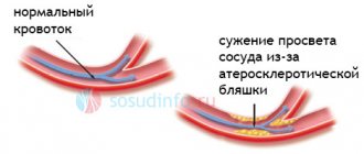
- Atherosclerosis. Develops against the background of insufficient lipid metabolism. Because of this, cholesterol plaques form on the walls of blood vessels, which clog their lumen. Their permeability also decreases, they become brittle and less elastic.
- Thrombosis of arteries and veins. An increase in blood viscosity leads to the formation of blood clots. Subsequently, the detached clot can block the flow of blood, which will cause a cessation of blood supply to the affected area of the brain.
The cause of insufficient cerebral blood supply may be cervical osteochondrosis - progressive damage to the intervertebral discs in the cervical spine. With this disease, due to the destruction of bone tissue, the arteries and veins that supply the brain are compressed.
Micronutrient deficiency leads to oxygen starvation, so cervical osteochondrosis in advanced form usually causes fainting and dizziness.
Compression of blood vessels and their destruction can be caused by tumors, cysts and TBI.
Causes and risk factors
In normal condition, the vessels of the head, which supply the brain, constantly narrow and expand in time with the contractions of the heart muscle. Due to this, oxygen-enriched blood reaches the brain structures as quickly as possible. Failure in these processes, namely vasospasm and other disorders of the circulatory system lead to oxygen starvation.
Any cerebrovascular disease develops under the influence of several factors. They may be: an increase in blood viscosity, metabolic disorders, hereditary predisposition or an unhealthy lifestyle.
The most common cause of cerebral bleeding, and as a consequence of damage to the brain substance, is a decrease in blood flow against the background of an increase in its viscosity. This is caused by hereditary diseases, due to which a person has a predisposition to sticking (aggregation) of platelets, insufficient drinking regimen, and alcohol abuse.
Mechanical damage to the walls of blood vessels is often a consequence of hypertension. In this case, they cannot withstand a sharp increase in blood pressure. This often results in spasm and paralysis of the arteries and veins, which can also lead to cerebral bleeding.
Disturbances in the metabolism of tissue in the walls of blood vessels can be caused by drinking alcohol and smoking. As a result, they lose their elasticity and become brittle, which, combined with surges in blood pressure, can lead to their rupture.
Also common causes of vascular diseases are:
- Excess weight;
- Insufficient physical activity;
- Inactive lifestyle;
- Disturbances in sleep-wake patterns;
- Unhealthy food.
Impaired elasticity of the walls of blood vessels may be a consequence of genetic predisposition. At the same time, an adequate approach: diet, healthy lifestyle, etc., can adjust the risk of developing vascular diseases of the brain.
General symptoms of cerebrovascular diseases
Although the above vascular diseases of the head and neck differ significantly from each other, they all lead to an insufficient supply of nerve tissue with nutrients.
Therefore, common symptoms and signs can be identified for them:
- Sleep disturbance;
- Dizziness, headaches;
- Deterioration of memory processes;
- Decreased intellectual abilities;
- Apathy, fatigue;
- Numbness of hands and limbs.
Often, against the background of impaired blood circulation in the brain, necrosis of the nervous tissue or its organic damage begins. This becomes especially noticeable in older people: they often cannot remember what happened some time ago, characteristic habits change, irritability appears, which is often replaced by apathy or even indifference.
In especially severe cases, vascular diseases of the brain can cause movement disorders: tremors of the limbs, unsteady or shuffling gait.
Chronic circulatory failure - DEP
Chronic cerebrovascular insufficiency is a common type of cerebrovascular disease (with primary brain damage and secondary damage to the vascular system).
Discirculatory encephalopathy is the first stage of chronic failure, characterized by small-focal brain damage due to a smaller volume of blood supply (by 15 ml/100 g per minute) and can cause micro-strokes, hypoxia, and brain atrophy.
As a rule, the development of dyscirculatory encephalopathy occurs as a result of vascular disorders in the brain. There are venous, mixed and atherosclerotic forms of pathology.
Based on an assessment of the degree of neurological disorders, dyscirculatory encephalopathy is divided into the following degrees:
- First stage . Stage 1 DEP is characterized by decreased memory and attention (with impaired memorization of new information), decreased performance, rapid fatigue, and difficulty switching attention from one event to another. Prolonged mental stress can cause dull headaches and sleep disturbances with periodic dizziness.
- Second stage . At the second stage of DEP, personal changes are revealed (viscosity of thinking, increasing memory impairment, resentment, selfishness, irritability, narrowing of interests, decreased ability of associative thinking, generalization and abstraction). There is intermittent, short sleep, dull headaches, instability and dizziness. In addition to anisoreflexia and pseudobulbar factors, vestibulo-cerebellar disorders, decreased social adaptation and work ability are determined.
- Third stage . Symptoms of stage three DEP worsen, manifesting weakness, impaired control of the sphincters of the pelvic organs, a combination of headaches with memory disorders and dizziness.
Diagnosis and treatment
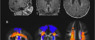
Diagnosis of DEP is based on clinical information and additional analyzes of the brain and vascular system. In the fundus, pallor of the optic nerve head, vascular atherosclerosis are determined, palpating of compacted and twisted temporal arteries, rheoencephalography, Doppler ultrasound, MRI, and ultrasound angiography are performed.
When initial signs of pathology appear, periodic therapeutic courses should be carried out. In accordance with the somatic status and manifestations of the pathologist, drug treatment is prescribed:
- vasoactive agents (Vinpocetine, Cavinton, Cynarizine, etc.);
- antiplatelet agents (Curantil, Acetylsalicylic acid);
- antisclerotic drugs;
- neuroprotectors and nootropics;
- tranquilizers;
- antihypoxants;
- vitamins E and B.
Blood pressure is controlled, ACE inhibitors are prescribed (Quadropril, Captopril), nicotinic acid drugs are recommended to improve blood circulation, and statins (Simvastatin, Atovastatin) are recommended to correct the lipid profile.
If you have cerebrovascular insufficiency, you should avoid overheating, climbing mountains, smoking and drinking alcohol, watching TV and using a PC for long periods of time.
Results
Local humoral (metabolic) autoregulatory mechanisms predominate in the control of cerebral circulation. Hypoxia and hypercapnia cause vasodilatory effects. Blood flow in the brain is determined by pressure gradient and peripheral resistance. Thanks to the effective self-regulating myogenic mechanism, the blood supply to the brain and its nutrition remain unchanged even with significant changes in pressure. This prevents dysfunction.
When the vascular system is disrupted, cerebrovascular diseases can develop, the symptoms and treatment of which are an important problem not only from a medical, but also from an economic, ethical and social point of view. Opportunities for improving the situation lie mainly in improving the organization of care, providing intensive care in the acute stage of the disease, clarifying and speeding up diagnosis, and understanding the importance of prevention (physical activity, quitting smoking and alcohol, diet).
Classification and types of vascular diseases of the brain
Vascular diseases of the brain are classified as follows:
- initial signs of circulatory failure;
- transient disorders (ischemic attacks, cerebral hypertensive crises, acute hypertensive encephalopathy);
- persistent circulatory disorders (hemorrhagic, ischemic strokes, consequences of a previous illness);
- progressive cerebral circulatory disorders (non-traumatic chronic subdural hematoma, dyscirculatory encephalopathy).
Acute circulatory disorders are classified as secondary diseases. The main causes of their manifestations are atherosclerosis and hypertension. Sometimes strokes can occur as a result of vasculitis (allergic, infectious, syphilitic), systemic lesions of connective tissues, congenital pathologies of cerebral vessels, systemic blood disorders (coagulopathy, erythmias, leukemia).
The course of many diseases is complicated by transient ischemic attacks, which often develop acutely or prolonged.
Stroke is treated in an inpatient setting to reduce mortality. Diagnosis is carried out using computed tomography of the brain. In this case, MRI better reveals brain swelling, recognizing the appearance of small infarcts. Echoencephaloscopy and angiography can be performed.
The basis for the treatment of strokes is a combination of thrombolytic treatment with TRAs and neuroprotectors; at later stages, therapy with antioxidants and antiplatelet agents is carried out.
Vascular diseases of the spinal cord: treatment and diagnosis
The diagnosis and treatment of vascular pathologies of the spinal cord is usually carried out by a neurologist. At the initial appointment, he collects a complete medical history, asks the patient about chronic and hereditary diseases, about troubling symptoms and the period in which they appeared. Based on the clinical picture, we can assume the presence of damage to any vascular system. The patient usually does not require laboratory examination. Most often they resort to the following instrumental diagnostic methods:
● magnetic resonance imaging (MRI) of the spinal column. It is recognized as the most effective (sensitive) imaging technique for spinal cord disease. With its help, it is possible to diagnose soft tissue lesions (for example, neoplasms or abscesses), compression fractures, spinal cord parenchyma and other pathologies;
● myelography (with radiopaque agents). This technique is used less frequently than MRI, since it is invasive and not as informative. With its help, it is possible to detect lesions of the bone apparatus;
● electrophysiological studies. They are based on recording impulses of bioelectrical activity of the brain and nerve trunks. It allows you to accurately assess the degree of damage to the nerve pathways;
● spinal angiography. This is a lengthy examination in which the patient is given a large dose of contrast agent. It allows you to detect vascular disorders (in particular, aneurysms, occlusions and compressions, aneurysms);
● aortography. This diagnostic method is required in exceptional situations when there is a suspicion of an aneurysm, aortic occlusion or aortic valve insufficiency;
● echocardiography (EchoCG). Additionally, this non-invasive technique may be prescribed if the specialist suspects cardiac pathologies. With its help, intracardiac hemodynamics are assessed and many disturbances in the functioning of the muscular organ are detected;
● lumbar puncture. This is a procedure in which a specialist inserts a puncture needle into the subarachnoid space of the spinal cord (in the area of the spinal column). Further examination of the taken biomaterial allows us to detect signs of hematomyelia, excluding the infectious-inflammatory nature of myelopathy.
Detailed diagnostics are required to differentiate vascular diseases of the spinal cord from multiple sclerosis, spinal cord neoplasms, compression and infectious myelopathy. After a thorough examination, we proceed to prescribing the most appropriate therapy. Attempts at self-medication can lead to a number of negative consequences, so you should trust in the hands of professionals. Vascular diseases of the spinal cord are treated with two main methods - conservative and surgical. Let's take a closer look at the features of each method separately.
General treatment concept
Treatment of vascular diseases of the brain and cerebrovascular disorders can be carried out by a therapist, neurologist and cardiologist. Complex therapy is often carried out, consisting of nutritional correction, the use of drugs and traditional methods. Sometimes surgery is prescribed.
Drug treatment
In each case, individual treatment is prescribed with certain drugs and their dosages are selected.
Popular means include:
- fibrates (Lipanor, Fenofibrate);
- statins (Simvastatin, Zocor);
- antioxidants;
- vasodilators (Eufillin, Papaverine);
- drugs to improve metabolic processes and blood circulation (Vasobral, Cavinton);
- Antiplatelet, decongestant, anti-inflammatory drugs, and glucocorticoids can also be prescribed.
For the treatment of atherosclerosis, angioprotectors (Anginin, Prodectin, Stugeron), anticoagulants (Pelentan, Sincumar, Heparin), antioxidants, vitamin-mineral complexes, cholesterol-lowering drugs (Thiamin, Pyridoxine, Diosponin) are prescribed.
Treatment and support of blood vessels using traditional methods
Among traditional methods, products based on garlic (oil, milk or alcohol tinctures), hawthorn and sea buckthorn oil will be effective.
Recipes for preparing medicinal compositions:
- To prepare garlic milk, take 2 tablespoons of peeled garlic cloves, put them in a saucepan and pour a glass of milk. Cook over low heat until the slices are soft. The milk is drained and taken one tablespoon at a time before meals.
- A large head of garlic is crushed with lemon , mixed and poured with a liter of water. The composition is infused and refrigerated, taking 50 grams before meals.
- A glass of hawthorn fruit is poured into 0.5 liters of boiling water, simmered over low heat for several minutes, filtered and mixed with 2 tablespoons of honey. Take 2 spoons at night and 1 spoon before meals.
- Sea buckthorn oil is taken every day, a teaspoon half an hour before meals, 3 times a day for 21 days.
Surgical intervention
Intravascular and intracranial operations are performed through the frontal-temporo-basal area of the skull using various clips and catheters.
To eliminate ischemic lesions, endarterectomy and thrombectomy are performed. Prevention of progressive ischemia due to narrowing or occlusion of the arteries is carried out by revascularization operations (anastomoses between arterial branches in the soft tissues of the head).
Diagnostics
Problems with the blood vessels of the head and neck can begin at any age, so everyone should know the main symptoms of the pathology. This information can subsequently help in diagnosing pathology.
Ultrasound. In this case, the standard examination procedure is ineffective, so specialists conduct the following types of examination of cerebral vessels: duplex scanning, Dopplerography, echotomography or transcranial Dopplerography. Neurosonography is performed in infants.
CT or MRI. With their help, a diagnostician can determine the area of damage to the brain substance, the size of the area of necrosis and the condition of the organ as a whole.
Angiography. Allows you to detect areas of obstructed cerebral circulation in the head, the degree and order of filling of blood vessels. Using it, you can obtain data on the presence of additional blood routes when the main arteries and veins are blocked.
Rheoencephalography. This method of studying the vascular system of the brain is based on recording the electrical resistance of nervous tissue when a weak high-frequency electric current is passed through it. After decoding, the specialist receives information about the elasticity of the walls of blood vessels, the presence of neoplasms and aneurysms.
Preventive measures
To avoid vascular diseases and concomitant cerebral circulatory disorders, preventive measures should be taken against atherosclerosis and hypertension.
It is necessary to adjust nutrition, remove fatty products of animal origin, smoked, fried and salty foods, sweets, processed foods, and carbonated drinks from the diet.
You need to add fish, seafood, fresh fruits, herbs, and vegetables to your diet. You should maintain a normal weight, stop smoking, alcohol, often stay in the fresh air, and exercise.
You should also follow a drinking regime (drink at least 1.5 liters of water per day) and limit the effects of stress.